
| Admissions | Catalog | Contact Us | Examinations | Grants | Instructors | Lecture | Membership | Students login | Schools | Colleges | Universities | Professional Examinations | Recommendations | Research Grants | Search | Librarians | Forms | Booksellers | Continents/States/Districts | Contracts | Volunteer |
|
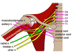
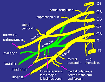
The Brachial Plexus is formed by the anterior division of the fifth, sixth, seventh, and eighth cervical nerves, and the greater part of the first thoracic nerve. The second thoracic nerve, although not part of the plexus, helps in the innervation of the arm through the intercosto-humeral nerve.
The nerves forming the plexus appear in the posterior triangle of the neck, and, passing between scalenus medius and anticus with the subclavian artery, they accompany the axillary artery to the shoulder and upper limb. As the nerves enter the posterior triangle they form the three primary cordsFirst primary cord : Fifth and sixth nerves joined together. Second primary cord : Seventh nerve alone. Third primary cord: Eighth cervical and first thoracic joined together. As soon as the three cords are formed they each divide into anterior and posterior divisions to form the secondary cords, which are named according to their relation to the axillary artery. Outer cord: Anterior divisions of first and second primary cords. Inner cord: Anterior division of third primary cord. Posterior cord : Posterior divisions of all three primary cords. The nerves supplying the shoulder and arm are derived from these three cord-viz. : Outer cord: Musculo-cutaneous, outer head of median, external anterior thoracic nerves. Inner cord: Ulnar, inner head of median, internal anterior thoracic, internal cutaneous, and lesser internal cutaneous nerves. Posterior cord: Circumflex, three subscapular and musculo-spiral nerves. Before the nerves join up to form the cords, a few branches are given off which are called Supraclavicular nerves to distinguish them from the branches derived from the secondary cords which are called the Infraclavicular nerves. Supraclavicular Nerves Muscular branches to scaleni, sub-clavius, and longus colli. Posterior scapular supplies the rhomboids and levator anguli scapulae. Long thoracic supplies serratus magnus. It pierces scalenus medius and enters the axilla between the artery and serratus magnus. This nerve is also called the respiratory nerve of Bell. Suprascapular supplies supra- and infraspinatus and articular branches to the shoulder-joint. It passes down to the superior border of the scapula, then through the suprascapular foramen and winds round the great scapular notch. Infraclavicular Nerves The anterior set from the inner and outer cords supply the chest and front of the limbs, the posterior set of nerves from the posterior cord supply the shoulder and the back of the limb. Anterior Thoracic Nerves The external anterior thoracic nerve arises from the outer cord, and the internal one from the inner cord. They pass down one on each side of the axillary artery, and are finally distributed to pectoralis major and minor. Musculo-Cutaneous nerve from the outer cord lies first between coraco-brachialis and the axillary artery; it then lies between biceps and brachialis to the bend of the elbow. It becomes cutaneous between biceps and brachio-radialis, and ends by supplying the skin on the outer side of the forearm. Branches Muscular to biceps, brachialis anticus and coraco-brachialis (this last nerve is not really a branch of musculocutaneous, but is an independent branch from the sixth and seventh cervical nerves incorporated with it). Cutaneous Anterior branch supplies the outer half of the anterior surface of the forearm as far as the ball of the thumb. The posterior branch supplies the upper three-fourths of the oute r half of the posterior surface of the forearm. Nerves of the Upper Limb The Axillary Nerve * This nerve passes to the posterior aspect of the arm through the quadrangular space in the company of the posterior circumflex vessels. * On emerging from the quadrangular space, the axillary nerve winds around the surgical neck of the humerus to supply the teres minor and deltoid muscles. * The axillary nerve ends as the upper lateral brachial cutaneous nerve. * It supplies skin over the inferior half of the deltoid and adjacent areas of the arm. The Main Nerves of the Forearm The Median Nerve * This nerve enters the forearm with the brachial artery. * It lies on the brachialis muscle and passes between the two heads of the pronator teres muscle, giving branches to both. * Afterwards, it descends deep to the flexor digitorum superficialis muscle, closely attached by the muscle's facial sheath. * Then is continues between the two flexor digitorum muscles. * Near the wrist, it becomes superficial, passing between the tendons of the flexor digitorum superficialis and the flexor carpi ulnaris muscles, deep to the tendon of palmaris longus muscle. * The median nerve enters the hand through the carpal tunnel, between the tendons of flexor digitorum superficialis and flexor carpi radialis. * It supplies motor fibres to the 3 thenar muscles and the 1st and 2nd lumbrical muscles. Branches of the Median Nerve * There are no branches in the arm. * Articular branches pass to the elbow joint as the median nerve passes it. * Muscular branches supply the pronator teres, pronator quadratus, all flexors except flexor carpi ulnaris and medial half of flexor digitorum profundus (other half is supplied by the ulnar nerve). The Anterior Interosseous Nerve * This nerve arises from the median nerve in the distal part of the cubital fossa. * It runs between the flexor digitorum profundus and the flexor pollicis longus muscles to reach the pronator quadratus muscle. * It supplies all of these but only the lateral half of flexor digitorum profundus. * It then passes deep to the pronator quadratus muscle and ends by sending articular branches to the wrist joint. The Palmar Cutaneous Branch * This arises from the median nerve just proximal to the flexor retinaculum. * It becomes cutaneous between the tendons of the palmaris longus and flexor carpi radialis muscles. * It passes superficial to the flexor retinaculum to supply skin of the lateral part of the palm. Median Nerve Injury * Note: occasionally, there are communications between the median and ulnar nerves in the forearm. Usually, there are several slender nerves. * These are clinically important, as even with complete lesion of the median nerve, some muscles may not be paralysed. * This may lead to the erroneous conclusion that the median nerve has not been damaged. When Severed at the Elbow Joint * There is loss of flexion of the proximal interphalangeal joints of all the digits. * There is loss of flexion of the distal interphalangeal joints of the 2nd and 3rd digits (but not 4th and 5th). * The ability to flex the metacarpophalangeal joints is also affected because the median nerve supplies the 1st and 2nd lumbrical muscles. Severed Proximal to the Flexor Retinaculum (Wrist Slashing) * There is the inability to oppose the thumb owing to the paralysis of the thenar muscles. * There is the weakening of flexion of the metacarpophalangeal joints and extension of the interphalangeal joints of the 2nd and 3rd digits (paralysis of the 1st and 2nd lumbrical muscles). The Ulnar Nerve * This nerve passes posterior to the medial epicondyle of the humerus. * It enters the forearm by passing between the two heads of the flexor carpi ulnaris muscle. * It then descends deep to this muscle on the flexor digitorum profundus, where it accompanies the ulnar artery near the middle of the forearm. * Then it passes on the medial side of this artery and the lateral side of the tendon of flexor carpi ulnaris. * In the distal part of the forearm, the ulnar nerve become relatively superficial covered only by fascia and skin. * It pieces the deep fascia and passes superficial to the flexor retinaculum with the ulnar artery, lateral to the pisiform, between this bone and the hook of the hamate. * This passage for the nerve and artery, covered with a slip of flexor retinaculum, is referred to clinically as the canal of Guyon. Branches of the Ulnar Nerve * There are no branches in the arm. * Articular branches pass to the elbow joint, where the nerve is in the groove between the olecranon of the ulna and medial epicondyle of the humerus. * Muscular branches supply the flexor carpi ulnaris and the medial half of the flexor digitorum profundus muscles. The Palmar Cutaneous Branch * This arises from the ulnar nerve near the middle of the forearm. * It pierces the deep fascia in its distal 1/3 to supply the skin on the medial part of the palm. The Dorsal Cutaneous Branch * It arises from the ulnar nerve from the distal half of the forearm. * It passes posteroinferiorly between the ulna and flexor carpi ulnaris muscle. * It supplies the posterior surface of the medial part of the hand. The Superficial Branch of the Ulnar Nerve * This branch supplies the cutaneous fibres to the anterior surfaces of the medial one and a half digits. The Deep Branch of the Ulnar Nerve * This supplies the motor fibres to the hypothenar muscles, the medial two lumbrical muscles, the adductor pollicis muscle, and the interosseous muscles. * The deep branch of the ulnar nerve also supplies several joints (wrist, intercarpal, carpometacarpal and intermetacarpal joints). * The ulnar nerve is referred to as the nerve of fine movements. Ulnar Nerve Injury * This commonly occurs where the nerve passes posterior to the medial epicondyle of the humerus. * Often the damage occurs when the elbow hits a hard surface and the medial epicondyle if fractured. * There tends to be extensive motor and sensory loss to the hand. * Adduction is impaired, and when an attempt is made to flex the wrist joint, the hand is drawn to the radial side by the flexor carpi radialis muscle. * There is difficulty making a fist as the distal interphalangeal joints cannot be flexed. * When an effort is made to straighten the fingers, the hand goes into the position known as "claw hand". The Radial Nerve * The radial nerve descends in the arm between the brachialis and brachioradialis muscles. * It crosses the anterior aspect of the lateral epicondyle. * Soon after it enters the forearm, it divides into superficial and deep branches. The Superficial Branch * This is the smaller of the two branches and is the direct continuation of the radial nerve. * It passes distally, anterior to the pronator teres muscle and under the cover of the brachioradialis muscle. * In the distal 1/3 of the forearm, the superficial branch passes posteriorly, deep to the tendon of the brachioradialis muscle, and enters the posterior fascial compartment of the forearm. * It pierces the deep fascia 3 to 4 cm proximal to the wrist and supplies skin on the dorsum of the wrist, hand, thumb, and the lateral 1 (or 2) and a half digits. The Deep Branch * This is the larger of the two terminal branches and is entirely muscular and articular in its distribution. * As it passes posteroinferiorly, it gives branches to the extensor carpi radialis brevis and supinator muscles. * It then pierces the supinator muscle, giving additional branches to it, and curving around the lateral side of the radius to enter the posterior fascial compartment of the forearm. * On reaching the posterior aspect of the forearm, the deep branch of the radial nerve gives many branches to the extensor muscles. * One of these branches, the posterior interosseous nerve, accompanies the posterior interosseous artery and supplies the deep extensor muscles. Radial Nerve Injury * This may occur in deep wounds of the forearm. * Severance of the radial nerve results in the inability to extend the thumb and the metacarpophalangeal joints of the other digits. * There is no loss of sensation because the deep branch of the radial nerve is entirely muscular and articular in distribution. |