| Admissions | Ambassadors | Accreditation | A to Z Degree Fields | Books | Catalog | Colleges | Contact Us | Continents/States | Construction | Contracts | Distance Education | Emergency | Emergency Medicine | Examinations | English Editing Service | Forms | Faculty | Governor | Grants | Hostels | Honorary Doctorate degree | Human Services | Human Resources | Internet | Investment | Instructors | Internship | Login | Lecture | Librarians | Languages | Manufacturing | Membership | Observers | Publication | Professional Examinations | Programs | Professions | Progress Report | Recommendations | Ration food and supplies | Research Grants | Researchers | Students login | School | Search | Software | Seminar | Study Center/Centre | Sponsorship | Tutoring | Thesis | Universities | Work counseling |
Here are guidelines from Doctor Asif Qureshi.
| What must the head of the state healthcare know about a vaccine? |
Annotation or definition
|
What is a vaccine? How do vaccines work? What is vaccination? What is immunization? Why is immunization important? How effective are vaccines? How safe are vaccines? Where to get immunized When to get immunized Children's Vaccination: What is the schedule? When was it last updated? Who updated it? How do vaccines work? Are there any vaccine side-effects? Why is vaccinating so important? What are the ingredients / additives of vaccines? What vaccines do adults need? What vaccines do children need? What diseases do vaccines prevent? What are some of the common misconceptions about vaccinating? What would happen if we stopped immunizations? Does your pharmacy give shots? How do you know vaccines are genuine? How do you know vaccines won't cause harm? How do you know it's not a placebo? How do you confirm genuineness? What medical history is necessary before a vaccine is adminstered? What follow-up is necessary to measure the effectiveness of a vaccine? What are the various vaccines available in 2012? How are vaccines manufactured? How is the effectiveness of a vaccine verified? What are the dose, age and route of administration of each vaccine? Which vaccine is administered in left or right deltoid? Which vaccine is administered orally? |
|
What is a vaccine? A vaccine is a substance that primes the body’s immune system to make antibodies, T-cells and memory cells which are the body’s defense against infection. When you are vaccinated you actually build up your immune system, making you stronger and more resistant to disease as you grow. Vaccines are the best way to protect you and your family against some very serious infections. How do vaccines work? A vaccine contains a killed or weakened part of a germ that is responsible for infection. Because the germ has been killed or weakened before it is used to make the vaccine, it can not make the person sick. When a person receives a vaccine, the body reacts by making protective substances called "antibodies". The antibodies are the body's defenders because they help to kill off the germs that enter the body. In other words, vaccines expose people safely to germs, so that they can become protected from a disease but not come down with the disease. What is vaccination? Vaccination means having the vaccine - actually getting the injection. What is immunization? Immunization means both receiving the vaccine and becoming immune to ward off a disease as a result of immunization. Like eating well and exercise, getting immunized is a foundation for a healthy life. Immunizations help save lives, prevent serious illnesses, and are recognized as one of the most effective public health interventions. Immunizations help the body make its own protection (or antibodies) against certain diseases. Why is immunization important? When children are immunized, their bodies make antibodies that fight specific infections. If they are not protected and come in contact with one of these infections, they may get very sick and potentially experience complications, or even die. How effective are vaccines? Vaccines are very effective in preventing disease when given as recommended. However, no vaccine will work for 100 per cent of the children who receive it. Studies of disease outbreaks show that although some immunized children can develop the infection, the illness is often less severe. How safe are vaccines? All vaccines have to be tested to make sure they are both safe and effective. The most common side effects are mild pain, swelling and redness where the injection was given. Some infant vaccines may cause a low-grade fever (approximately 38°C) or fussiness for a day or two after the injection. Physicians may recommend acetaminophen to prevent fever and pain. Serious side effects from immunizations are rare. Please report any side effects or severe vaccine reactions to your health care provider or local public health unit. You should always discuss the benefits and risks of any vaccine with your health care provider. Children's Vaccination: What is the schedule? When was it last updated? Who updated it? Your child should get the first shots at 2 months of age (or in some cases before leaving the hospital after birth), then at 4 months, 6 months and 12-15 months of age. Remember, each of these visits is important! Your child must complete the series to be fully protected. More immunizations are recommended at 2 years and before school entry. Adolescents also need immunizations at 11-12 years of age, before entering 7th grade. 2 months old 4 months old 6 months old 12 months old 15 months old 18 months old 4 - 6 years old Grade 7 students Grade 8 females 14 -16 years old (10 years after 4-6 year old booster) Every year (in autumn) Vaccines that protect against the following diseases are available free of charge, and are required for attendance at school (unless there is a valid written exemption) : •Diphtheria is a very serious bacterial infection. It can cause breathing problems, heart failure, nerve damage and death in about 10% of cases. •Tetanus (Lockjaw) causes painful muscle spasms, breathing failure and can lead to death. It is caused by bacteria and spores in the soil that can infect wounds. •Polio can cause paralysis (loss of control over muscles in the body), inflammation of the brain and death. People get polio from drinking water or eating food with the polio virus in it. It is no longer common in Canada because of high immunization rates, but cases do occur elsewhere in the world and polio may be acquired when traveling if you are not fully immunized. •Measles causes rash, high fever, cough, runny nose and watery eyes. It can cause middle ear infection, pneumonia (lung infection), inflammation of the brain, hearing loss, brain damage and death. •Mumps causes fever, headache, painful swelling of the glands in the mouth and neck, earache and can cause inflammation of the brain. It can cause temporary or permanent deafness and swelling of the ovaries in women and testes in men, possibly leading to sterility. •Rubella (German Measles) causes fever, rash, swelling of the neck glands and swelling and pain in the joints. It can cause bruising and bleeding. If a pregnant woman gets rubella, it can be very dangerous for the unborn baby. Vaccines against the following diseases are recommended but not required for attendance at school. These vaccines are available free of charge : •Pertussis (Whooping Cough) causes severe coughing spells for weeks or months. It can also cause pneumonia (lung infection), middle ear infection, convulsions (seizures), inflammation of the brain and death. The risk of complications is greatest in children younger than one year of age. •Hepatitis B is a virus that can cause serious liver problems that can be fatal, such as liver failure and liver cancer. The vaccine is free for grade 7 students and certain high-risk groups (including infants born to mothers who are infected with hepatitis B and can pass the disease on to their babies). •Influenza (the Flu) is a viral infection that causes cough, high fever, chills, headache and muscle pain. It can cause pneumonia (infection of the lungs), middle ear infections, heart failure and death. The danger of this infection varies from year to year depending on the strain and can be mild to life-threatening. •Varicella (Chickenpox) is a highly contagious viral infection. It can cause fever, headache, chills, muscle or joint aches a day or two before the itchy, red rash appears. A pregnant woman with chickenpox can pass it on to her unborn baby. Mothers with chickenpox can also give it to their newborn baby after birth. Chickenpox can be very severe or even life-threatening to newborn babies. •Meningococcal disease is a very serious bacterial infection and a common cause of meningitis (infection of the lining of the brain and spinal cord) and meningococcaemia (severe infection of the blood) that can cause severe complications and death. •Human Papillomavirus (HPV) is a very common virus transmitted through sexual activity. HPV has been found to cause cervical cancer, some other rare cancers and genital warts. (About 75 per cent of adults will have at least one HPV infection in their lifetime.) The vaccine is free for grade 8 females. Vaccines against the following diseases are recommended for younger children. These vaccines are available free of charge : •Rotavirus is one of the leading causes of severe diarrhea in infants and children. Rotavirus is a very common and is easily spread from person to person. Rotavirus causes inflammation of the stomach and intestines (gastroenteritis). This vaccine is recommended for infants between 6 to 24 weeks of age. •Haemophilus Influenzae type b (HIB) is a bacteria that can infect any part of the body. It can cause middle ear infections, breathing problems, damage to joints, pneumonia (lung infection), inflammation of the brain leading to brain damage and death. This vaccine is recommended for children less than 5 years of age. •Pneumococcal disease is a bacterial infection that can cause serious illnesses such as pneumonia, blood infection and meningitis. The pneumococcal conjugate vaccine is now available free of charge in Ontario for the routine immunization of children less than 2 years old as well as high-risk children 2 to 59 months of age. Because of changes in the influenza (flu) strains, adults also need to receive the flu shot each year. Adults should continue to get the tetanus and diphtheria vaccine every 10 years throughout life, to be protected against these diseases. Thinking about getting pregnant? Be sure you are protected against rubella before pregnancy to protect your future baby from serious problems during its development. |
| Vaccinations/Injections |
| Vaccines |
| Vaccine |
| Adult Vaccines |
Signs and symptoms of flu
What are the symptoms of the flu? Here are further guidelines. |
Flu Vaccine
What are its constituents? Who manufactured these Flu Shots? How will this help to prevent Flue? Have there been reports of flue after Flue Shots? How is flu vaccine manufactured? How is the effectiveness of flu vaccine measured? Here are further guidelines. |
Types of vaccines
Types of microscopes
|
What are the types of microscopes? What is a magnification power? What is a light microscope? What is an electron microscope? What are the types of microscopes? Optical microscope Electron microscope Scanning probe microscope (SPM) Others Optical microscope Binocular stereoscopic microscope Brightfield microscope Polarizing microscope Phase contrast microscope Differential interference contrast microscope Fluorescence microscope Total internal reflection fluorescence microscope Laser microscope (Laser scanning confocal microscope) Multiphoton excitation microscope Structured illumination microscope
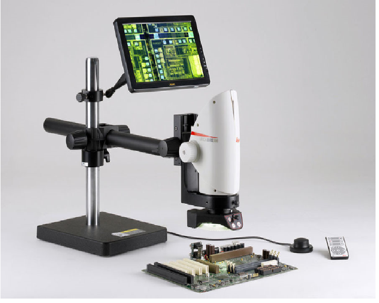
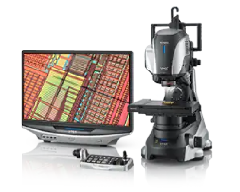
Exploring the Top Four and More There are several different types of microscopes used in light microscopy, and the four most popular types are Compound, Stereo, Digital and the Pocket or handheld microscopes. Some types are best suited for biological applications, where others are best for classroom or personal hobby use. Outside of light microscopy are the exciting developments with electron microscopes and in scanning probe microscopy. The Compound Light Microscope Commonly binocular (two eyepieces), the compound light microscope, combines the power of lenses and light to enlarge the subject being viewed. Typically, the eyepiece itself allows for 10X or 15X magnification and when combined with the three or four objective lenses, which can be rotated into the field of view, produce higher magnification to a maximum of around 1000X generally. The compound light microscope is popular among botanists for studying plant cells, in biology to view bacteria and parasites as well as a variety of human/animal cells. It is a useful microscope in forensic labs for identifying drug structures. Compound light microscopes are one of the most familiar of the different types of microscopes as they are most often found in science and biology classrooms. For this reason, simple models are readily available and are inexpensive. As well, several microscopy imaging techniques benefit scientists and researchers using the compound microscope and are worth exploring. The Stereo Microscope The Stereo microscope, also called a dissecting microscope, has two optical paths at slightly different angles allowing the image to be viewed three-dimensionally under the lenses. Stereo microscopes magnify at low power, typically between 10X and 200X, generally below 100x. With this type of microscope you generally have the choice of purchasing the fixed or zoom variety from a manufacturer and are relatively inexpensive. Uses for this type of microscope include looking at surfaces, microsurgery, and watch making, plus building and inspecting circuit boards. Stereo microscopes allow students to observe plant photosynthesis in action. The Digital Microscope Step into the 21st century with a digital microscope and enter a world of amazing detail. The digital microscope, invented in Japan in 1986, uses the power of the computer to view objects not visible to the naked eye. Among the different types of microscopes, this kind can be found with or without eyepieces to peer into. It connects to a computer monitor via a USB cable, much like connecting a printer or mouse. The computer software allows the monitor to display the magnified specimen. Moving images can be recorded or single images captured in the computer’s memory. An advantage of digital microscopes is the ability to email images, as well as comfortably watch moving images for long periods. The USB Computer Microscope Although not well suited to the same scientific applications as other light microscopes, the USB Computer microscope, among the different types of microscopes, can be used on almost any object and requires no preparation of the specimen. It is essentially a macro lens used to examine images on a computer screen plugged into its USB port. However, the magnification is restricted and is not comparable to your standard compound light microscope at only up to 200X with a relatively small depth of field. The Pocket Microscope In examining the different types of microscopes available on the market, the pocket microscope may be tiny but its abilities are impressive. This is a device which is a great gift for a child or your student. It is used by scientists for hand-held imaging of a variety of specimens/objects in the field or in the laboratory. It is small, durable and portable with a magnification ranging from 25x to 100x. There are many different models available. You may even want to check out the portable digital microscopes that are available now as this is an invaluable tool to aid in image sharing and analysis. The Electron Microscope v Among the different types of microscopes, the Electron Microscope(EM) is a powerful microscope available and used today, allowing researchers to view a specimen at nanometer size. v The transmission electron microscope(TEM), the first type of EM, is capable of producing images 1 nanometer in size. The TEM is a popular choice for nanotechnology as well as semiconductor analysis and production. A second type of electron microscope is the scanning electron microscope(SEM)are approximately 10 times less powerful than TEMs, they produce high-resolution, sharp, black and white 3D images. The Transmission Electron Microscopes and Scanning Electron Microscopes have practical applications in such fields as biology, chemistry, gemology, metallurgy and industry as well as provide information on the topography, morphology, composition and crystallographic data of samples. The Scanning Probe Microscope (SPM) Among the different types of microscopes and microscopy techniques, scanning probe microscopy is used today in academic and industrial settings for those sectors involving physics, biology and chemistry. These instruments are used in research and development as standard analysis tools. v Images are highly magnified and are observed as three-dimensional-shaped-specimens in real time. SPMs employ a delicate probe to scan the surface of the specimen eliminating the limitations that are found in electron and light microscopy. The Acoustic Microscope The Acoustic Microscope is less about resolution and more about finding faults, cracks or errors from samples during the manufacturing process. With the use of high ultrasound, this type of microscope is the easiest intra-cavity imaging tool available. It is a microscope that is under used primarily due to the fact that it is less known for its capabilities. Scanning acoustic microscopy, or SAM, is the most current type of acoustic microscopy available to today's scientists. They can use it to view a sample internally without staining it or causing it any damage thanks to point focusing technology, which relies on a beam to scan and penetrate the specimen while it is in water. What is a light microscope? •A light microscope is a biology laboratory instrument or tool, that uses visible light to detect and magnify very small objects, and enlarging them. •They use lenses to focus light on the specimen, magnifying it thus producing an image. The specimen is normally placed close to the microscopic lens. Microscopic magnification varies greatly depending on the types and number of lenses that make up the microscope. Depending on the number of lenses, there are two types of microscopes i. e Simple light microscope (it has low magnification because it uses a single lens) and the Compound light microscope (it has a higher magnification compared to the simple microscope because it uses at least two sets of lenses, an objective lens, and an eyepiece). The lenses are aligned in that, they can be able to bend light for efficient magnification of the image. •The functioning of the light microscope is based on its ability to focus a beam of light through a specimen, which is very small and transparent, to produce an image. The image is then passed through one or two lenses for magnification for viewing. The transparency of the specimen allows easy and quick penetration of light. Specimens can vary from bacterial to cells and other microbial particles 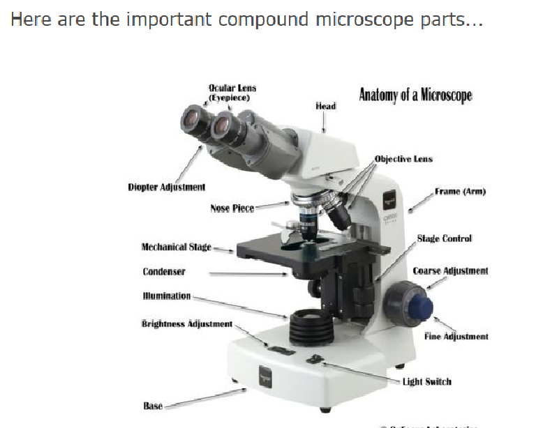
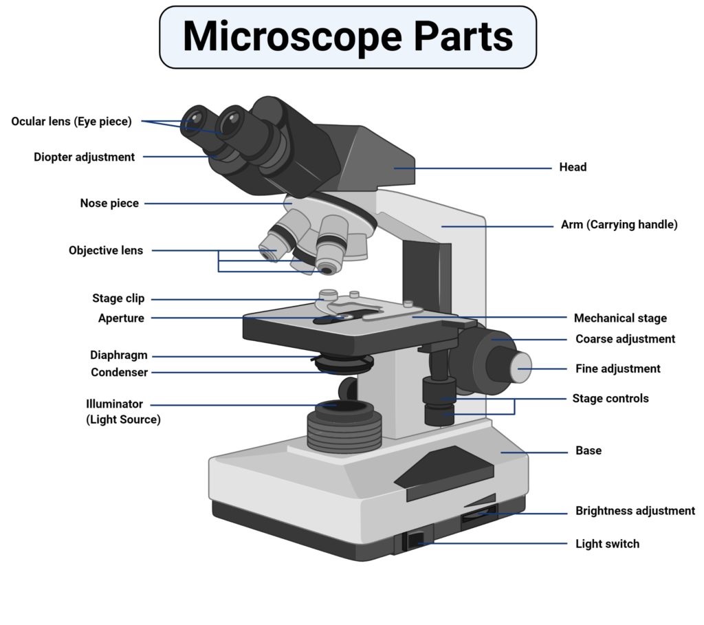 Types of light microscopes (optical microscope)
 The modern types of Light Microscopes include: 1.Bright field Light Microscope 2.Phase Contrast Light Microscope 3.Dark-Field Light Microscope 4.Fluorescence Light Microscope What is an electron microscope? Electron microscopy (EM) is a technique for obtaining high resolution images of biological and non-biological specimens. An electron microscope is a microscope that uses a beam of accelerated electrons as a source of illumination. As the wavelength of an electron can be up to 100,000 times shorter than that of visible light photons, electron microscopes have a higher resolving power than light microscopes and can reveal the structure of smaller objects. A scanning transmission electron microscope has achieved better than 50 pm resolution in annular dark-field imaging mode and magnifications of up to about 10,000,000× whereas most light microscopes are limited by diffraction to about 200 nm resolution and useful magnifications below 2000×. 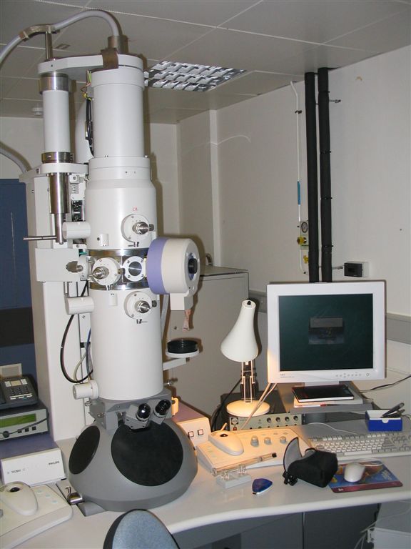 How do electron microscopes work? If you've ever used an ordinary microscope, you'll know the basic idea is simple. There's a light at the bottom that shines upward through a thin slice of the specimen. You look through an eyepiece and a powerful lens to see a considerably magnified image of the specimen (typically 10–200 times bigger). So there are essentially four important parts to an ordinary microscope: The source of light. The specimen. The lenses that makes the specimen seem bigger. The magnified image of the specimen that you see. In an electron microscope, these four things are slightly different. The light source is replaced by a beam of very fast moving electrons. The specimen usually has to be specially prepared and held inside a vacuum chamber from which the air has been pumped out (because electrons do not travel very far in air). The lenses are replaced by a series of coil-shaped electromagnets through which the electron beam travels. In an ordinary microscope, the glass lenses bend (or refract) the light beams passing through them to produce magnification. In an electron microscope, the coils bend the electron beams the same way. The image is formed as a photograph (called an electron micrograph) or as an image on a TV screen. That's the basic, general idea of an electron microscope. But there are actually quite a few different types of electron microscopes and they all work in different ways. The three most familiar types are called transmission electron microscopes (TEMs), scanning electron microscopes (SEMs), and scanning tunneling microscopes (STMs). Applications Semiconductor and data storage Circuit edit Defect analysis Failure analysis Biology and life sciences Cryobiology Cryo-electron microscopy Diagnostic electron microscopy Drug research (e.g. antibiotics) Electron tomography Particle analysis Particle detection Protein localization Structural biology Tissue imaging Toxicology Virology (e.g. viral load monitoring) Materials research Device testing and characterization Dynamic materials experiments Electron beam-induced deposition In-situ characterisation Materials qualification Medical research Nanometrology Nanoprototyping Other Chemical/Petrochemical Direct beam-writing fabrication Food science Forensics Fractography Micro-characterization Mining (mineral liberation analysis) Pharmaceutical QC |
Measurements that scientists, researchers, and manufacturers of a vaccine must know.
Start with millimeters, micrometers, nanometers, and picometers. Measure bacteria in micrometers Measure a virus in nanometers Measure atoms in picometers 1 meter = 100 centimeters 1 meter = 1000 millimeters 1 centimeter = 10 millimeters 1 millimeter = 1000 micrometers 1 micrometer = 1000 nanometers 1 nanometer = 1000 picometers What is the size of human cells, bacteria, viruses, and atoms? Human cell diameter = 25 micrometers or 25,000 nanometers Cell nucleus (control center of cell) diameter = 5 micrometers or 5,000 nanometers Bacteria cell length = 2 micrometers or 2,000 nanometers Mitochondria (powerhouse of cell) length = 2 micrometers or 2,000 nanometers Ribosome diameter (protein factory in cell) = 0.03 micrometers or 30 nanometers Cell membrane thickness = 0.08 micrometers or 80 nanometers Silver nanoparticle diameter = 0.04 micrometers or 40 nanometers Atomic size, atomic mass, atomic number: What is the difference? Atomic size: Picometers are used to measure the size of atomic structures. Atomic number: Take a look at this: https://www.qureshiuniversity.com/elements1.html Atomic mass: Take a look at this: https://www.qureshiuniversity.com/atomicmass.html How many oxygen atoms lined up in a row would fit in a one nanometer space? Seven. What is the diameter of one oxygen atom? The diameter of one oxygen atom is approximately 0.14 nanometers What is the diameter of one hydrogen atom? You can look up the covalent radius of hydrogen (37 pm) and double it for the diameter. Helium has the smallest atomic radius. What are some other useful links for vaccines? If you know any other useful resources for vaccines, email admin@qureshiuniversity.com |
New vaccine for a specific virus
|
How do you make a new vaccine for a specific virus? Get answers to all questions relevant to the specific virus. Decide the category of vaccines by manufacturer that needs to be manufactured. Verify safety and effectiveness of the new vaccine. No question relevant to a new vaccine for a specific virus can remain unanswered. Vaccine manufacture and approval for human use process. Questions that need to be answered. https://qureshiuniversity.com/healthcarelaws.html#Vaccine manufacture and approval for human use process. Vaccine manufacture and approval for human use process. 2019–2020 viral pandemic vaccine
|
Vaccine Issues
|
New vaccine approval process: What are various findings? On or before December 2, 2020, the Medicines and Healthcare products Regulatory Agency (MHRA) at 10 South Colonnade, London, did not get relevant questions answered before approving the vaccine for coronavirus. What must happen next? An interview with Vanessa Birchall-Scott, Director of Human Resources Medicines and Healthcare products Regulatory Agency (MHRA), 10 South Colonnade, London December 2, 2020. What questions do they need to answer? Is it correct that your resource has approved the coronavirus vaccine? Have they answered all questions relevant to the virus? Have all the findings been proven using scientific methods? What are the electron microscopic findings related to the virus? Who revealed the electron microscopic findings related to the virus? Who verified these findings? How will they scientifically differentiate among influenza, rhinovirus, and coronavirus? There are 10 patients presenting with a fever, cough, and sore throat. A few have influenza, a few have rhinovirus, and others have coronavirus. What tests will you run to prove the existence of a specific virus? Who received grants for this vaccine without answering these questions on or before November 22, 2020? Was it necessary to verify these findings before approving grants for the vaccine? Is it justified? Is the vaccine safe for human beings? Is the vaccine effective for human beings? Should the mentioned vaccine be approved for human use on or after November 22, 2020? What evidence proves the safety and efficacy of the mentioned vaccine? Have they answered all questions relevant to this vaccine? Who was the exact individual or team of individuals who approved the coronavirus vaccine from your resource? Where are the answers to the questions relevant to the virus and vaccine that were asked on December 2, 2020, before the approval of the specific vaccine? What must happen if they are not able to answer these relevant questions? On or before December 7, 2020, you need to answer these questions or the full team must resign. All states must be reminded to not use this vaccine until all questions are answered. What have previous research findings been? The Tuskegee Syphilis Study still haunts many Americans. On October 14, 2020, there was discussion about this issue in Chicago, Illinois. In 1974, specific legislation had to be approved to prevent these issues. As part of the settlement of a class action lawsuit subsequently filed by the NAACP on behalf of study participants and their descendants, the U.S. government paid $10 million ($51.8 million in 2019 dollars) and other resources to surviving participants and surviving family members infected as a consequence of the study. If all questions relevant to these issues are not answered, grant-grabbing scandals will unfold. Marketing a coronavirus vaccine that is actually something else will unfold. What else must happen? The FDA is likely to have deliberations on December 10, 2020, and December 17, 2020. Make sure all questions are answered ahead of any process of approval. Do not approve the vaccine if all questions are not answered. All state public health resources worldwide should circulate an alert. Not all questions have been answered. Do not use the vaccine until all questions are answered. |
Vaccine Issues on or after December 13, 2020
|
On December 10, 2020, FDA advisers recommended the authorization of the Pfizer COVID-19 vaccine without getting answers to relevant questions: Is this justified? No. Why must this process be compared with the Tuskegee Syphilis Study? The Tuskegee Syphilis Study end date: 1972 still haunts many Americans. This became a scandal while a similar process was used. Not all questions were answered. Similarly, on or after December 12, 2020, the current situation can become a scandal if all questions are not answered by those who have the responsibility to do so before circulating the vaccine. You are given a vial and told that it is an mRNA vaccine: How do you prove this is really an mRNA vaccine? How do you scientifically prove that the mRNA vaccine is really generating specific antibodies to this specific virus? Is this a real vaccine or a saline shot? Does the vial contain the mentioned ingredients? Does the vial contain the right concentration of the vaccine? How did you verify the facts? Are they falsely claiming the vaccine is more than 94% effective? Did Pfizer unfairly grab 1 billion dollars for research that could be completed for less than 50,000 dollars? Do you know that the emergency use authorization before the final-stage testing of a vaccine is complete is not justified? What are examples of dummy shots and placebos? Is it justified to approve any vaccine without getting answers to the relevant questions? Who has the responsibility to remind Pfizer, Moderna, AstraZeneca, and similar entities to answer questions relevant to the vaccine before vaccine circulation? Is there a focus on public service or an effort to grab research grants and millions in vaccine money without answering the relevant questions? If you give a placebo or saline shot to a person, there will not be any adverse reaction: Does that mean the ingredients of the vaccine are genuine? Who has the responsibility to prove that the vial provided has an mRNA vaccine? How do you prove this is really an mRNA vaccine? Who has responsibility to prove scientifically that this mRNA vaccine is really generating specific antibodies to this specific virus? What must happen after December 13, 2020, relevant to these issues? The offices of attorney general of all states: If these questions are not answered ahead of time, remind the vaccine manufacturers of the impending FDA complaints of consumer fraud. On or after December 13, 2020, replace the head of the FDA and 17 of the 22 members of the Vaccines and Related Biological Products Advisory Committee with new nominees who can answer these questions. The Vaccines and Related Biological Products Advisory Committee should hold a virtual meeting or deliberations via the Internet that includes the participation of all state attorney generals or their representatives. The meeting must proceed under Chapter 410 Public Health statute of the Illinois compiled statutes or the equivalent. All states must hold similar meetings under the equivalent of Chapter 410 Public Health statute of the Illinois compiled statutes. We may need to have global public health statutes relevant to these issues. A question-and-answer presentation will be appreciated, as circulated by Doctor Asif Qureshi. |
Last Updated: December 13, 2020

