
What Do Ophthalmologists Check During Eye Exams?
Assessment of the patient with eye problems
|
Introduction Eye problems are common presentations in emergency care areas. Adults and children frequently attend with minor injuries to the eye, visual problems, and other painful/red eye conditions. Assessment of patients with eye problems requires the clinician to have some basic eye assessment skills, e.g. lid eversion (Ophthalmological emergencies see Eyelid eversion, p. [link]), and access to a slit lamp to enable a complete assessment of the eye/s to be made. If a slit lamp is not available or the clinician is not familiar with how to use it, the patient will need referral to a centre with comprehensive eye assessment facilities. Assessment of the patient with eye problems ► Assessment of visual acuity (VA) is mandatory and should be undertaken at the point of triage for any patient presenting with an eye problem. A VA should be established before any other investigations or treatment (except irrigation or instillation of LA) are carried out. An accurate triage category cannot be assigned without an accurate VA and an assessment of whether this is normal for the patient. History taking An accurate history is important. Questions should include the following. Questions that must be answered. Where is the patient now? How old is the patient? What is the gender of the patient? Who is reporting this emergency? What seems to be the complaint? What seems to be the problem? Management of a corneal injury Foreign bodies Superglue injuries Ultraviolet radiation injury (welder’s flash, arc eye) Chemical injury Blunt trauma Major trauma Open trauma The red eye Conjunctivitis Subconjunctival haemorrhage Anterior uveitis (iritis, anterior cyclitis, or iridocyclitis) Corneal ulcers Glaucoma Loss of vision: history Profound monocular loss of vision Segmental loss of vision and loss of central vision Blurring of vision and transient loss of vision Other What is your identification mark? How do I identify you? How long have the symptoms been present? Are they getting better/worse/stable? How did the problem begin and over what period of time? The degree, type, and location of any pain? Whether vision is reduced and to what degree? Is there any discharge or watering? Is the patient photophobic? Has the patient had this, or a similar problem, before? Are there any concurrent systemic problems? Examining the eye It is very easy to assume a diagnosis from the history and miss less obvious problems. Eye examination, therefore, must be systematic, starting at the outside (eye position and surrounding structures) and working inwards to consider the globe itself. The clinician should consider the following points. Eyes • Position normal for the patient? • Enophthalmos (may indicate orbital fractures) or exophthalmos (may indicate orbital bleeds in trauma, thyroid eye disease)? • Restriction of movement or double vision in any of the eight gaze positions? Lids • Integrity. Lacerations of the lid and lid margin? • Position. Entropion/ectropion? • Lash line. Intact? Ingrowing lashes/infestation/crusting? • Swelling. Whole or part of the lid/one or both lids? • Puncta visible and correctly sited? Conjunctiva • Integrity. • Structure. Smooth? Follicles or papillae? • Other features. Conjunctival cysts, pingueculae, pterygia? • Inflammation. Generalized or local? • Subconjunctival haemorrhage? • Discharge. Type, frequency? • Fornices. Both lower and subtarsal areas. Concretions or FBs may be visible. Cornea • Integrity. Lacerations, abrasions, ulcers? • Clarity? • FBs? Anterior chamber • Depth. The distance between the curved cornea and the iris. Generally equal in both eyes. • Contents, e.g. WBCs or RBCs; any cells are abnormal. Iris and pupil • Colour. Similar to the other eye? • Position. Should be round and regular but may be slightly off centre (check both eyes). • Integrity. Normal for patient? Previous surgery may change its appearance. • Size and shape. Smaller or larger than the other eye; round or oval? • Reaction to light and to near. Always compare both eyes. What appears to be an abnormality may be bilateral and normal for the patient. Top Previous Next The injured eye By far, the commonest ophthalmic problem to present to EDs and minor injury units is the injured eye. Adults and children frequently sustain minor injuries to the surface of their eye from FBs or ‘poking’-type mechanisms. A carefully elicited history should enable the assessing clinician to determine if the injury is likely to be ‘minor’, and assessment can proceed without specialist intervention. Many minor eye injuries are successfully managed by ENPs. Obtaining a clear minor mechanism is key to ensuring that a potentially more serious injury is not missed. History It is crucial to identify and document the following. • What actually happened, e.g. poke in the eye or felt FB go in the eye? • Where did it happen—was the injury at work, home? • Was any eye protection used? • Was any first aid administered at the time? • When did the injury occur? • How was the injury sustained? Is there a risk of a penetrating eye injury? • Past ophthalmic history. Has the patient any previous or current eye problems? Blunt injuries to the eye, e.g. a punch to the face, can cause significant facial fractures, as well as eye injuries. The orbit tends to protect the eye from the force of larger objects. However, small balls, e.g. squash balls or golf balls, can cause significant globe trauma. Is there a history of a high-velocity injury? Hammer and chisel use typically causes small fragments to travel at high speed. Glass, knives, darts, and pencils are other causes of penetrating eye trauma and will require urgent ophthalmic referral. • Common mechanisms for minor eye injuries include being poked in the eye with finger, hairbrush, plants, bushes, and twigs. • Grinding injuries can be caused by metallic or brick FBs. • Other common FBs are dust, grit, and flakes of metal/paint. Symptoms The following are the commonest symptoms of corneal injury or corneal FB. • Pain. Injury to the cornea is acutely painful. • Large superficial abrasions are intensely painful (Ophthalmological emergencies see Fig. 13.1). • Deep lacerations with little epithelial loss are only mildly painful (Ophthalmological emergencies see Fig. 13.2). • Visual disturbance. • FB sensation. Fig. 13.1 Large superficial abrasions. Click to view larger Fig. 13.1 Large superficial abrasions. Fig. 13.2 Deep laceration. Click to view larger Fig. 13.2 Deep laceration. Other symptoms include redness, watering, blurred vision, and discharge. Examination VA. • Examine the eye systematically (Ophthalmological emergencies see Assessment of the patient with eye problems. • Slit lamp examination to identify the depth of the injury and presence of any FB. • Lid eversion to identify any subtarsal FBs. • Fluorescein staining to reveal the extent of corneal injury. Treatment of corneal injuries is based on the management of three areas: pain, prevention of infection, and optimization of healing. Management of a corneal injury Prevention of infection Any corneal damage requires treatment with a topical prophylactic antibiotic (chloramphenicol or fusidic acid). Fusidic acid is useful for children, as it only requires a twice-daily application. If perforation is suspected or confirmed, a single drop of unpreserved, single-dose chloramphenicol may be instilled before transfer to the ophthalmic unit. Both preservatives and ointment are toxic to ocular tissues and should not be used. Pain • Topical anaesthesia is for examination purposes only and obtaining an accurate VA assessment. Repeated instillation will result in dose-related toxicity and delay in epithelial healing. • Cyclopentolate 1% will relieve ciliary spasm and associated pain. • Topical NSAIDs four times daily (qds) also provide a significant degree of effective pain relief. Padding • Is for comfort only. • There is no evidence that it aids healing. • Pad those patients who have significant pain, advising that, if the pad makes the pain worse, they should remove it. Double eye pad A double eye pad should always be used, one pad folded over the closed lids and the other open on top of it. The whole is taped firmly to the face, so that the patient cannot open the eye underneath the pad. If comfortable, the pad should be left intact for 24h and then removed, and instillation of medication commenced. There is no need to pad the eye just because a topical anaesthetic has been instilled. Optimization of healing Reviewing simple corneal abrasions depends very much on the individual clinician. Large abrasions can be reviewed to ensure that healing is taking place and that there is no loose epithelium that needs debriding. Small abrasions heal very quickly. At review, if there is any loose epithelium visible or any sign of infection, ↑ in pain, or reduction in VA, it is safer to refer the patient for an ophthalmic opinion. Recurrent abrasion syndrome is common in those patients who have an animal- or vegetation-based cause for their abrasion (e.g. plants or fingernail). This can be prevented by using ointment at night before sleeping to keep the eye lubricated. ‘Simple’ or another lubricating ointment, or Lacri-Lube® (ointment based without drugs) should be used for a period of up to 3 months. Referral of corneal injuries Each emergency care area will have guidelines about which types of corneal injuries should be referred for ophthalmic assessment and follow-up. Foreign bodies Conjunctival foreign body These are usually superficial. • Instil topical anaesthetic. • Remove from the conjunctiva by wiping with a dampened cotton bud (any swab/cotton bud used on the eye should be pre-moistened with Saline Minims® or the residue of an anaesthetic Minims®—otherwise, epithelial tissue sticks to the swab, rather than the eye, and significant injury can result). • Stain after the FB has been removed to identify the extent of damage. • If there is minimal stain, a single application of chloramphenicol ointment may be instilled. • If there is significant stain, chloramphenicol ointment should be prescribed qds until the eye feels back to normal. Deeper FBs on the conjunctiva may be removed using a 21G hypodermic needle, often mounted on the end of a cotton bud to form a longer and more easily manipulated tool. Corneal foreign body • Assess depth using a slit lamp. The depth of the cornea may be seen within the slit lamp beam. • If the FB is anything other than superficial, it should be referred to the local eye unit for further assessment and removal. Ophthalmological emergencies The cornea is only 1mm thick at its thickest. Accidental perforation does occur and can be devastating. FB removal should always take place at a slit lamp to provide magnification and stability for the patient’s head. If this is not possible, consideration should be given to referral to a more appropriate setting. • Instil topical anaesthetic. • Use a 21G needle; bore upwards to gently lift off the FB from the cornea. • Metallic FBs may need slightly more forceful removal. • Rust must be removed at some point. This can be facilitated by giving the patient chloramphenicol ointment or drops to use for 2 or 3 days, and then reviewing in the ED or in the eye unit. Rust is much easier to remove at this stage. Again, an antibiotic or ointment should be prescribed until the eye feels back to normal. Once an FB is removed, treatment is the same as for a corneal abrasion and should be focused on preventing infection, relieving pain, and optimizing healing. Superglue injuries History Superglue may be instilled into the eye, as its containers often resemble eye drop containers. Signs and symptoms • Pain. • Eyelids stuck together. As the eye is permanently wetted by the tear film, the glue does not generally stick to the tissues of the eye but hardens, forming a plaque inside the lids and abrading the cornea as the eye and lids move. The glue usually glues the eyelashes together and therefore holds the lids together. Management • Instil a topical anaesthetic to the lids, allowing it to drain between them to act on the cornea. This relieves pain and allows examination and treatment. • Trim the eyelashes very close to the lid margin. • Pick off the remaining glue to allow the lids to open. This must be done very carefully and may take some time. A very fine pair of scissors is required, and a laceration of the lid margin must be avoided. This may be a lengthy procedure, necessitating repeated topical anaesthetic and much patience. • When the lids are open, remove the plaque of glue and all particles of glue. • VAs. • Treat resulting abrasions as corneal abrasions. Children may be much less cooperative than adults and may need a general anaesthetic for this procedure. Referral to an ophthalmic unit should be considered. Ophthalmological emergencies Superglued eyelids will take a considerable time to open on their own, and practitioners should not be tempted to ‘let nature take its course’. The abrasions are likely to be extremely painful, the glue plaque will abrade as long as it is in the eye, and the loss of corneal epithelium provides an entry point to the eye for pathogens. Ultraviolet radiation injury (welder’s flash, arc eye) History • Exposure to welding arcs. • Exposure to ultraviolet (UV) ‘sunlamps’. The UV radiation is absorbed by the corneal epithelial cells, some of which are damaged or destroyed. There is a latent period before symptoms are experienced, which depends on the amount of exposure and explains the typical late-night presentation of these patients. Signs and symptoms • Gritty sensation to one or both eyes. • Intensely painful eye(s). • Photophobia. • Watering and blurred vision. • Lid swelling. • Redness. Examination • Instil a topical anaesthetic. • VA, and examine the eye systematically (Ophthalmological emergencies see Assessment of the patient with eye problems, pp. [link]–[link]). • Instil fluorescein. • Fluorescein reveals punctate staining on the surface of the cornea where some cells have been destroyed. Management • Treatment is as for a corneal abrasion. • A mydriatic drop will provide comfort. • Antibiotic ointment as prophylaxis and for comfort. • Padding may help, but both eyes should not be padded simultaneously. • Advise complete recovery is usually within 24–36h. Chemical injury History It is almost irrelevant what chemical is splashed into the eye, and no time should be wasted in the ED trying to find out. Chemical splashes in the eye can result in devastating injury, and the time to irrigation is the most important factor in minimizing ocular problems. Irrigation The initial treatment of ocular chemical injuries involves irrigation to dilute the chemical and remove any solid debris (Ophthalmological emergencies see Eye irrigation, p. [link]). Examination • VA. • Examine the eye systematically (Ophthalmological emergencies see Assessment of the patient with eye problems, pp. [link]–[link]). Management Superficial injury may be treated with chloramphenicol ointment qds until the eye feels back to normal. • All but the most trivial chemical injuries should be referred to an ophthalmologist. • Solvent injuries are generally much less damaging than those due to acid and alkaline chemicals. Blunt trauma Blunt trauma may result in disruption to any, or all, of the ocular tissues. Any patient presenting with blunt trauma to the eye or surrounding tissues with any reduction in vision should be referred to an ophthalmologist for specialist assessment. Traumatic hyphaema Signs and symptoms • Blood in the anterior chamber, detected by slit lamp or visible with the naked eye, sometimes to the extent of filling the whole of the anterior chamber. • ↓ VA. • ↑ in intraocular pressure, as RBCs block the trabecular network. • Severe pain. • Irregular or sluggish pupil. Management • Urgent referral to an ophthalmologist is required. • Sit the patient upright in order to allow the blood cells to settle and absorb away from the visual axis. This will reduce any staining of the corneal endothelium with haem pigment, which may affect vision. Regular review is undertaken to monitor the intraocular pressure and treat any rise in pressure. Traumatic uveitis Is a common effect of blunt trauma and may be the only sign of trauma. Treatment is as for any uveitis, with pupil dilatation and topical steroids. Iris and pupil abnormalities Traumatic mydriasis or miosis may occur as a consequence of blunt trauma, and the pupil may be irregular, when compared with the fellow eye, due to partial or complete rupture of the iris sphincter. Lens abnormalities The impact of the iris on the lens, as the eye distorts and then moves back into shape, may leave a circle of iris pigment, which can be seen after dilatation (Vossius ring). Traumatic rupture of the zonules holding the lens in place may occur, and luxation or subluxation of the lens may take place. ↑ intraocular pressure may occur if the lens blocks the pupil. Iridodonesis (iris tremble) may be visible. ► Urgent referral to an ophthalmic unit is required for any iris, pupil, or lens abnormalities, or if the patient reports any reduction in VA. Major trauma Orbital injury Both facial and skull trauma can result in orbital injury. Medial orbital fractures The lacrimal secretory system (especially the nasolacrimal duct) may be damaged, and the medial rectus muscle may be trapped within the fracture. Orbital floor fractures Often referred to as blowout fractures, because they are produced by transmission of forces through the bones and soft tissues of the orbit by a non-penetrating object such as a fist or a ball. These fractures may be complicated by the entrapment of muscles and orbital fat, which limits ocular motility (Ophthalmological emergencies see Orbital floor fractures, p. [link]). Signs and symptoms These include: • diplopia; • enophthalmos; • infraorbital anaesthesia. A classic presentation involves an injured patient, who perhaps would not have presented to the ED otherwise, blowing their nose and then attending because their eyelids have swollen alarmingly as air from the sinus has been driven into the tissues of the lid. The patient should be given advice about the avoidance of the Valsalva manoeuvre such as blowing the nose or straining at stool. Any patient with an orbital fracture and any degree of double vision should be referred to an ophthalmic unit. Orbital apex trauma All clinicians in the ED must be alerted to the possibility of ocular involvement from indirect trauma, such as base of skull fractures, as well as from more direct trauma where the eyes themselves do not appear to be involved. Fractures of the orbital apex may result from direct, non-penetrating blunt trauma or from penetrating trauma, e.g. with large orbital FBs. Orbital apex fractures present differently, depending on the degree of injury to the vascular and neural structures within the orbital apex, and a number of different presentations are possible. • Optic nerve injury may occur, commonly due to traumatic optic neuropathy from indirect trauma (such as fractures of the base of the skull). A haematoma may compress the nerve, or it may be damaged by an FB or from a fracture, which can result in a spectrum of injuries from minor trauma to the nerve to complete transection. • Injury to the cranial nerves present in the orbit (CN III, IV, and VI) may present as extraocular muscle palsy with double vision. • Injury to the trigeminal nerve (CN V) presents as sensory disturbance to areas it supplies. Collaboration of ophthalmic units with the ED is important in order to ensure that patients with this type of injury do not lose vision unnecessarily. Ophthalmological emergencies Patient complaints of loss of, or reduction in, vision must be taken very seriously. In order to quantify this, VA must be checked on arrival, as a baseline and then hourly. Ophthalmology opinion must be obtained immediately if vision is involved. Retrobulbar haemorrhage This may occur from direct or indirect trauma to the orbit and can progress rapidly. Signs and symptoms • Pain. • Proptosis of the globe. • Congested conjunctival vessels. • Lid and conjunctival swelling. • Subconjunctival haemorrhage may be dense and may extend beyond the visible conjunctiva. Management • Regular observation of the appearance of the eye patient with direct or indirect trauma to that orbit is required to minimize complications of the injury. • VA should be measured as a baseline, and the patient should be encouraged to report new symptoms and any reduction in vision. • ►► If the globe begins to proptose after trauma, an ophthalmologist should be involved immediately. A CT or MRI scan may be required urgently, and the patient’s VA should be checked very frequently (every 10min). If VA reduces, emergency decompression by lateral canthotomy (a horizontal incision at the lateral canthus, through the skin and conjunctiva, and then through the lateral canthal tendon, under LA) will be required to relieve pressure on the optic nerve. Equipment for this procedure will not be needed very often but should be readily available in the ED, so that avoidable loss of vision may be prevented. Open trauma An open eye requires immediate assessment. No attempt should be made to remove any retained materials protruding from the globe. Management • Stabilize any protruding material as far as possible, perhaps by taping it to the cheek or by covering the whole area with a plastic shield or small receiver. • No pressure should be put on to the eye, and an eye pad should only be used if absolutely no other method of covering the eye exists. The pad must be loose and taped well away from the globe—any pressure on the globe may result in further injury and/or loss of ocular contents. • Manage pain and nausea. Vomiting with an open eye is likely to lead to loss of the ocular contents. • Lie the patient flat or propped up at around a 30° angle. This can minimize any rise in intraocular pressure. • Do not cover both eyes, unless they are both extensively damaged. A patient with one damaged eye is unlikely to be relaxed, comforted, or reassured by being unable to see anything or anyone around them. ►► Immediate referral to an ophthalmologist is required. Small penetrating injuries Penetrating injuries and intraocular FBs may cause damage to the globe by: • tissue disruption at the time of injury; • formation of scar tissue causing long-term damage—retinal scars may contract and cause retinal detachment (RD); corneal scars will distort or disrupt vision; • introduction of foreign material to which the eye reacts; • allowing pathways for infection to enter the globe. Large penetrating eye injuries are very obvious, but small perforations may be easily missed, as the eye may look completely intact. The wound may have self-sealed or may be obscured by the upper lid. Examination Must always include all aspects of the anterior part of the globe by asking the patient to look in each different direction, so that all segments may be examined. As the patient looks down, the upper lid should be retracted, so that the upper portion of the globe may be seen. All penetrations of the lid should lead to a high index of suspicion about the state of the globe. • Corneal perforations always leave a full-thickness scar, even if very small. • Scleral perforations may be masked by an overlying subconjunctival haemorrhage. • A small hole in the iris may mark the passage of an FB. ► Analgesia and anti-emetics should be considered, as vomiting may lead to expulsion of the contents of a perforated globe. ►► Patients with penetrating trauma should be referred urgently to an ophthalmologist. The red eye Another common eye problem that presents frequently to emergency care areas is the ‘red eye’. Patients cannot identify any specific injury and can complain of varying symptoms, e.g. redness, pain, itch, watering, discharge, swelling, headache, and visual disturbance. Allergies Allergic conjunctivitis presents in several ways. • Red eyes with itching and watering and an appearance of large bumps (papillae) on the subtarsal conjunctiva are particularly common during the ‘hay fever season’ and may therefore be associated with a runny nose, sneezing, and other allergic symptoms. Treatment is with systemic antihistamines and/or topical treatment such as emedastine or olopatadine. Sodium cromoglicate is of no value in allergic reaction. It is useful only when the allergic phase has been controlled, as a mast cell stabilizer. • A more severe chronic atopic reaction, most often seen in children, is the appearance of ‘cobblestone papillae’. The appearance of the subtarsal conjunctiva is of massive papillae arranged in a cobblestone fashion. This is intensely irritating and often requires complex management. Patients presenting in this way should be referred to an ophthalmologist urgently. • An acute atopic reaction involves massive chemosis or swelling of the conjunctiva which the patient or parent often describes as ‘jelly’ on the eye. This is due to an allergen transferring directly to the eye on the fingers or by blowing in. This is completely self-limiting. It resolves quickly (over a period of hours), and reassurance and information are all that is required. Conjunctivitis Inflammation of the conjunctiva is by far the commonest cause of red eyes. Common organisms involved in infective conjunctivitis include viruses, bacteria, and Chlamydia (Ophthalmological emergencies see Table 13.1). Almost all conjunctivitis in adults is caused by a virus, often a type of adenovirus, and, unless there are large amounts of green/yellow discharge present all day, the infection should be presumed not to be bacterial. Conjunctivitis in children is more likely to be bacterial. Table 13.1 Types of conjunctivitis Viral Bacterial Chlamydial Bilateral and acute Rare, bilateral Unilateral and chronic Lids May be swollen; follicles May be swollen Unlikely to be swollen Conjunctiva Injected Injected Injected Cornea May have keratitis (punctate staining of cornea) No involvement No involvement Discharge Watery, sticky in morning Green/yellow discharge all day Watery, stuck together in mornings Vision May be affected by corneal involvement Unlikely to be affected Unlikely to be affected Pain Gritty, dry; may be intensely irritating Gritty and very sticky Mild irritation Investigation None: clinical diagnosis only. Identification of organism does not change management None: clinical diagnosis only. Identification of organism does not change management If clinical appearance suggestive of Chlamydia, swab for Chlamydia Treatment Artificial tear regime; information; reassurance Antibiotic drops Artificial tear regime; on positive identification, appointment with sexual health services Ophthalmological emergencies Eye pads should never be suggested for patients with conjunctivitis. The warm, damp atmosphere underneath an eye pad will allow further organism growth and exacerbate the condition. Viral conjunctivitis Signs and symptoms • Gritty sensation to the eye/s. • Profuse watering. • Dry feeling to the eye/s. • Stickiness often only in the morning when the watery discharge has dried and the lids are stuck together. • Some types of adenovirus also cause URTI and general malaise. Examination • On lid eversion, the conjunctiva covering the lids will appear very bumpy, rather than smooth. This roughness of the conjunctiva is what makes the eye feel gritty and irritable. • There may be punctate erosions (small staining areas) on the cornea when stained with fluorescein. Management • Symptom relief with artificial tears, and advice about very frequent use (hourly or even more frequently). • Advice about the self-limiting nature of the condition. The patient should be aware that viral conjunctivitis may persist for 3–6wk, and the symptoms of dryness may last much longer. There is no point in prescribing antibiotic eye drops. Ophthalmological emergencies Adenovirus is highly infectious, and infection control is of paramount importance, both for the patient and the emergency care setting. Handwashing is vital to stop the spread of viral conjunctivitis. Major epidemics of viral conjunctivitis associated with ophthalmic units have been linked with poor handwashing. All equipment should also be cleaned between each patient. It is not necessary to take eye swabs for culture, and the patient should not be followed up. Bacterial conjunctivitis Signs and symptoms • Redness. • Irritable gritty eye. • Profuse, purulent discharge; the lashes may be coated with discharge. Management A broad-spectrum antibiotic, such as chloramphenicol or fusidic acid, applied topically in the form of drops. The condition is self-limiting, and no investigations are required. Chlamydial conjunctivitis Signs and symptoms • Unilateral. • Chronic (the patient may have had a red and irritable eye for some time which has reached an irritating stage but not progressed to a severe viral infection). • Large pearly follicles are present on lid eversion. If Chlamydia is suspected from the clinical picture (Ophthalmological emergencies see Table 13.1), a chlamydial swab should be obtained from the eye. The patient’s details should be checked to ensure that they are contactable, should the result be positive. Patients should be referred to sexual health services for treatment. Subconjunctival haemorrhage Patients may present with a spontaneous subconjunctival haemorrhage with a deep red patch of blood under the conjunctiva, which may be quite small and circumscribed or may be severe enough for the conjunctiva to appear like a ‘bag of blood’. Providing that there is no history of trauma, no treatment is needed, unless the haemorrhage is severe or the eye irritable, in which case artificial tears are useful to provide comfort and lubrication. Subconjunctival haemorrhage may occasionally be associated with hypertension, so it might be useful to check the patient’s BP. Patients with clotting disorders or those on anticoagulants may be prone to repeat episodes. Subconjunctival haemorrhages may take some weeks to resolve, and, because the conjunctiva is an elastic membrane, the blood may spread under it and the haemorrhage appears worse, before it begins to resolve. Anterior uveitis (iritis, anterior cyclitis, or iridocyclitis) Uveitis is an inflammatory condition, which may be associated with systemic disease or as a response to trauma, but is often idiopathic. It is unusual to encounter uveitis (especially as a 1° attack) in an elderly patient. Signs and symptoms (Ophthalmological emergencies see Box 13.1.) • Photophobia. • Pain (due to ciliary spasm). • Conjunctival redness (injection), which may be more marked around the limbus. • ↓ vision due to protein and WBCs in the anterior chamber. • Small pupil that reacts sluggishly because of spasm and inflammation. Box 13.1 Characteristics of uveitis • Lids normal. • Conjunctiva injected. • Cornea normal. • Anterior chamber deep. • Iris may look ‘muddy’. • Pupil: slight miosis (compared with fellow pupil); sluggish. • Pain: deep pain in eye. • Discharge: may water. • Photophobia. • Systemically well. Examination When the cornea is illuminated with a torch, the reflection will be bright, and there will be no staining with fluorescein. Treatment The patient must be referred on to an ophthalmic unit for investigation and treatment. Corneal ulcers There are three main types of corneal ulcer that are likely to be seen in the emergency care settings. All should be referred to an ophthalmologist urgently. Differentiation between the different types of corneal ulcer is sometimes difficult, and the treatment is completely different. A careful history will usually identify symptoms of a ‘red eye’: pain; redness; visual disturbance; and no history of injury. Ulcers are seen in contact lens wearers, and the clinician should always be alert to the possibility of a corneal ulcer if there has been a history of lens wearing. On staining, a defect is seen on the cornea, which may have a characteristic shape. ► Any corneal defect that cannot be explained by a history of injury should be discussed with an ophthalmologist. Bacterial ulcers On staining, bacterial ulcers occur as ‘fluffy’, white demarcated areas. They are caused by a number of organisms, e.g. Pseudomonas, which is very difficult to treat, and are most commonly, but by no means exclusively, seen in contact lens wearers. The patient should be urged to leave their contact lens out of their eye, whilst waiting to be seen in the ophthalmic unit, but should keep it with them, as it is likely to be sent for culture. ►► Delay in treatment of infected corneal ulcers can result in a devastating intraocular infection. Viral ulcers (dendritic ulcers) These are caused by the herpes simplex virus. They are known as ‘dendritic’ ulcers because of their branching, tree-like shape when stained with fluorescein. They are treated with aciclovir eye ointment, and should be referred to an ophthalmologist for treatment and follow-up. Marginal ulcers Appear as ulcerated areas that stain with fluorescein and are usually close to the limbus. They are part of a hypersensitivity response by the eye to staphylococcal exotoxins and are treated with steroidal or nonsteroidal anti-inflammatory eye drops by an ophthalmologist. Glaucoma Glaucoma describes a rise in the pressure inside the eye, causing damage to the neural tissue that makes up the retina and resulting in permanent reduction in the field of vision. There are two main types of glaucoma: • chronic, open-angle glaucoma where the rise in pressure is small, damage is done to the retina over a large period of time, and visual field loss is insidious; • acute, closed-angle glaucoma, which is an ophthalmic emergency. Chronic open-angle glaucoma This type of glaucoma is only likely to be encountered in the ED if the patient has run out of eye drops or as an incidental part of history taking. Although it cannot be recommended that patients manage without the appropriate medication for their condition, it cannot be considered an emergency in the same way as perhaps a diabetic without insulin. The patient should be referred to their own ophthalmic clinic or GP at the earliest opportunity. There is no danger associated with dilating the eye of a patient who has chronic, open-angle glaucoma. Indeed, on each visit to the ophthalmic outpatient department, this procedure will be undertaken to evaluate the optic disc. Acute glaucoma (angle-closure glaucoma) In acute glaucoma, the outflow of aqueous in the eye is obstructed by the peripheral iris covering the trabecular meshwork at the drainage angle in the anterior chamber. As aqueous continues to be produced, the pressure inside the eye ↑ rapidly. Patients are usually elderly. A key thing to remember is that patients with acute glaucoma often present with a 1° complaint other than their eye. Severe abdominal pain is often the presenting symptom. Nausea and vomiting may also be the main presenting feature. Any red eye that is noticed may be assumed to be a 2° and unimportant issue, unless the clinician keeps a high index of suspicion. Dealing with the eye problem will cure the abdominal pain/nausea/vomiting. Ignoring the eye problems will result in irreversible (and preventable) visual loss. Signs and symptoms (Ophthalmological emergencies See Box 13.2.) • Severe pain (due to ↑ intraocular pressure). • Blurred vision (due to corneal oedema). • Haloes seen around lights. • Headache. • Nausea and vomiting, and abdominal pain. Box 13.2 Characteristics of acute glaucoma • Lids normal. • Conjunctiva injected. • Cornea very hazy. • Anterior chamber shallow or flat. • Iris may be difficult to see. • Pupil fixed, oval, semi-dilated. • Pain: severe pain in and around eye and head. • Discharge: none. • Photophobia: none. • Systemically: • nausea and vomiting; • severe abdominal pain; • dehydration. Examination • Red eye. • Reflection of light from the cornea will be very diffuse—showing that the cornea is oedematous. • Semi-dilated oval pupil that responds to light sluggishly, if at all. ►► Acute glaucoma is an ophthalmic emergency, and the patient should be referred to an ophthalmologist urgently, including emergency ambulance transportation, if necessary. Initial treatment—in emergency care • Analgesia and anti-emetics may be required. • Treatment of dehydration if vomiting has been prolonged. • Reassurance and careful explanation are needed by these ill, and often terrified, patients. Loss of vision: history An accurate history of visual loss is crucial to the identification of possible causes. This is especially the case in the emergency setting where specialist examination may not be immediately available. History—important features • Are there patches or areas of absolute vision loss, or is the vision blurred? • Sudden or gradual loss? If sudden, is it possible that it has been there for a while but has only just been noticed? If the loss was gradual, over what period of time has it occurred (days, weeks, or even months)? • Does the loss of vision involve some, or all, of the vision? Are there sectors of the field of vision that are missing? Is the loss worse centrally or peripherally? • Was the loss transient—has it come back now or is it recovering (for how long was vision affected), or does it seem to be permanent? • Is the vision now getting better or worse, or is it staying the same? • Are there any other symptoms that the patient is experiencing (such as headache, weakness, or pain elsewhere)? Monocular versus binocular loss of vision Ocular pathology, or optic nerve problems, will cause monocular loss of vision. A problem at, or posterior to, the optic chiasma in the brain will cause binocular loss of vision. Conditions include migraine (with visual symptoms, such as fortification spectra, hemianopia, and blurring, that may not be accompanied by headache), vascular lesions, tumour, and stroke. A generalization, but one that works in practice, is that, if a patient complains of binocular loss of vision, the problem is likely to be of neurological, rather than ophthalmic, origin, and a neurological opinion should be sought. Two rare exceptions to this are bilateral blurring of vision that has appeared over a small number of days—this is characteristic of papilloedema and can be discovered by eye examination. Bilateral transient blurring may be caused by a rise in blood glucose (causing an oedematous lens) in diabetes (known or unknown). Profound monocular loss of vision This is defined as complete or severely diminished vision affecting the whole of the visual field that may occur suddenly or gradually over a period of days. Sudden, profound loss of vision suggests a vascular cause, and the most likely of these are central retinal artery occlusion and vitreous haemorrhage. Vitreous haemorrhage Is the most likely cause if there is an associated history of diabetes. Signs and symptoms • The patient may be aware of the haemorrhaging. • Floaters can be seen by the patient, becoming denser over a short period and resulting in a profound loss of vision. Any attempt by the clinician to visualize the back of the eye will be unsuccessful due to haemorrhage between the clinician and the retina. Management The patient should be referred to an ophthalmologist, although it is unlikely that treatment (LASER) will take place until the haemorrhage has cleared sufficiently for the retina to be visualized. Central retinal artery occlusion The patient may describe the vision disappearing ‘like someone switching the light off’. The loss may be absolute. Examination • The retina is likely to be pale due to swelling, and the macular area is seen as a ‘cherry red spot’. • An embolus in the central retinal artery may be seen. ►► This condition is an ophthalmic emergency, and immediate treatment must start in the emergency setting. Investigations • ESR, CRP. • FBC, lipid, and clotting profile. • USS of coronary arteries and ECG—to identify the site of embolus. Treatment • IV acetazolamide 500mg ↑ perfusion of the retina by reducing the intraocular pressure. • Ocular massage to encourage the outflow of aqueous. • Rebreathing exhaled air by breathing into a paper bag. This ↑ the CO2 concentration in the body, dilating blood vessels, and perhaps allowing the embolus to move further into the retinal circulation. If the latter occurs, a sector of visual loss, rather than profound loss, may be a good outcome for the patient. An anoxic retina is irreversibly damaged in 90min, and the visual outcome of this condition is often poor. Segmental loss of vision and loss of central vision Segmental loss of vision The most likely causes of the loss of an area of the visual field in one eye are vascular causes such as occlusions of branches of the retinal artery or vein (branch retinal artery or vein occlusions) or RD. • If onset is sudden and the loss stable, the cause is likely to be vascular. • If the area of visual loss changes over time, the cause is more likely to be an RD. Branch retinal artery and vein occlusions These may be seen with an ophthalmoscope. • A branch retinal artery occlusion will lead to a segment of the retina being paler than the rest; all the vessels will appear in the correct location, and an embolus may be seen in one of the vessels. • There may be multiple retinal haemorrhages if the cause of the loss of vision is a branch retinal vein occlusion. The haemorrhages will be in the area of the retina that is served by the blocked vein. Retinal oedema may be seen, and an occlusion may be visible. There is no immediate treatment for either of these conditions, although follow-up by an ophthalmologist will be necessary. Retinal detachment Spontaneous RD affects 1 in 10 000 of the population each year and is commoner in ♂ and in short-sighted (myopic) eyes. It most commonly occurs due to collapse of the vitreous gel in middle age, resulting in traction on a weak area of the retina causing a hole to form. Serous fluid may get between the retina and its basement membrane, pushing them apart and resulting in an RD. Signs and symptoms • Flashing lights. The only way that the brain can interpret movement of the retina is in terms of light, so, as the retina moves, the brain interprets and the patient ‘sees’ flashes of light. • Floaters. The appearance of a large circular floater is due to the detachment of the vitreous gel from its ring-shaped adhesion to the optic disc. A shower of tiny floaters is due to haemorrhage into the vitreous, as a small retinal blood vessel is damaged. • A sector of loss of vision that tends to enlarge over a period of hours or days may be noticed. The patient may complain of seeing a ‘shadow’ that tends to move or a ‘curtain’ descending over the eye. • Central vision may be lost due to macular detachment. The detached retina will appear grey and may seem slightly wrinkled. ►► Patients with RD need an urgent ophthalmic opinion. Surgery may be almost immediate in order to preserve macular function. Loss of central vision Common causes of loss of central vision include age-related macular degeneration (AMD), optic neuritis, central serous retinopathy (CSR), and macular burns. Age-related macular degeneration Refers to a degeneration of the macula. It is the commonest cause of visual loss in the over 75s and affects ~20% of individuals. There is usually a very gradual loss of central vision, although different types of AMD may proceed at different speeds, and marked, sudden reduction in central vision should be referred urgently, as effective treatments to preserve, and even retrieve, vision are readily available. Optic neuritis Is inflammation of the optic nerve. Episodes are usually monocular, and it is commonest in ♀ aged 20–40y. It is strongly associated with multiple sclerosis (MS) and is the presenting problem in 25% of cases. The patient is likely to present with loss of central vision that may progress to a generalized loss of vision and can become severe. Other symptoms include pain that is worse on ocular movement (due to the inflamed optic nerve moving as the eye moves) and altered colour perception. The pupil reactions will be abnormal, and the optic nerve head may be swollen. Referral to a neurologist or neuro-ophthalmologist for further assessment is required. Central serous retinopathy Usually occurs in young adult ♂. The cause is unknown. Symptoms usually include a unilateral blurring of central vision and a generalized darkening of the visual field with some distortion. VA is usually only mildly reduced. Referral to an ophthalmologist is necessary, although most episodes of CSR resolve spontaneously within 3–6 months. Macular burns May be caused by MIG (metal inert gas) welding equipment, which produces high-intensity white light that the eye transmits, rather than absorbs. It can cause macular burns, resulting in loss of central vision. The patient may notice a black mark in the centre of the visual field that stays in the same place when they move the eye. Macular burns may be caused by the patient looking at the sun without adequate eye protection. Blurring of vision and transient loss of vision Blurring of vision Blurring of vision may be due to problems anywhere in the visual pathway, between the cornea and visual cortex. Many patients will have problems in differentiating between generalized blurring and loss of central vision—therefore, careful questioning is required. Some causes of blurring (vitreous haemorrhage, CSR, optic neuritis, vessel occlusion) have already been described. • Patients with papilloedema often present with blurring of vision. This may be worse in one eye and may be exacerbated by a change in position (lying to standing). Patients with bilateral swollen optic discs need urgent neurological referral. • Opacities in any of the clear structures of the eye will result in blurring of vision, as less light reaches the retina. • The commonest opacity is a cataract—lens opacity. Cataract causes glare and reduction in vision, and lens opacities may be seen on examination with a slit lamp or, if severe, with a pen torch. Reassurance and referral to an optometrist (for optimization of vision) and GP for referral to an ophthalmologist are appropriate. If the lens opacity has occurred after trauma or is in a younger person, more urgent referral to an ophthalmologist should be considered. • ►► Corneal opacities or irregularities resulting in blurring should be referred to an ophthalmologist urgently. Transient loss of vision The commonest causes of transient visual loss are vascular, and not ophthalmic. Carotid artery disease and giant cell arteritis require referral to a physician. If transient loss of vision is accompanied by pain in an elderly person, particularly when light levels are low and especially if accompanied by a red eye, intermittent angle-closure glaucoma may be suspected, and urgent referral to an ophthalmologist is required. |
|
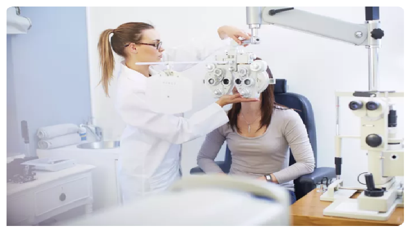
What Do Ophthalmologists Check During Eye Exams? A comprehensive eye exam is simple and comfortable. It shouldn't take more than 45 to 90 minutes. Your doctor may have a staff member do portions of this exam. Here is what the exam should include: Your medical history Your doctor will ask you about your vision and your general health. They will ask about: your family's medical history, what medications you take, and whether you wear corrective lenses. Your visual acuity Are all eye charts the same? How to use the Snellen eye chart When should you get an eye exam? A "Tumbling E" eye chart from the 1950s. The chart has capital E's facing in different directions, so people being tested can indicate which direction the letter is pointing, instead of having to read different letters. This is the part of an eye exam people are most familiar with. You will read an eye chart to determine how well you see at various distances. You cover one eye while the other is being tested. This exam will determine whether you have 20/20 vision or not. Your prescription for corrective lenses Your doctor will ask you to look at an eye chart through a device called a phoroptor. The phoroptor contains different lenses. It will help determine the best eyeglass or contact lens prescription for you. Your pupils Your doctor may check how your pupils respond to light by shining a bright beam of light into your eye. Pupils usually respond by getting smaller. If your pupils widen or don't respond, this may reveal an underlying problem. Your side vision Loss of side vision (peripheral vision) may be a symptom of glaucoma. This test can find eye problems you aren't aware of because you can lose side vision without noticing. Your eye movement A test called ocular motility evaluates the movement of your eyes. Your ophthalmologist looks to see if your eyes are aligned. They also check that your eye muscles are working properly. Your eye pressure Eye pressure testing, called tonometry, measures the pressure within your eye (intraocular eye pressure, or IOP). Elevated IOP is one sign of glaucoma. The test may involve a quick puff of air onto the eye or gently applying a pressure-sensitive tip near or against your eye. Your ophthalmologist may use numbing eye drops for this test for your comfort. The front part of your eye Your ophthalmologist uses a slit-lamp microscope to light up the front part of the eye. This includes the eyelids, cornea, iris and lens. This test checks for cataracts or any scars or scratches on your cornea. Your retina and optic nerve Your ophthalmologist will put dilating eye drops in your eye to dilate, or widen, your pupil. This will allow them to examine your retina and optic nerve for signs of damage from disease. Your eyes might be sensitive to light for a few hours after dilation. Other Tests During an Eye Examination Your ophthalmologist may suggest other tests to further examine your eye. This can include specialized imaging techniques such as: optical coherence tomography (OCT) fundus photos fluorescein angiography (FA) topography, which is a scan of the surface of your cornea automated visual field What you can expect Before the exam If you're seeing a new eye doctor or if you're having your first eye exam, expect questions about your vision and general health history. Your answers help your eye doctor understand your risk of eye disease and vision problems. Questions might include: Are you having eye problems now? Have you had eye problems in the past? Do you wear glasses or contacts? If so, are you satisfied with them? What health problems have you had in recent years? Were you born prematurely? What medications do you take? Do you have allergies to medications, food or other substances? Have you had eye surgery? Does anyone in your family have eye problems, such as macular degeneration, glaucoma or retinal detachments? Do you or does anyone in your family have diabetes, high blood pressure, heart disease or any other health problems that can affect the whole body? During the exam A clinical assistant or technician might do part of the examination, such as taking your medical history and giving the initial eye test. An eye exam usually involves these steps: Measurement of your visual acuity to see if you need glasses or contact lenses to improve your vision. Measurement of your eye pressure. You'll be given a numbing drop in your eyes. To make it easier for your doctor to examine the inside of your eye, he or she will likely give you eyedrops to dilate your eyes. Evaluation of the health of your eyes. After the dilating drops take effect, your eye doctor might use several lights or imaging to evaluate the front of the eye and the inside of each eye. Your doctor might use several tests to check your vision and the appearance and function of all parts of your eyes. After the exam At the end of your eye exam, you and your doctor will discuss the results of all testing, including an assessment of your vision, your risk of eye disease and preventive measures you can take to protect your eyesight. Different types of eye exams Eye muscle test This test evaluates the muscles that control eye movement. Your eye doctor watches as your eyes follow a moving object, such as a pen or small light. He or she looks for muscle weakness, poor control or poor coordination. Visual acuity test This test measures how clearly you see. Your doctor asks you to identify different letters of the alphabet printed on a chart or a screen positioned some distance away. The lines of type get smaller as you move down the chart. Each eye is tested separately. Your near vision also may be tested, using a card with letters held at reading distance. Refraction assessment Light waves are bent as they pass through your cornea and lens. If light rays don't focus perfectly on the back of your eye, you have a refractive error. That can mean you need some form of correction, such as glasses, contact lenses or refractive surgery, to see as clearly as possible. Assessment of your refractive error helps your doctor determine a lens prescription that will give you the sharpest, most comfortable vision. The assessment can also determine that you don't need corrective lenses. Your doctor may use a computerized refractor to estimate your prescription for glasses or contact lenses. Or he or she may use a technique called retinoscopy. In this procedure, the doctor shines a light into your eye and measures the refractive error by evaluating the movement of the light reflected by your retina back through your pupil. Your eye doctor usually fine-tunes this refraction assessment by having you look through a masklike device that contains wheels of different lenses (phoropter). He or she asks you to judge which combination of lenses gives you the sharpest vision. Visual field test (perimetry) Your visual field is the full extent of what you can see to the sides without moving your eyes. The visual field test determines whether you have difficulty seeing anywhere in your overall field of vision. Types of visual field tests include: Confrontation exam. Your eye doctor sits directly in front of you and asks you to cover one eye. You look straight ahead and tell the doctor each time you see his or her hand move into view. Manual testing, including tangent screen and Goldmann exams. You sit a short distance from a screen and focus on a target at its center. You tell the doctor when you can see an object move into your peripheral vision and when it disappears. Automated perimetry. As you look at a screen with blinking lights on it, you press a button each time you see a light. Using your responses to one or more of these tests, your eye doctor determines the fullness of your field of vision. If you aren't able to see in certain areas, noting the pattern of your visual field loss can help your eye doctor diagnose your eye condition. Color vision testing You could have poor color vision without realizing it. If you have difficulty distinguishing certain colors, your eye doctor might screen your vision for a color deficiency. To do this, your doctor shows you several multicolored dot-pattern tests. If you have no color deficiency, you'll be able to pick out numbers and shapes from within the dot patterns. If you do have a color deficiency, you'll find it difficult to see certain patterns within the dots. For most people, color blindness that's present at birth (congenital) is red-green, meaning you can't distinguish those colors. Most people who develop color blindness as a result of disease, such as glaucoma or optic nerve disease, can't distinguish blue-yellow. Slit-lamp examination A slit lamp is a microscope that magnifies and illuminates the front of your eye with an intense line of light. Your doctor uses this device to examine the eyelids, lashes, cornea, iris, lens and fluid chamber between your cornea and iris. Your doctor may use a dye, most commonly fluorescein (flooh-RES-een), to color the film of tears over your eye. This helps reveal damaged cells on the front of your eye. Your tears wash the dye from the surface of your eye fairly quickly. Retinal examination This examination — sometimes called ophthalmoscopy or funduscopy — allows your doctor to evaluate the back of your eye, including the retina, the optic disk and the retinal blood vessels that nourish the retina. Having your pupils dilated with eyedrops before the exam keeps your pupils from getting smaller when your doctor shines light into the eye. After administering eyedrops and giving them time to work, your eye doctor may use one or more of these techniques to view the back of your eye: Direct exam. Your eye doctor uses an ophthalmoscope to shine a beam of light through your pupil to see the back of the eye. Sometimes eyedrops aren't necessary to dilate your eyes before this exam. Indirect exam. During this exam, you might sit up or be reclined in the exam chair. Your eye doctor examines the inside of the eye with the aid of a condensing lens and a bright light mounted on his or her forehead. This exam lets your doctor see the retina and other structures inside your eye in great detail and in three dimensions. Screening for glaucoma Tonometry measures the fluid pressure inside your eye (intraocular pressure). This is one test that helps your eye doctor detect glaucoma, a disease that damages the optic nerve. Several methods to measure intraocular pressure are available, including: Applanation tonometry. This test measures the amount of force needed to temporarily flatten a part of your cornea. You'll be given eyedrops with fluorescein, the same dye used in a regular slit-lamp examination. You'll also receive eyedrops containing an anesthetic. Using the slit lamp, your doctor moves the tonometer to touch your cornea and determine the eye pressure. Because your eye is numbed, the test doesn't hurt. Noncontact tonometry. This method uses a puff of air to estimate the pressure in your eye. No instruments touch your eye, so you won't need an anesthetic. You'll feel a momentary pulse of air on your eye, which can be startling. If your eye pressure is higher than average or your optic nerve looks unusual, your doctor might use a pachymeter, which uses sound waves to measure the thickness of your cornea. The most common way of measuring corneal thickness is to put an anesthetic drop in your eye, then place a small probe in contact with the front surface of the eye. The measurement takes seconds. You might need more-specialized tests, depending on your age, medical history and risk of developing eye disease. Results Results from an eye exam include: Whether you need vision correction, either through glasses, contact lenses or surgery Whether your eyes are healthy, or you have cataracts, glaucoma or retinal disorders, such as macular degeneration or diabetic retinopathy If you need corrective lenses, your doctor will give you a prescription. If your eye exam yields other abnormal results, your doctor will discuss with you the next steps for further testing or for treating an underlying condition. |
|
When Should You Have an Eye Exam? Childhood vision screening Baseline eye exams for adults Seniors and eye exams When Should You Have an Eye Exam? Childhood vision screening From birth through the teenage years, children's eyes are growing and changing quickly. The American Academy of Ophthalmology and the American Association for Pediatric Ophthalmology and Strabismus have developed specific childhood eye screening guidelines. Follow these guidelines to get your child screened at the right times. These screenings help identify when your child might need a complete eye exam. Baseline eye exams for adults If your eyes are healthy and vision is good, you should have a complete exam by your ophthalmologist once in your 20s and twice in your 30s. There are some exceptions: If you have an infection, injury, or eye pain, or you notice sudden floaters and flashes or patterns of light, call your ophthalmologist. If you wear contact lenses, see your eye specialist every year. If you have diabetes or have a family history of eye disease, talk with your ophthalmologist about how often your eyes should be examined. The American Academy of Ophthalmology recommends that adults get a complete eye examination at age 40. This is when early signs of disease or changes in vision may appear. It is important to find eye diseases early. Early treatment can help preserve your vision. Not everyone should wait until age 40 for an eye exam Some adults shouldn't wait until they are 40 to have a complete eye exam. See an ophthalmologist now if you have an eye disease or risk factors such as: diabetes high blood pressure family history of eye disease. After an exam, your ophthalmologist can tell you how often you should have your eyes checked in the future. It's important to follow the schedule your ophthalmologist gives you, especially as you age. Your risk for eye disease increases as you get older. Seniors and eye exams If you are 65 or older, make sure you have your eyes checked every year or two. Your ophthalmologist will check for signs of age-related eye diseases such as: cataracts diabetic retinopathy age-related macular degeneration glaucoma Remember, always follow the schedule your ophthalmologist recommends for future eye exams. |
Exam Room
 An examination room at an eye doctor's office usually consists of an exam chair, a phoropter, an eye chart, a slit lamp and a stool for the eye care practitioner. The 8-Point Eye Exam The key to any examination is to be systematic and always perform each element. 1. Visual acuity In the clinic, visual acuity is typically measured at distance. Otherwise, in a consult setting outside of the clinic, it’s measured at near. Don’t forget to have a near card with you. Make sure the patient is wearing his or her correction. Always have a pair of +3.00 readers with you, as many people in the emergency room won’t have their glasses with them. A pinhole occluder will also reduce the impact of uncorrected refractive error. If the patient is unable to see the biggest optotype on the card, the progression (from better to worse) is counting fingers (CF), hand motions (HM), light perception (LP) with projection, LP without projection and no light perception (NLP). For children who are too young to use Allen pictures, employ the “central, steady, maintain (CSM)” approach. Central: Is the corneal light reflex in the center of the pupil? Steady: Can the patient continue fixating when the light is slowly moved around? Maintain: Can the patient maintain fixation with the viewing eye when the previously covered eye is uncovered? 2. Pupils Look for anisocoria. If present, carefully check the pupil size in both well-lit and dark conditions. Check the reactivity of each pupil with a penlight or Finoff transilluminator. Use the swinging flashlight test to look for a relative afferent pupillary defect. 3. Extraocular motility and alignment Have the patient look in the six cardinal positions of gaze. Test with both eyes open to assess versions — repeat monocularly to test ductions. Figure 1 below shows which muscle is tested in each position. Use the cover/uncover test to assess for heterotropias. Use the alternate cover test to assess for the total amount of deviation. This amount minus any heterotropia is the amount of heterophoria. 4. Intraocular pressure Goldmann applanation tonometry is the gold standard and should be used in the clinic whenever possible. Outside of the clinic, Tono-Pen tonometry is much more practical. If you suspect a ruptured globe, skip this part of the exam. 5. Confrontation visual fields Assess each quadrant monocularly by having the patient count the number of fingers that you hold up. If acuity is particularly poor, have the patient note the presence of a light. Use the colored lid of an eyedrop bottle to define the position of a scotoma more accurately. 6. External examination Look for any ptosis by measuring the margin-to-reflex distance, which is the distance from the corneal light reflex to the margin of the upper lid. Look for lagophthalmos. Note any unusual growths or lesions that may require a biopsy. Palpate lymph nodes and the temporal artery if indicated by the history or exam. Measure proptosis or enophthalmos with an exophthalmometer. Perform a full cranial nerve exam for patients with diplopia or other neurologic symptoms. 7. Slit-lamp examination Lids/lashes/lacrimal system: Normal anatomy and contours? Any lesions? Conjunctiva/sclera: White and quiet? Injection? Lesions? Cornea: Clear? Epithelial disruptions? Stromal opacities? Endothelial lesions? Anterior chamber: Deep? Cell or flare? Iris: Round pupil? Transillumination defects? Nodules? Lens: Clear? Nuclear, cortical or subcapsular cataract? Anterior vitreous: Inflammation? Hemorrhage? Pigmented cells? 8. Fundoscopic examination Optic nerve: Cup-to-disc ratio (see Figure 2 below)? Focal thinning? Pallor? Symmetric? Macula: Foveal light reflex? Drusen, edema or exudates? Vessels: Contour and size? Intraretinal hemorrhage? Periphery: Tears or holes? Lesions? Pigmentary changes?  What Do All Those Abbreviations and Numbers Mean on Your Eye Prescription?
 |