
|
Are you having any problems with your vision? How long have you had these problems? When do these problems occur? When was your last eye examination? Do you have any family history of eye problems? How is your general health? What medications are you taking? Do you have any allergies? Do you wear glasses/contacts now? Have your glasses/contacts become stronger over the years? Do you work with a computer? Do your eyes ever tire (burn, itch) when reading? Do you ever see double images or halos around images? Do you suffer from eye strain and/or tension headaches? |
|
Visual Acuity Visual Acuity: What is 20/20 Vision? What should the illumination be? Does 20/20 mean perfect vision? Is 15/15 vision better than 20/20 Vision? Why do some people have less than 20/20 vision? Will clarity of vision vary with distance? If my vision is less than 20/20, what can I do? What are its limitations? What are visual acuity tests used for? How is visual acuity defined and recorded? What is the reference standard? What should be the measurement steps? What is the Preferred Numbers series? How can I convert between different notations? How can I compare and average the functional consequences? What is the preferred chart layout? What should the viewing distance be? What symbols should be used? What display method should be used? Can colored filters improve Visual Acuity? What is a Snellen chart? Q. Visual Acuity: What is 20/20 Vision? A. 20/20 vision is a term used to express normal visual acuity (the clarity or sharpness of vision) measured at a distance of 20 feet. If you have 20/20 vision, you can see clearly at 20 feet what should normally be seen at that distance. If you have 20/100 vision, it means that you must be as close as 20 feet to see what a person with normal vision can see at 100 feet. 20/20 does not necessarily mean perfect vision. 20/20 vision only indicates the sharpness or clarity of vision at a distance. There are other important vision skills, including peripheral awareness or side vision, eye coordination, depth perception, focusing ability and color vision that contribute to your overall visual ability. Some people can see well at a distance, but are unable to bring nearer objects into focus. This condition can be caused by hyperopia (farsightedness) or presbyopia (loss of focusing ability). Others can see items that are close, but cannot see those far away. This condition may be caused by myopia (nearsightedness). A comprehensive eye examination by a doctor of optometry can diagnose those causes, if any, that are affecting your ability to see well. In most cases, your optometrist can prescribe glasses, contact lenses or a vision therapy program that will help improve your vision. If the reduced vision is due to an eye disease, the use of ocular medication or other treatment may be used. Q. What should the illumination be? A. Visibility of objects is determined by their size, their illumination and their contrast. In visual acuity measurement we want to determine a size threshold. We therefore have to make sure that illumination and contrast are at levels where a ceiling effect is reached, so that small variations in illumination and contrast do not influence the measurement. This leaves a fairly broad range of acceptable values. Q. Does 20/20 mean perfect vision? A. No. 20/20 vision only indicates the sharpness or clarity of vision at a distance. There are other important vision skills, including peripheral awareness or side vision, eye coordination, depth perception, focusing ability and color vision that contribute to your overall vision ability. Q. Is 15/15 vision better than 20/20 Vision? A. No. 15/15 vision means normal sharpness of vision at 15 feet just as 20/20 indicates normal acuity at 20 feet. For consistency, optometrists in the United States use 20 feet as the standard to express sharpness of vision. Other ________ express visual acuity in their own way. In England, for example, optometrists express visual acuity in meters (6/6 is considered normal). Q. Why do some people have less than 20/20 vision? A. Visual acuity is affected by many factors. Less than optimum clarity may result from vision conditions like nearsightedness, farsightedness or astigmatism or from eye diseases. Q. Will clarity of vision vary with distance? A. Some people can see well at a distance, but are unable to bring nearer objects into focus. This condition can be caused by hyperopia (farsightedness) or presbyopia (loss of focusing ability). Others can see items that are close, but cannot see those far away. This condition may be caused by myopia (nearsightedness). Q. If my vision is less than 20/20, what can I do? A. A comprehensive eye examination by a doctor of optometry can diagnose those causes, if any, that are affecting your ability to see well. In most cases, your optometrist can prescribe glasses, contact lenses or a vision therapy program that will help improve your vision. If the reduced vision is due to an eye disease, the use of ocular medication or other treatment may be used. Traditional Snellen chart used for visual acuity testing 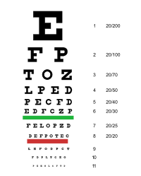
|
|
Visual Acuity, Vision Impairments and Low Vision
What are the medical terms for nearsightedness and farsightedness? What is visual acuity? What is legal blindness? What is visual impairment? What is low vision? Can eyes be transplanted? What does 20/20 vision mean? What are the medical terms for nearsightedness and farsightedness? The medical term for nearsightedness is myopia, correctable with glasses, contact lenses or, in some cases, refractive surgery (LASIK or Corneal Ring Implants). The medical term for farsightedness is hyperopia, correctable with glasses, contact lenses or, in some cases, refractive surgery. Related conditions (also correctable with glasses or contact lenses) include astigmatism and presbyopia. What is visual acuity? Acuity is the measure of the eye's ability to distinguish the smallest identifiable letter or symbol, its details and shape, usually at a distance of 20 feet. This measurement is usually given in a fraction. The top number refers to the testing distance measured in feet and the bottom number is the distance from which a normal eye should see the letter or shape. So, perfect vision is 20/20. If your vision is 20/60, that means what you can see at a distance of 20 feet, someone with perfect vision can see at a distance of 60 feet. What is legal blindness? You are legally blind when the best corrected central acuity is less than 20/200 (perfect visual acuity is 20/20) in your better eye, or your side vision is narrowed to 20 degrees or less in your better eye. Even if you are legally blind, you may still have some useful vision. If you are legally blind, you may qualify for certain government benefits. What is visual impairment? If neither of your eyes can see better than 20/60 without improvement from glasses or contacts, you may be defined as visually impaired. In addition, poor night vision, limited side vision, double vision and loss of vision in one eye may also determine visual impairment. What is low vision? Low vision is a term describing a level of vision below normal (20/70 or worse) that cannot be corrected with conventional glasses. Low vision is not the same as blindness. People with low vision can use their sight. However, low vision may interfere with the performance of daily activities, such as reading or driving. Can eyes be transplanted? No. Currently, there is no way to transplant a whole eye. However, corneas have been successfully transplanted for many years. What does 20/20 vision mean? 20/20 vision is a term used to express normal visual acuity (the clarity or sharpness of vision). 20/20 basically means that you can see clearly at 20 feet what should normally be seen at that distance. Therefore, the first number (20) is always the same, but the second number changes to indicate your visual acuity. For example, if your doctor tells you that you have 20/100 vision, it means that you must be as close as 20 feet to see what a person with normal vision (normal being 20/20) can see at 100 feet. However, 20/20 does not necessarily mean perfect vision � it only indicates the sharpness or clarity of vision at a distance and does not tell you how well you see things that are close up. |
| Eye Examination Report |
| Eye exams for children |
| Adult Eye Exams |
|
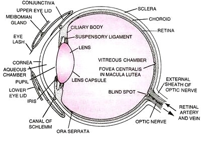
How does the eye work? When you take a picture, the lens in the front of the camera allows light through and focuses that light on the film that covers the back inside wall of the camera. When the light hits the film, a picture is taken. The eye works in much the same way. The front parts of the eye (the cornea, pupil and lens) are clear and allow light to pass through. The light also passes through the large space in the center of the eye called the vitreous cavity. The vitreous cavity is filled with a clear, jelly-like substance called the vitreous or vitreous gel. The light is focused by the cornea and the lens onto a thin layer of tissue called the retina, which covers the back inside wall of the eye. The retina is like the film in a camera. It is the seeing tissue of the eye. When the focused light hits the retina, a picture is taken. Messages about this picture are sent to the brain through the optic nerve. This is how we see. |
The Human Eye
|
| Avoiding Eye Injuries |
| Corrective Eye Surgeries for Refractive Errors |
| Cosmetic Safety for Contact Lens Wearers |
| Eye Care Glossary |
| Eye Glasses and Contact Lenses |
| Eye Safety at the Computer |
|
Eye Disorders A - D Age-Related Macular Degeneration Allergic Conjunctivitis Amblyopia Anatomy of the Eye Astigmatism Atopic Keratoconjunctivitis Blepharitis Cancer (Ocular Melanoma) Cataract Chalazion (Stye) Cone Dystrophy Conjunctivitis Contact Lens Corneal Erosions Corneal Ulcer (Keratitus) Cystoid Macular Edema Cytomegalovirus Retinitis Detached Retina Diabetic Retinopathy Diplopia (Double Vision) Dry Eye E - M Ectropion Endophthalmitis Entropion Esotropia Exotropia Eye Examinations Eye Injuries Eye Spasm (Eye Twitch) Floaters and Flashes Glaucoma Graves' Disease Seasonal Allergic Conjunctivitis (Hayfever) Herpes Simplex Herpes Zoster How the Eye Works Hyperopia (Farsightedness) Hypertension Iritis (Uveitis) Itchy Eyes Juvenile X-linked Retinoschisis (Retinoschisis) Keratitis (Corneal Ulcer) Keratoconus Low Vision Macular Degeneration Melanoma (Ocular Melanoma) Myopia (Nearsightedness) N - Z Nail-Patella Syndrome Ocular Melanoma (cancer) Optic Atrophy Optic Neuritis Overflow Tearing Pingueculum Pink Eye Presbyopia Pterygium Ptosis Refractive Errors Reiters Syndrome Retinal Detachment Retinitis Pigmentosa Retinoblastoma Retinopathy of Prematurity (ROP) Retinoschisis Stargardt Disease Stye (Chalazion) Strabismus Thyroid Eye Disease (Graves' Disease) Twitch Ushers Syndrome Uveitis (Iritis) Vernal Keratoconjunctivitis (VCK) |
|
Common Eye Disorders
Allergic Conjunctivitis (Eye Allergies): a condition occurring when eyes react to something irritating them. The eyes become red, swollen, watery and itchy. The condition is usually temporary, associated with seasonal allergies. Severe allergic eye symptoms can be extremely annoying and are a common reason for visits to an allergist, ophthalmologist, and even the emergency room. Amblyopia (Lazy Eye): loss or lack of development of central vision in an eye that has no health related problems and is not correctable with glasses. It is most often due to crossed eyes or a large difference in the amount of nearsightedness or farsightedness between the two eyes. Astigmatism: a defect of an ocular structure (most commonly the cornea or the crystalline lens) causing rays from a point to fail to meet in a single focal point, resulting in an imperfect blurred or smeared image. For example, astigmatism may result if the cornea is more oval than round. Blepharitis: inflammation, redness, burning, and itching of the eyelids, most commonly in adults, that can be associated with a low grade bacterial infection or a generalized skin condition. Cataract: a clouding of the crystalline lens of the eye or its surrounding transparent membrane. It causes light to be obstructed or scattered throughout the eye resulting in blurry, hazy, or distorted vision. Chalazion: an inflamed lump on the eyelid formed by retention of Meibomian gland secretions (oil). The inflammation usually subsides, but may need surgical removal. Choroiditis (posterior uveitis): inflammation of the choroid (the underlying bed of the retina). Color Deficiency: an inability to distinguish some colors and shades. It occurs when the color-sensitive cone cells in the retina do not properly pick up or send normal color signals to the brain. Conjunctivitis (Pink Eye): infection and inflammation of the conjunctiva (the mucous membrane that lines the inner surface of the eyelids and the sclera on the front of the eyeball), usually from an allergy, a virus, or a bacterium. Crossed Eyes (Strabismus): a visual disorder where the eyes are misaligned due to some type of muscle imbalance in one eye, causing that eye to turn in, out, up, or down relative to the other eye. Diplopia: double vision, usually caused by a strabismus (a visual disorder where the eyes are misaligned due to some type of muscle imbalance in one eye, causing that eye to turn in, out, up, or down relative to the other eye). Diabetic Retinopathy: changes in the retina due to uncontrolled diabetes. There are two types of diabetic retinopathy. Non proliferation diabetic retinopathy (NPDR) is an early stage where retinal blood vessels weaken which leads to microaneurisms, causing vision loss. Proliferative diabetic retinopathy (PDR) involves the growth of new blood vessels out of the retina, which leads to the formation of scar tissue and/or the leaking of blood into the eye, causing severe vision loss or blindness. Dry Eye: a chronic lack of sufficient lubrication and moisture in the eye, causing sensations of dryness, scratchiness or burning. It can be caused by many things that are all related to tear film abnormalities. It occurs more often in women and becomes more common as you age. Endophthalmitis: an inflammation of tissues inside the eye, usually caused by bacteria or fungi. Episcleritis: an inflammation (usually localized) of the episclera, a thin layer of tissue covering the sclera and containing many blood vessels that nourish the sclera which is the white, fibrous, protective, external layer of the eye. Eye Allergies (Allergic Conjunctivitis): a condition occurring when eyes react to something irritating them. The eyes become red, swollen, watery and itchy. The condition is usually temporary, associated with seasonal allergies. Severe allergic eye symptoms can be extremely annoying and are a common reason for visits to an allergist, ophthalmologist, and even the emergency room. Farsightedness (Hyperopia): a condition in which visual images come to a focus behind the retina of the eye and vision is better for distant than for near objects. Flashes (Photopsia): the brief perception of light that is purely subjective and accompanies a pathological condition, especially of the retina or brain. Floaters (Muscae Volitantes): bits of optical debris (such as dead cells or fibrils), usually in the vitreous humor, that may be perceived as spots, spiders or mesh before the eyes. Glaucoma: a progressive disease of the optic nerve, resulting in a reduction in the visual field and even blindness. The most significant risk factor is elevated intraocular pressure (IOP), which results from less fluid leaving the eye than is entering the eye. Graves' Eye Disease: an autoimmune thyroid condition that often attacks the eye muscles and connective tissue within the eye socket. The disease is found 5 or 6 times more frequently in women than men. Hemorrhage, subconjunctival: leakage of blood from blood vessels underneath the conjunctiva, often due to a sudden jolt from blunt trauma, coughing, or sneezing. Normal reabsorption of blood usually takes 1-2 weeks. Herpes Zoster Ophthalmicus (Shingles): involves the orbit of the eye and is caused by the virus reactivating in the ophthalmic division of the trigeminal nerve. In some people, symptoms may include conjunctivitis, keratitis, uveitis, and optic nerve palsies that can sometimes cause chronic ocular inflammation, loss of vision, and debilitating pain. Hordoelum (Sty or Stye): an inflamed swelling of a sebaceous gland at the margin of an eyelid. Hyperopia (Farsightedness): a condition in which visual images come to a focus behind the retina of the eye and vision is better for distant than for near objects. Hyphema: a hemorrhage in the anterior chamber of the eye. Iridocyclitis (intermediate uveitis): inflammation of the iris and the ciliary body. Iritis (anterior uveitis): inflammation of the iris. Keratitis: inflammation of the cornea. It often occurs after a corneal trauma with a foreign body (including contact lenses), and with dry eyes or an eyelid disease which allows bacteria or fungi to enter the cornea. Keratoconus: a degenerative corneal disease characterized by generalized thinning and cone-shaped protrusion of the central cornea. It is hereditary and usually occurs in both eyes. Lazy Eye (Amblyopia): loss or lack of development of central vision in an eye that has no health related problems and is not correctable with glasses. It is most often due to crossed eyes or a large difference in the amount of nearsightedness or farsightedness between the two eyes. Legal Blindness / Low Vision: a loss of eyesight (usually indicated by vision of less than 20/200) that can't be improved with eyeglasses, medicine or surgery. Everyday tasks become more difficult to accomplish, causing a person to find new ways to do them. Low vision aids and low vision rehabilitation can empower a person to maintain much of their independence. Macular Degeneration: a degenerative process of the cells in the central area of the retina (the macula). It reduces the central part of the field of vision as opposed to the peripheral vision. There are two common types of macular degeneration. Dry macular degeneration affects 90% of those with the disease, and wet macular degeneration affects about 10%, but can be more devastating than dry. Migraine (Classic): a chronic neurovascular disorder characterized by moderate to severe headaches usually affecting one half of the head, lasting 2 - 72 hours and often associated with nausea. The headache is associated with a visual aura that occurs before or during the headache. It is characterized by partial alterations in the field of vision which can flicker, zig zag, appear as flashing lights, sometimes accompanied by sensations of smoked glass or tunnel vision. Migraine (Retinal ): a retinal migrane is a symptom of a retinal disease typically affecting only one eye. It is caused by a vascular spasm in or behind the affected eye. (see below: Retinal Migrane) Migraine (Silent): a silent migrane is a classic migrane including a visual aura symptoms and without the headache symptoms. Muscae Volitantes (Floaters): bits of optical debris (such as dead cells or fibrils), usually in the vitreous humor, that may be perceived as spots before the eyes. Myopia (Nearsightedness): a condition in which visual images come to a focus in front of the retina of the eye and vision is better for near than for far objects. Nearsightedness (Myopia): a condition in which visual images come to a focus in front of the retina of the eye and vision is better for near than for far objects. Nystagmus: involuntary, rhythmical, repeated oscillations of one or both eyes, in any or all fields of gaze. Optic Neuritis: inflammation of the optic nerve within the eyeball (papillitis) or behind the eyeball (retrobulbar optic neuritis). Photophobia: an abnormal sensitivity to light, sometimes causing discomfort. It often results from an inflammation of the cornea or iris. Photopsia (Flashes): the brief perception of light that is purely subjective and accompanies a pathological condition, especially of the retina or brain. Pinguecula: a yellowish nodule in the conjunctiva at the front of the eye, usually but not always on the nasal side. It is thought to represent degeneration in the conjunctiva as a result of dryness, as well as of exposure to wind and dust. Pink Eye (Conjunctivitis): infection and inflammation of the conjunctiva (the mucous membrane that lines the inner surface of the eyelids and the sclera on the front of the eyeball), usually from an allergy, a virus, or a bacterium. Posterior Vitreous Separation (PVS): pulling away or shrinking of the vitreous (the gel-like substance that fills the interior of the eye) from the retinal surface causing flashes and floaters. Presbyopia: a visual condition which becomes apparent most often after the age of 45 in which loss of elasticity of the crystalline lens causes reduced accommodation and the inability to focus sharply at a near distance without the use of magnifying reading glasses. Protanopia: a type of color deficiency in which the spectrum is seen in tones of yellow and blue with confusion of red and green and reduced sensitivity to monochromatic lights from the red end of the spectrum. Pterygium: a wedge-shaped fleshy mass of thickened conjunctiva occurring usually on the inner aspect of the eyeball, covering part of the cornea, and often causing a disturbance of vision due to corneal distortion. They are benign growths most commonly caused by exposure sunlight. Ptosis: a paralytic drooping of an eyelid. Red Eye: a term used to describe an eye that appears red due to illness, injury, or some other condition. There are many possible causes of a red eye. The most common is conjunctivitis. Others include blepharitis, acute glaucoma, injury, subconjunctival hemorrhage, keratitis, iritis, episcleritis, scleritis, inflamed pterygium, inflamed pinguecula, dry eye syndrome, airborne contaminants, a burst blood vessel, tick borne illnesses like Rocky Mountain spotted fever, high stress levels and drug use including cannabis. Retinal Detachment: a separation of the retina from the underlying supportive tissue, often initiated by a retinal tear. Observation of sudden spots, floaters, flashes of light, and/or a web moving in your field of vision indicate the possibility of retinal detachment. Retinal Migrane: a migraine where there are repeated attacks of visual disturbances preceding the headache phase. Attacks begin with monocular visual symptoms, where each eye is used separately,like scintillations (sparks or flashes), scotoma (areas of depressed vision) or temporary blindness. The headache phase begins within 1 hour of the original symptoms and lasts 4 to 72 hours. Retinitis Pigmentosa: degeneration of the retina manifested by night blindness and gradual loss of peripheral vision, eventually resulting in tunnel vision or total blindness. Shingles (Herpes Zoster Ophthalmicus): involves the orbit of the eye and is caused by the virus reactivating in the ophthalmic division of the sensory nerve supplying the eye and surrounding structures. In some people, symptoms may include conjunctivitis, keratitis, uveitis, and optic nerve palsies that can sometimes cause chronic ocular inflammation, loss of vision, and debilitating pain. Strabismus (Crossed Eyes): a visual disorder where the eyes are misaligned due to some type of muscle imbalance in one eye, causing that eye to turn in, out, up, or down relative to the other eye. Sty or Stye (Hordoelum): an inflamed swelling of the sebaceous glands of Zeis, located in an eyelash follicle on the margin of an eyelid. Subconjunctival Hemorrhage: leakage of blood from blood vessels underneath the conjunctiva, often due to a sudden jolt from blunt trauma, coughing, or sneezing. Normal reabsorption of blood usually takes 1-2 weeks. Tritanopia: a type of color deficiency marked by confusion of blue and yellow. Uveitis: inflammation of any or all of the structures contained in the uvea (including the iris, ciliary body and choroid). Here are further guidelines. |