
State Department of Emergency Medicine
Emergency Medical Services Training Programs
Emergency Medicine Specialist Training Programs
Annotation or definition of Emergency Medical Services.
|
What are Emergency Medical Services in the state or outside the state? Why was there a need to explain definition of Emergency Medical Services in the state or outside the state? Some consider emergency medical services as providing out-of-hospital acute medical care. Emergency medical services are out-of-hospital on the spot, on the spot to medical emergency room, in medical emergency room, in an operating room or in an intensive care unit. Assessment by medical emergency specialist in emergency medical situation. What should be your focus of assessment in an emergency medical situation? 1. Glasgow coma scale. 2. Vital signs, including consciousness. 3. No pain, wounds, or abnormal findings that needs emergency treatment. 4. Mobility assessment (Is individual able to walk or make limb movements relevant to age?) 5. Survival needs assessment. 6. Emotion: Anger, sadness, fear or emotions not relevant to situation need further evaluation 7. Psychiatric evaluation if required: Normal What are other names for emergency medical specialist? Emergency physician How are emergency medical Services abbreviated in various states? EMS (emergency medical services). EMS also is elaborated as Express Mail Service. To avoid confusion, elaborate emergency medical services fully. Who should do evaluation or treatment of patient on the spot, on the way to the hospital, and in the emergency room? A physician should do this job. On the spot and on the way to the hospital, one physician should attend. If the patient needs emergency room treatment, the case should be handed over to another physician in the medical emergency room. What are other names for emergency medical services in various states? Emergency squad Rescue squad Ambulance squad Ambulance service Ambulance corps Life squad Paramedic service, a first aid squad, is not an appropriate term for emergency medical service. Emergency medical services have to be provided by a physician on the spot, on the way to the hospital, and in the hospital. How do you contact an emergency medical service in the state? Every state has an emergency telephone number. An emergency telephone number operator, who should also ideally be a physician, puts them in contact with a control facility, which will then dispatch a suitable resource to deal with the situation. In some parts of the world, emergency medical service also encompasses the role of moving patients from one medical facility to an alternative one. What have been findings of emergency medical services in various states in North America? A physician should take care of patient on the spot and on the way to hospital. Paramedics who are not able to reach to correct diagnosis and treatment and education of only basic life support (BLS) or advanced life support (ALS) personnel, including paramedics and nurses, were only available up to March 1, 2015. What is the job title of a physician who has the duty to evaluate and treat various emergency medical complaints? Emergency medicine specialist. A junior emergency medicine specialist attends on the spot. A senior emergency medical specialist provides guidance through the Internet from a distance, for example the guidelines displayed here. How many emergency medical specialists (first responder) are required in every state? We will take example of Chicago, Illinois. On March 8, 2015, there were 60 ALS ambulances and 15 BLS ambulances in Chicago, Illinois. 300 emergency medical specialists (first responder) were required in Chicago, Illinois on March 8, 2015. How many emergency medical specialists (first responder) were there in Chicago, Illinois on March 8, 2015? Almost negligible. Is there a difference between an emergency medical specialist (first responder) and an emergency medicine specialist (medical emergency room)? Yes. What is the difference between an emergency medical specialist (first responder) and emergency medicine specialist (medical emergency room)? Emergency medical complaint evaluation, diagnosis and treatment of out-of-hospital acute medical situation is the job of an emergency medical specialist (first responder) up to the medical emergency room. The job location of an emergency medicine specialist (medical emergency room) is in a medical emergency room. An emergency medicine specialist (medical emergency room) can receive walk-in patients; others can bring the patient, referrals, or via ambulance. Emergency medicine specialist or senior emergency medical specialist guide communicates through Internet from a distance, for example the guidelines displayed here. Why is there a need for an emergency medical specialist (first responder)? Is this medical situation undetermined, good, fair, serious, or critical? What is the diagnosis? What is the treatment? An emergency medicine specialist has to answer these questions. What staff is associated with emergency medical services in the state or outside the state? First responder (ideally should be emergency medicine specialist) Emergency medicine specialist (medical emergency room) Guide for emergency medicine specialist Ambulance driver Ambulance care assistant Emergency medical technician Emergency medical dispatcher Paramedic What should an emergency medicine specialist know? Here are further guidelines http://www.qureshiuniversity.com/medicalemergencyworld.html |
Assessment of a patient in medical emergency by an emergency medicine specialist
|
Assessment in medical emergency situation. | ||||||||||||
|
Assessment in medical nonemergency situation. | ||||||||||||
|
Is there a difference between analysis of complaints, incidents, issues, problems, and assessment of a patient? Yes. What is the difference between analysis of complaints, incidents, issues, problems, and assessment of a patient? Assessment of patient includes:
Analysis of complaints, incidents, issues, and problems. What should be your first question in case a patient is referred to you? Assessment in medical emergency situation. Questions that must be answered. Where is the patient now? How old is the patient? What is the gender of the patient? Who is reporting this emergency? What seems to be the complaint? What seems to be the problem? Glasgow Coma scale analysis. First, analyze Glasgow Coma scale, then analyze vital signs including consciousness. When was the patient normal? Can the patient open both eyes spontaneously? Can the patient talk or make noise relevant to age? Can the patient walk or move extremities relevant to age? If yes, Glasgow Coma scale is 15. Glasgow Coma scale of 15 means the patient is not in a coma. The patient can have less serious medical issues. Go ahead with vital signs, including consciousness. When did it start? How did it start? Where did it start? How much time has elapsed from the start of the emergency until now? What do you think causes it? What do I think caused it? What needs to be done to verify what caused it? Has patient taken any medication or substance before this issue? Has any specific thing happened that led to this issue? Is this troubling your everyday activity? How is this troubling your everyday activity? Does one individual or many individuals have medical emergencies at this location? How many individuals have medical emergencies at this location? Is it a medical emergency? ___________________________ In what type of setting does this patient need treatment? ___________________________ Do any recent causes lead to this problem; for example, trauma, missed medication, inadequate survival needs, stress, or other issue? ___________________________ What are further details? ___________________________ Does any past medical history lead to this problem? ___________________________ Is there any recent history within past few minutes or hours of any of the following: 1.Unconsciousness at a public location. 2.Sudden unconsciousness at home. 3.Trauma. 4.Survival needs issues. 5.Seizures. 6.Burns. 7.Drowning. ___________________________ If there is even one recent history of the above, on the spot diagnosis and treatment is required. Is the victim's condition life or limb threatening? ___________________________ Could the victim's condition worsen and become life or limb-threatening on the way to the hospital? ___________________________ Could moving the victim cause further injury? ___________________________ Would distance or traffic conditions cause a delay in getting the victim to the hospital? ___________________________ What have been his activities for the last 10 years? ___________________________ Does the individual use or abuse any of these substances? Alcohol. Drugs. Tobacco. ___________________________ Is the individual on any medication? ___________________________ Questions doctor on duty needs to answer. Is it a medical emergency? ___________________________ Does this need on-the-spot diagnosis and treatment? ___________________________ What is the most likely diagnosis? ___________________________ What do you think causes it? ___________________________ Why do you think this happened? ___________________________ What is the diagnosis? ___________________________ In what setting/location does this medical condition need treatment? Treatment required on the spot. Treatment required in the medical emergency room. Treatment required in the intensive care unit. Treatment required in the ward. Treatment required in the operating room. Treatment required at home. Treatment required Internet health care. Treatment required in OPD consultation. ___________________________ What treatment do you recommend for this patient? ___________________________ What are other treatment options for this patient? No other treatment option. Other treatment options are enumerated. ___________________________ Here are further guidelines.
If it is a multiple causality incident, the guidelines are different. What problems, complaints, incidents, and issues need on-the-spot diagnosis and treatment? Can the patient talk? Can the patient respond to verbal questions accurately? Can the patient do spontaneous eye opening? Does the patient respond to painful stimulus? Is the patient conscious, oriented in time, space, and person? Conscious means able to see, hear, and talk. In pediatric patients younger than six months of age, the ability to make any verbal noise or cry is equivalent to talking. What best describes you in a medical emergency?
What is wrong with existing physicians around the world? Existing physicians around the world are not able to do proper health care assessment in various human healthcare settings. Existing physicians are not able to reach correct diagnoses and treatment in various human healthcare settings. How many types of human health care assessment are there? There are 18 types of human health care nonemergency assessment and 15 types of human health care, emergency assessment in various human healthcare settings. What is an assessment of a patient? Patient assessment is the term used to describe the process of identification of the condition, needs, abilities, and genuine preferences of a patient. Identify possible solutions and/or remedies. Prepare a plan. Who should ideally do an assessment of a patient? The physician should ideally do an assessment of the patient. There is only one best doctor on this planet.  His name: Dr. Asif Qureshi. His focus: The planet. Questions relevant to the patient. Here are further guidelines. PATIENT ASSESSMENT DEFINITIONS Emergency Medical Services. Here are further guidelines. Emergency medical room in a hospital Here are further guidelines. Primary care physician consultation New patient relevant to primary health care (nonemergency). Here are further guidelines. What is a Medical Emergency? Who are the people behind 911 or emergency call responders? Ideally, an experienced competent medical doctor should be behind 911 or emergency call responders. What is the role of www.qureshiuniversity.com/medicalemergencyworld.html in a medical emergency? Guide the following: Doctor in a medical emergency. Emergency responder in a medical emergency. Emergency call center in a medical emergency. Watcher, relative, or acquaintance of the victim in a medical emergency. Victim himself or herself in a medical emergency. Guide the state department of health worldwide. | ||||||||||||
| Administrative Issues | ||||||||||||
| Emergency Diagnosis Code | ||||||||||||
| Assessment of the newborn infant | ||||||||||||
| Annual Physical Examinations | ||||||||||||
| OUTLINE FOR PEDIATRIC HISTORY & PHYSICAL EXAM |
Assessment in medical nonemergency situation.
|
How should you do assessment in a medical nonemergency situation? Is it a human healthcare complaint or a nonhuman healthcare complaint? Why is the answer to this question essential? A complaint can be relevant to more than 40 departments in the state or outside the state. A human healthcare complaint is one of them. What should an emergency medical specialist verify in the community? Are there competent primary care physicians in the community? Is it safe to live in the community? Assessment in a medical nonemergency situation Consultation through Internet. Doctor Consultation Patient Profile Individualized consultation nonemergency patient Comprehensive patient assessment How is a comprehensive patient assessment done? Take a look at this. Questions you need to answer. How should you answer these questions? Answer to the best of your ability and knowledge. What should you write if a question is not applicable to you? This is not applicable to me at this point. What seems to be the issue? What is the name and date of birth of the patient? What is your Email address? What is the name of the individual who needs doctor consultation? What is the date of birth of the individual who needs doctor consultation? Address What is your mailing address? What was your mailing address from birth until now? Where is the patient now? Where do you live now? How long have you lived at this address? How long do you plan to live at this address? What is your contact information including current mailing address, telephone, e-mail, and any other details, and person to contact in case of emergency? What is the gender of the patient? What best describes the patient? Child Adolescent girl Adolescent boy Woman Man In general, how is your physical and mental health? Excellent Good Fair Poor What is your telephone number? Have you been in the hospital in the last month? Yes No Do you have health problems that you need help with right away? Yes No Do you have any appointments scheduled with doctors or other specialists? Yes No Screening for survival needs Do you have enough of these resources from the state? Food Clothing Housing Health care Transportation Security Education Consumer goods Communication Do you need any of these resources to be enhanced? What are the issues? Do you need extra help to access services, such as a wheelchair ramp, a computer screen reader or large print materials? Yes No What is the number on your medical card? A medical card number is usually a nine digit number. What state or entity has issued this medical card? What is troubling you? How old is the patient? What languages can you understand? What are the sources of medical history? Where are you located now? Is your complete medical history ready? Do you have a physician referral? Yes No Don't know Who is writing answers to these questions? The patient. Someone else on behalf of patient. If someone else is answering these questions on behalf of the patient, how are you related to the patient? Sister Cousin Brother Mother Father Case manager Relative Primary care physician Nurse If other, specify. Have you gone through the Internet human healthcare guidelines? Take a look at this http://www.qureshiuniversity.com/internethealthcareservices.html, public health guidelines, http://www.qureshiuniversity.com/publichealthworld.html patient education guidelines http://www.qureshiuniversity.com/patienteducation.html at mentioned resource? Do you think your issue or issues have not been answered at this resource and need individualized doctor consultation? What type of doctor consultation is required? Ambulatory human health care Adolescent girls’ consultation Community health center evaluation Critical care consultation (anesthesiology) Coroner investigations Cardiology consultation Dermatology consultation Disability consultation Dental consultation Emergency medicine consultation Endocrinology consultation Forensic medicine consultation Gastroenterology consultation Geriatrics consultation Hematology consultation Internet healthcare consultation Medical negligence consultation Nephrology consultation Neurology consultation Oncology consultation Ophthalmology consultation Orthopedics consultation Otorhinolaryngology consultation Obstetrics & Gynecology consultation Primary care physician consultation Pediatrics consultation Psychiatry consultation Pulmonary medicine consultation Physical medicine & Rehabilitation consultation Public health guidelines Radiology & nuclear medicine consultation Surgical consultation Women's health consultation Brain & central nervous system (nervous system) Circulatory System Digestive System Endocrine System Integumentary system Lymphatic (immune) system Muscular system Reproductive System Respiratory System Skeletal System Urinary system Impairment Rating and Disability Determination Health status How would you describe your health status relevant to your age? 100% mentally fit. 100% physically fit. Do you have any problems with activities mentioned below relevant to your age? Walking Seeing Hearing Speaking Breathing Learning Working Caring for oneself (eating, dressing, toileting, etc.) Performing manual tasks Getting started after sleep Sitting Sleeping Face-to-face, in person consultation. Why are you here today? What best describes your existing medical conditions? In the list below you will find many of the most common diseases and pre-existing conditions. ACID REFLUX (GERD) ADDISON’S DISEASE ALCOHOL ABUSE AND RECOVERY ANXIETY ARTHRITIS ASTHMA ATTENTION DEFICIT HYPERACTIVITY DISORDER (ADHD) AUTISM BASAL CELL CARCINOMA BERGER’S DISEASE BIPOLAR DISORDER BLOOD CLOT BREAST CANCER BRONCHITIS (CHRONIC) CANCER CARDIOVASCULAR DISEASE CHOLESTEROL CHRONIC OBSTRUCTIVE PULMONARY DISEASE (COPD) CONGESTIVE HEART FAILURE (CHF) CROHN’S DISEASE DEPRESSION (MAJOR DEPRESSIVE DISORDER, CLINICAL DEPRESSION AND CHRONIC DEPRESSION) DIABETES TYPE 2 DIABETES TYPE 1 DUI/DWI DUODENAL ULCER EMPHYSEMA EPILEPSY EROSIVE ESOPHAGITIS FIBROMYALGIA GASTRIC ULCER GASTROESOPHAGEAL REFLUX DISEASE (GERD) GLAUCOMA GOUT HEART ATTACK HEARTBURN HEART DISEASE HEART MURMUR HEPATITIS HERPES HIGH CHOLESTEROL HYPERTENSION HYPERTHYROIDISM HYPOTHYROIDISM INSOMNIA KIDNEY STONES MARIJUANA MIGRAINES NARCOLEPSY NASAL POLYPS OBSESSIVE COMPULSIVE DISORDER OSTEOPOROSIS OVERWEIGHT PAGET’S DISEASE PANIC DISORDER PEPTIC ULCER PREMENSTRUAL DYSPHORIC DISORDER POST TRAUMATIC STRESS DISORDER (PTSD) PROSTATE (ENLARGED) RESTLESS LEG SYNDROME (RLS) SCHIZOPHRENIA SEASONAL AFFECTIVE DISORDER (SAD) SEIZURES SLEEP APNEA SLEEP DISORDERS STENT (CARDIAC) STROKE SQUAMOUS CELL CARCINOMA THROMBOSIS TRANSIENT ISCHEMIC ATTACK (TIA) ULCERS ZOLLINGER-ELLISON SYNDROME Did you have any of these medical conditions in the past? What is a pre-existing medical condition? Pre existing condition is a medical condition or healthcare problem that existed before enrolling in a healthcare plan. The "objective standard" of the definition states that a pre-existing condition is anything for which you've received medical advice, care, diagnosis, and treatment prior to your enrollment. By contrast, the "prudent standard" of the definition states that a pre-existing condition is any condition for which symptoms were present although medical advice, care, diagnosis, or treatment may not have been received prior to enrollment. Under the "prudent standard," an "ordinarily prudent" individual surely would have sought medical advice, care, diagnosis, or treatment for any symptoms that were present prior to enrollment. Most states implement the prudent standard of the definition. Here are further guidelines. |
Blood Alcohol Concentration (BAC)
|
What is BAC? BAC refers to the percent of alcohol in a person's blood stream. A BAC of .10% means that an individual's blood supply contains one part alcohol for every 1000 parts blood. In ______, a person is legally intoxicated if he/she has a BAC of .08% or higher. Factors that determine BAC •Number of standard drinks (see below) •Amount of time in which drinks are consumed •Body weight •Gender •Race/Ethnicity •Medications •Food (to lesser extent) One standard drink: •One 12 oz. regular beer (4.5% alcohol) •One 7 oz. malt liquor (7% alcohol) •One 4.5 oz. glass of wine (12% alcohol) •One-third jigger (.5 oz.) of Everclear (95% alcohol) More than one standard drink: •One 16 oz. cup of beer = 1.4 drinks •One 40 oz. beer = 3.6 drinks •One 22 oz. malt liquor = 3 drinks •One 12 oz. glass of wine = 2.9 drinks •One 12 oz. margarita = 2-4 drinks, depending on ingredients •One 12 oz. cup of trashcan punch = 4-10 drinks, depending on ingredients How is Blood Alcohol Concentration Determined? Q: What different methods are used to measure blood alcohol concentration (BAC)? A: There are five different bodily samples that can be screend for a DUI suspect's BAC level: urine, saliva, hair follicles, blood and breath. However, only breath analysis (i.e. breathalyzer) and blood screening are broadly used by law enforcement for gathering evidence. As blood screening is considered more invasive, this method is used less often than breath analysis (mainly after a serious accident or where the suspect has refused a breath test). Q: How do breathalyzer tests work? A: There are different machines on the market used for analyzing a DUI suspect's blood alcohol concentration. Some, though rarely used today, employ what is called a "wet chemical" technique to compare one's breath to a sample. The most current generation of breath analysis machines (still commonly referred to as "breathalyzers"), analyze the alcohol content of exhaled vapor through a method called infrared spectroscopic analysis. The latter method of analysis is based on the scientific principal that captured alcohol vapor absorbs light waves of a particular frequency in the presence of light, depending on the amount of alcohol present. A computer translates this data into the more familiar BAC measurement used to determine the level of alcohol in a person's bloodstream (for example, 0.08 percent BAC). Q: How accurate are they? A: While they are not as accurate as blood tests, breathalyzers have been considered acceptably accurate by most courts as tools for gathering evidence. However, some independent studies have determined that breath readings can vary by 15 percent from actual BAC levels (as measured by a blood draw). Some courts have even thrown out breathalyzer results, calling into question the reliability of the machines. In 1988, a New Jersey court cited the following scientific evidence: (1) high readings for 14 percent of the population due to design flaws; (2) variance in results based on the temperature of the machine itself; (3) different results from the varying body temperatures of test subjects and (4) variances in the presence of hematocrit in the blood also affecting test results. Accuracy also can be hampered by the use of an improperly calibrated machine. Q: Can you beat a breathalyzer test if you're intoxicated? A: No. Some popular methods thought to help individuals fool a breathalyzer test, including the ingestion of breath mints, mouthwash, onions and even pennies, have been shown not to work. Mouthwash (which often contains alcohol) may actually have a tendency to raise a person's BAC. Q: Is it possible to successfully challenge breathalyzer results in court? A: Absolutely. Breathalyzer machines must be tested routinely to make sure they are properly calibrated. A skilled DUI lawyer will look into the maintenance records of the device used to test his or her client and otherwise determine the validity of the evidence. Q: How accurate are those small, inexpensive BAC-testing devices sold to consumers? A: It depends. There is a wide range of products available, some more accurate than others, but it's always best to assume they may be a little off (and they may not be used as as a defense to DUI charges). Saliva alcohol test strips are considered to be among the most accurate consumer BAC tests, since the amount of alcohol in one's bloodstream closely corresponds to the concentration of alcohol in the saliva. Blood alcohol concentration in an emergency room Case report of blood alcohol concentration in an emergency room If blood alcohol concentration results in an emergency room are greater than expected, what should an emergency room physician do? Here is a case report. This case report will make you understand. This happened in Chicago, Illinois, North America, before the year 2004. A person had never consciously consumed alcohol because of being religious Muslim. Suddenly, he had an accident. There was no injury but he felt something was wrong and he needed to go to a medical emergency room. He was taken to Swedish Covenant Hospital in Chicago, Illinois. In the medical emergency room, his blood tests show a high concentration of alcohol. What negligence was the emergency physician guilty of? He did not ask him these questions. He did not verify answers to these questions. Do you consume alcohol? No. Have you ever consciously consumed alcohol? No. How did alcohol concentration come up in his blood? Here are further facts. Before taking a vehicle, he asked for a glass of water from a restaurant while having a meal near Swedish Covenant Hospital. Instead of giving him water he was intentionally given alcohol by others so that sabotage occurred and he was harmed. Sabotage occurred due to the accident. He had never consciously consumed alcohol. This was a scenario of intentional harms and sabotage against him. The emergency room physician should have reported this as medicolegal case and that the patient was a victim of sabotage. What is the best scenario about this case? The victim or patient in this case was himself an emergency room specialist with seven years of hospital experience at that point. |
Urine Drug Test
|
Screening urine for drugs of abuse in the emergency department: Do test results affect physicians' patient care decisions? What is a Urine Drug Test? A urine drug test, also known as a urine drug screen or a UDS, is quick and painless. It tests your urine (pee) for the presence of certain illegal drugs and prescription medications. The urine drug test usually screens for alcohol, amphetamines, benzodiazepines, marijuana, cocaine, and opioids (narcotics). A urine drug test can catch possible substance abuse problems. Once these problems are identified, doctors can help you start a treatment plan. Urine drug tests throughout the treatment help ensure that the plan is working and that you are no longer taking drugs. When Is the Test Used? There are several scenarios where a urine drug test might be ordered. Your primary care doctor may order this test if he or she suspects you have a problem with drugs or alcohol. An emergency room doctor may also request this test if you are confused and your behavior seems strange or dangerous. Many Departments in the state require potential workkers to get a urine drug test before hiring them. One benefit of the urine drug screen is that it can be used to keep people with drug problems out of jobs that require the ability to be alert and focused. A drug-addicted air traffic controller or semi-truck driver, for instance, could put the safety of many people at risk. Drug and alcohol rehabilitation centers test residents on a regular basis. This helps ensure that people receiving treatment for drug or alcohol abuse are staying sober. If you are on probation or parole for a drug- or alcohol-related offense, the officer in charge of your case may request random drug tests to verify sobriety. Finally, the tests can be used in home settings. For instance, parents may ask teenagers to take this test to prove that they are not using drugs or alcohol. If you are planning to test your teen at home, it’s a good idea to consult with your family doctor or another health professional beforehand. He or she can advise you on how to follow up if the test is positive. Types of Urine Drug Tests There are two types of urine drug screens. The first, called the immunoassay, is cost-effective and gives results quickly. However, it has drawbacks. For example, it does not pick up on all opioids (narcotics). Also, it sometimes gives false positives. A false positive occurs when the test results come back positive for drugs, but no drugs have been taken. If your first test does come back positive, but you deny drug use, you will be given a second type of test: a gas chromatography or a mass spectrometry. The procedure for getting the specimen is no different from the procedure for an immunoassay described above. These tests are more expensive and take longer to give results, but they rarely produce a false positive. Other weaknesses common to both types of tests include: •false negatives (the test reports a negative result even if drug use has occurred) •vulnerability to cheating the system •failure to capture same-day drug use How to Take the Test You may take the urine drug test anywhere a bathroom is available—in a doctor’s office or hospital, a place of business, or at home. The test is carried out as follows: •The person administering the test will give you a specimen cup. •You will be required to leave your purse, briefcase, or other belongings in another room while you take the test. You may also be asked to change from your street clothes into a hospital gown. •In some cases, the nurse or technician administering the urine drug screen will accompany you into the bathroom so that you cannot do anything to skew the test results. •Clean your genital area with a moist cloth that the technician will provide. •Begin to urinate into the toilet. •While the urine is in midstream, place the specimen cup in the urine stream. Do not allow the cup to touch your genital area. •When you have finished urinating, put a lid on the cup and deliver it to the technician. Urine Drug Test Results The technician or nurse should be able to inform you of the test results almost immediately. Immunoassays, the most common type of urine drug screening, do not measure drugs themselves. Rather, they measure how drug use interferes with the body’s ability to form antigen-antibody complexes. Results of this test are expressed in ng/mL (nanograms per milliliter). The test is based on a cutoff point so that any number below the cutoff is recorded as a negative screen and any number above the cutoff point is interpreted as a positive screen. The people who are responsible for administering the drug test usually provide results in terms of positive or negative as opposed to numeric values. Many immunoassay tests do not even display the ng/mL measurements. Rather, the results are shown on a test strip that turns different colors to indicate the presence or absence of various substances. If the results are positive for illegal drugs that you have not taken, you should immediately request a gas chromatography. Urine drug test case report What are the possibilities if a urine drug test result is positive? Prescription drug detection. Lab error. Drug abuse. Here is a case report. A person is on prescription medication and is asked to take drug test. Natural prescription medication is detected in the urine. This is not a case of drug abuse. This is a case of legal prescription medication detected in urine that is normal. An incompetent physician secretly tried to report a case of drug abuse, causing embarrassing for all. This happened in Chicago, Illinois, North America, before 2006. |
Criteria for emergency medical services
|
Who needs emergency medical services? Do you feel very sick? Have you been hurt? Do you know someone else who is been harmed? Do you think someone else is very sick or has been hurt? Do you think this situation needs help now within 2-10 minutes? If yes, you should use emergency medical services. Is this medical emergency undetermined, good, fair, serious, or critical? What is the diagnosis? What is the treatment? Emergency medicine specialist has to answer these questions. |
Criteria for discharge from medical emergency room
|
1. Vital signs including consciousness: Normal 2. No pain, wounds, or abnormal findings that needs emergency treatment. 3. Glasgow coma scale: 15. 4. Mobility assessment: Normal. 5. Survival needs assessment: Yes. 6. Emotions: Normal relevant to situation. 7. Psychiatric evaluation if required: Normal 8. Relationship Emergency Emergency diagnosis and treatment and disability assessment are two different assessments. For example, a person can be 100% mentally fit and 95% physically fit. This is relevant to the individual’s age. 1. Vital signs including consciousness: Normal Are vital signs including consciousness normal? Yes. 2. No pain, wounds, or abnormal findings that needs emergency treatment. 3. Glasgow coma scale: 15. Is Glasgow coma scale 15? Yes. 4. Mobility assessment Is individual able to walk or make limb movements relevant to age? Normal 5. Survival needs assessment. Does the individual have survival needs form the state? Yes. Is it safe for the individual at the residence? 6. Emotions: Anger, sadness, fear, or emotions not relevant to situation needs further evaluation. Normal. What best describes the individual’s emotions at this point? Are emotions expression normal relevant to situation? 7. Psychiatric evaluation if required. Is this individual mentally fit relevant to age? Advice on discharge from medical emergency room. This depends on original complaint and diagnosis with relevant treatment. Treatment depends on the underlying cause. 8. Relationship Emergency What is a relationship emergency? Sometimes a relationship emergency is a medicolegal case. Sometimes a relationship emergency can be without medicolegal issues. Is everything normal with your family relationships? Has your relationship become a victim of a criminal conspiracy? If yes, this is relationship emergency. It is a medicolegal case. Where is the women spouse at this point? Who all are involved in this criminal conspiracy? No question can remain unanswered. |
Types of ambulances (Road)
|
Why do you need to mention road ambulance?
Nowadays there are air ambulances as well. What are the types of road ambulance? ALS ambulances BLS ambulances How many total road ambulances are there in the state? What are the types of ambulances in the state? What is the area-wide number of ambulances in the state? For example Chicago, Illinois, on March 8, 2015, had 60 ALS ambulances and 15 BLS ambulances. What type of ambulance will be associated with you? What are salient features of emergency medical ambulances? All ALS and BLS ambulances, and ALS engines, are equipped with state-of-the-art telemetry radio equipment, which allows emergency medical responders to radio important information from the scene of the incident directly to a nearby hospital, where physicians can review the patient’s condition and provide further medical instructions and transport information. |
License Status
|
What should be included in the license status in the state, outside the state, or internationally in this situation?
Active Active-probation Active-provisional Active-restricted Administrative error Approved Deactivated Deleted Denied Expired Expired -reapply Expired-probation Inactive Lapsed Lapsed-probation Lapsed-provisional Lapsed-restricted License in this situation not required Null and void Pending Reinstatement pending Rescinded provisional license Revoked Surrendered Suspended Transfer pending Withdrawn What are examples of situations in which a license is not required? Take a look at this. http://www.qureshiuniversity.com/healthcareworld.html Who has established these guidelines for emergency medical specialist (first responder) and emergency medicine specialist (medical emergency room)? Doctor Asif Qureshi Everything is displayed publicly. Worldwide healthcare professionals including emergency medical specialists (first responder and emergency room) are scrutinizing these guidelines publicly. License or license renewal is not required in this situation. |
Emergency Diagnosis and Treatment
Examples of emergency medical diagnosis and treatment
| Here are further guidelines. |
Glasgow Coma Scale
| Best eye response (E) - 4 grades |
Spontaneous (4 points) To verbal command (3 points) To pain (2 points) None (1 point) |
Score 4 3 2 1 |
Oriented conversation (5 points) Disoriented conversation (4 points) Inappropriate words (3 points) Incomprehensible words (3 points) Incomprehensible sounds (2 points) None (1 point) | 5 4 3 2 1 |
Obeys verbal command (6 points) Localizes painful stimuli (5 points) Flexion withdrawl from painful stimuli (4 points) Decorticate response to painful stimuli (3 points) Decerebrate response to painful stimuli (2 points) None (1 point) |
6 5 4 3 2 1 |
3-14 Points: Abnormal
There is a Paediatric Glasgow Coma Scale applicable to infants too young to speak - and the equivalent infant responses are given in the various sections below.
Three types of response are independently assessed and are recorded on an appropriate chart (and the overall score is made by summing the scores).
|
Best eye response (E) - 4 grades •No eye opening; •Opening to response to pain to limbs as above •Eye opening in response any speech (or shout, not necessarily request to open eyes); •Spontaneous eye opening. |
1 pt - No eye opening 2 pts - Eye opening in response to pain 3 pts - Eye opening in response to speech 4 pts - Spontaneous eye opening Record best level of speech. If patient is intubated, a "derived verbal score" is calculated via a linear regression prediction. •No verbal response. •Incomprehensible speech: Moaning but no words. Infant: Inconsolable, agitated. •Inappropriate speech: Random or exclamatory articulated speech, but no conversational exchange. Infant: Inconsistantly inconsolable, moaning. •Confused conversation: Patient responds to questions in a conversational manner but some disorientation and confusion. Infant: Cries but consolable, inappropriate interactions. •Orientated: Patient 'knows who he is, where he is and why, the year, season, and month. Infant: Smiles, orientated to sounds, follows objects, interacts. |
UNABLE TO ASSESS (eg Intubated) 1 pt - None 2 pts - Incomprehensible speech 3 pts - Inappropriate speech 4 pts - Confused conversation 5 pts - Orientated Apply varied painful stimulus: trapezius squeeze, earlobe pinch, supraorbital pressure, sternal rub, nail-bed pressure etc: •No response to pain. •Extensor posturing to pain: The stimulus causes limb extension (abduction, internal rotation of shoulder, pronation of forearm, wrist extension) - decerebrate posture. •Abnormal flexor response to pain: Stimulus causes abnormal flexion of limbs (adduction of arm, internal rotation of shoulder, pronation of forearm, wrist flexion - decorticate posture. •Withdraws to pain: Pulls limb away from painful stimulus. Infant: withdraws from pain. •Localizing response to pain: Purposeful movements towards changing painful stimuli is a 'localizing' response. Infant: withdraws from touch •Obeying command: The patient does simple things you ask (beware of accepting a grasp reflex in this category). Infant: moves spontaneously or purposefully |
1 pt - No response to pain 2 pts - Extensor posturing to pain 3 pts - Abnormal Flexor response to pain 4 pts - Withdraws to pain 5 pts - Localizing response to pain 6 pts - Obeying commands |
Emergency Surgeon and Emergency anesthetist
|
Why has training for emergency surgeon and emergency anesthetist been mentioned together? Emergency surgeon and emergency anesthetist services in an operating room are interdependent at a specific time duration and location. What medical emergency cases go to an emergency surgeon or emergency anesthetist in a hospital operating room? Out of 1,150 human medical emergencies, only 27 go to an emergency surgeon and/or an emergency anesthetist in operating room. What are examples of emergency cases that go to an emergency surgeon and/or emergency anesthetist in a hospital operating room?
What should an emergency surgeon and emergency anesthetist know? An emergency surgeon and emergency anesthetist should know all types of human diagnosis and treatment in various healthcare settings. Then, he or she should be allowed to manage cases listed after proven expertise. What has experience revealed up to now? Experience has shown that an individual who can do an 8-inch incision closing in three layers without knowing about correct human diagnosis and treatment claims to be surgeon specialist. Another individual who can give IV anesthesia with ventilation claims to be an anesthesia specialist without knowing correct human diagnosis and treatment. A technician knows these techniques. These techniques can be learned in 4-6 weeks. Surgical procedure Questions to be answered before the surgery. Questions to be answered in postoperative notes. Questions to be answered in follow-up consultations. If the expected procedure or surgery is likely to harm the patient, do not go ahead with surgery. If all the questions are not answered, do not go ahead with surgery. What is your Email address? What is the name of the individual who needs doctor consultation? What is the date of birth of the individual who needs doctor consultation? What is your mailing address? What was your mailing address from birth until now? Where is the patient now? Where do you live now? How long have you lived at this address? How long do you plan to live at this address? What is your contact information including current mailing address, telephone, e-mail, and any other details, and person to contact in case of emergency? Questions to be answered before the surgery. Who will do the expected procedure? What is the expected procedure? What is the expected date, time, and location of surgery? What is the diagnosis of the patient? Who verified the diagnosis of the patient? What is the profile of the patient’s primary care physician? How will this procedure help or enhance the life of the patient? Is the surgery really required? If surgery is really indicated, these questions must be answered. Why do you recommend this operation? What operation are you recommending? Is there more than one way to do this operation? Are there alternative to surgery? What are the details of the operation? What are the advantages of this operation? What are the risks of having this operation? What will happen if this operation is not done? Who can give a second opinion? What kind of anesthesia is required? How long is the operation? How long will it take to recover from the operation? How much experience has the doctor had in diagnosing and treating such cases? How much experience does the doctor have in this specific operation? Has this type of operation been discussed publicly? At what hospital will the operation be done? How long will the doctor be available in the hospital? Has the surgeon marked the site where he or she will operate with all the preoperative, operative, and postoperative guidelines? What is the gender of the patient? What best describes the patient? Child Adolescent girl Adolescent boy Woman Man What best describes the surgery? Cardiothoracic surgery Eye surgery General surgery Neurosurgery OB/GYN surgery Oral and maxillofacial surgery Orthopedic surgery Otolaryngology Pediatric surgery Plastic surgery Urology Is this emergency surgery, urgent surgery, or elective surgery? What are examples of emergency surgery, urgent surgery, and elective surgery? Emergency surgery www.qureshiuniversity.com/emergencysurgery.html Urgent surgery href://www.qureshiuniversity.com/urgentsurgery.html Elective surgery href://www.qureshiuniversity.com/electivesurgery.html Questions to be answered in postoperative notes. Here are further guidelines. Questions to be answered in follow-up consultations. How did the patient improve or was helped by the specific procedure or surgery? In general, how is your physical and mental health? Excellent Good Fair Poor I have read and agree to the Terms & Conditions. These are basic questions. There are many more. Surgery: Is it really indicated? A statement mentions that the surgery department lacks equipments and infrastructure. What type of equipment do you need? If you audit existing surgeries, you will discover that most of them are not required, and there has been wrong clinical diagnosis. A diagnosis of appendicitis: on operation, no findings of appendicitis. A diagnosis of cholecystitis or cholelithiasis: on operation, no findings of cholecystitis or cholelithiasis. A medical doctor is required to make correct clinical diagnoses. A surgeon is basically a medical doctor. They ask for number of unwanted investigations but after that they still cannot reach a correct diagnosis and treatment. Q: Who is a surgeon? A: A surgeon is a medical doctor with additional training in specific medical procedures. Getting the title of surgeon does not mean he or she is a competent medical doctor. Not all surgeons can perform all medical procedures. Not all medical doctors can perform all medical procedures. Making an eight-inch incision and closing in three layers does not prove you are a surgeon or a medical doctor. Doing a burr hole and closing does not prove you are a surgeon. This is a medical or surgical procedure that can be taught in a few weeks. Doing medical or surgical procedures does not prove you are a competent medical doctor. The ability to reach to a correct diagnosis and provide treatment is a requirement of all medical doctors while maintaining good character and good behavior. | |
| Surgical Skills | |
| What type of suggestions should a medical doctor (MD) forward to improve training programs in health care and medical education? | |
| What do you have to do in case you need to be a surgeon? | |
| What questions should a medical doctor or surgeon ask an anesthetist? | |
|
What are the different types of surgery? What are the surgical specialties? | |
Neurosurgery
Q: What is a neurosurgeon? Q: Who sees a neurosurgeon? Q: What might neurological care involve? Q: What areas of care are available? Q: Who is a neurosurgeon? Q: What does neuroscience care involve? Q: Where is the neuroscience patient cared for? Q: What medical conditions require brain surgery? Q: What risks are associated with brain surgery? Q: How is brain surgery done? Q: What are other names for brain surgery? Here are further guidelines. | |
Cardiothoracic surgery
Q: What is an MCh in cardiovascular and thoracic surgery? Q: How many MCh's in cardiovascular and thoracic surgery are required in the state? Q: What skills and knowledge are needed for an MCh in cardiovascular and thoracic surgery? Q: What are the duties and responsibilities of a person with an MCh in cardiovascular and thoracic surgery? Q: What equipment does cardiovascular and thoracic surgery need? Q: What other resources does cardiovascular and thoracic surgery need? Here are further guidelines. | |
| Oral and maxillofacial surgery | |
| Otolayrngology | |
| Eye Surgery | |
| OB/GYN Surgery | |
| Paediatric surgery | |
| Plastic Surgery | |
| Orthopaedic surgery | |
| Urology | |
General surgery
Do all cases of cholecystitis or gallstones need surgery? Dr. Qureshi's technique Q: What are the advantages of laparoscopy? A: It is less invasive, cost effective, results in fewer infections, and shorter hospital stay. Also, early return to work, minimal postoperative complications, and cosmetic advantages, too. Can appendicitis be managed with endoscopic/Laparoscopy removal without general anesthesia? Q: What does the surgeon use to close the wound? Q: What is the difference between sutures, staples and Steri-Strips? Q: Do all sutures dissolve? Q: Is it painful to have sutures and staples removed? Q: How is the wound bandaged? Q: How should I care for my wound? Q: Is it normal for the wound to itch? Q: How do I take care of my wound at home? Q: When can I take a shower? Q: Does it take a long time for the wound to heal? Do you have a question? Can you make me wiser? How? Can you make us wiser? How? Would you like to add anything? Who among you has done laparoscopic surgery? How many surgeries have you done so far? What was the diagnosis? What were the indications? What were the results? Were there any post- procedure complications? What were these complications? What is been done to prevent these complications? Who is the manufacturer of the equipment? What is the material of the existing equipment? What is been done to enhance the efficiency of a laparoscopy? What is been done to train others? Who has the responsibility to fund this research and development? | |
|
Surgical Skills
Do you know various surgical skills? What are various surgical skills? What is a surgical technique? A systematic surgical procedure by which a medical condition is treated. What questions should you answer in case you introduce new surgical technique? Is this a new surgical technique or already listed in surgical skills practiced by others on human beings? New Surgical Technique Is there any specific name for this new surgical technique? What is the name of this new surgical technique? Have you discussed with other doctors the benefits, complications, and harms due to this new surgical technique? For what type of patients is diagnosis and treatment with this new surgical technique useful? How is this surgical technique going to improve the condition of the patient? How is this surgical technique performed, from beginning to end? For what medical condition is this surgical technique the only option of treatment? What issues is this medical condition causing the patient? What complications can occur due to this surgical technique? Why was there a need to elaborate on these facts? On September 12, 2013, Department of Surgical Gastroenterology SKIMS started sophisticated pancreatic surgery, pancreaticoduodenectomy with portal venous resection and later reconstruction. A team of surgeons headed by Prof. Omar Javed Shah was the first of its kind in Kashmir. The above questions were not answered in the academic deliberations. |
General anesthesia skills
|
How long will it take you to learn general anesthesia skills? Maximum six months if you have desire to learn. Who should learn general anesthesia skills? Anesthetist Emergency medical specialist who occasionally has to practice these skills. What should you know about general anesthesia as an anesthetist or medical specialist? Preoperative assessment Premedication Induction Airway management Maintenance of anesthetic Complications Recovery Safety Historical perspective References Premedication What premedication is used before surgery? Temazepam generally is given 30 minutes to 2 hours before surgery. Midazolam is used in the anaesthetic room, both as an anxiolytic and to reduce the amount of induction agent used. Drugs also may be used to reduce gastric acidity - generally ranitidine but sometimes sodium citrate for rapid sequence pre-induction. IV induction. What are the types of IV induction? Sodium thiopentone was commonly used until recently. Today, propofol and fentanyl or alfentanil are often used. This is followed by use of a laryngeal mask airway (LMA) or an ordinary BOC mask (all sized appropriately). Inhalational induction Inhalational induction uses a volatile anesthetic such as sevoflurane to induce anaesthesia over 3-5 minutes, followed by either placement of an LMA or muscle relaxant and intubation. Sevoflurane has superseded isoflurane, halothane, and others as the agent of choice for inhalational induction of anaesthesia because it has a more rapid onset of action. The patient maintains his/her own airway as anesthesia progresses; thus, if there are difficult airway issues, laryngoscopy can be performed with the patient still breathing. Fentanyl or alfentanil are normally given at induction to obtund the stress response from direct laryngoscopy. Other available Suxamethonium Rocuronium Maintenance of anaesthetic What are aims of general anaesthesia? Controlled unconsciousness. Pain relief. Muscle relaxation. Reducing the response of the autonomic nervous system. How are these aims of general anaesthesia achieved? How should monitoring be done during anaesthesia? What are the complications of general anaesthesia? How long does it take to recover from anaesthesia? What types of intravenous anaesthesia are available in the hospital? What types of inhalation anaesthesia are available in the hospital? How many emergency surgeons and emergency anesthetists are there in the state? How many intensivists for intensive care units are there in the state? Here are further guidelines. Controlled unconsciousness is due to general anesthesia. |
Intensivist
|
What medical emergency cases go to the intensive care unit in a hospital? Out of 1,150 human medical emergencies, 18 of them go to a hospital’s intensive care unit. What are examples of emergency cases that go to Intensive care unit in a hospital? http://www.qureshiuniversity.com/criticalcareworld.html |
Director of the state emergency medical services
|
Questions you need to answer. How many emergency medicine specialists (first responder) and emergency medicine specialists (Medical Emergency room) are there in the state? Where is their biodata publicly displayed? |
Survival needs monitoring
|
Deprivation of human survival needs (not having human survival needs):Is it an emergency? Yes, it is. Do you know whether the deprivation of human survival needs is a criminal offense? Do you know whether the deprivation of human survival needs (not having survival needs) is a medical emergency/administrative emergency? What is the deprivation of human survival needs (not having human survival needs)? What are basic/normal human survival needs?
Medical history relevant to this medical condition Has the person been provided essential survival needs? Does the person need any help in getting survival needs? What are the details of the survival needs of the person? Who has a duty to provide the basic human survival needs? The state of which a person is a resident has a duty to provide for the survival needs for the person. Ocean-going vessels and aircrafts in the atmosphere are connected to a specific state. That state has a duty to arrange for the survival needs regardless of the actual location of the vessels. Is the deprivation of human survival needs (not having human survival needs) a public health issue? Yes, it is. Do basic human rights and human survival needs refer to the same idea? Yes, they do. What should you do if you do not have survival needs? Cal 211; make a statement: I do not have survival needs. Prevention How can this medical emergency/situation be prevented? Through the state's proper administration. The second, third, fourth, and fifth in command should always be ready to take over administration of the state in case the existing governor is incompetent or becomes influenced to do harm. How should the distribution of human survival needs in every state progress? http://www.qureshiuniversity.com/humanservicesworld.html Survival needs monitoring Who has the duty to provide and monitor survival needs of residents in the state? The state department of human services. The state department of food and supplies. The state department of law. The state department of defense. Other similar departments in the state. What will happen if the survival needs of residents are not properly provided and monitored in the state? Acute stress reaction. Starvation. Dehydration. Malnutrition. Premature death. Other harms. What concept of law is applicable to this situation? Human rights violation. Failure to provide necessities. Deprivation of rights under the color of law. Exclusion. Other harms. How should you monitor survival needs of residents in the state? Circulate relevant questions to the residents. What are the sources of your survival needs? The state department of human services. The state department of food and supplies. Etc. What best describes your survival needs for the one year from January 1, 2015, to January 1, 2016? 1. Water 2. Food 3. Building needs 4. Everyday sleeping/living location 5. Health care 6. Clothes 7. Transportation 8. Safety 9. Education (a lack of education leads to long-term consequences) 10. Caregiver 11. Communication (etc.) 12. Air (oxygen) (Properly ventilated living room/Not properly ventilated living room) What options can you add to each survival need? 1. Enough 2. Not enough 3. Need more 4. I do not have survival needs from the state for the next 24 hours or one month. This is an emergency. In various regions, specific numbers have been displayed to call if you do not have survival needs. What number should an individual call if the individual does not have survival needs in the state for next 24 hours or the next one month? http://www.qureshiuniversity.com/justiceworld.html You can update the relevant date every year. These are monitoring details for survival needs. Harms and other health care monitoring issues have other details. Here are further guidelines. Here are further guidelines. http://www.qureshiuniversity.com/survivalneeds.html |
Adult
Basic Life Support
|
What is Basic Life Support (BLS)? Basic Life Support (BLS) refers to the act of supporting an unconscious patient's breathing and circulation in order to preserve their life and buy time for professional emergency medical attention. What should you know about basic life support? Basic life support is meant for an unconscious patient. All unconscious patients need basic life support. Not all unconscious patients need cardiopulmonary resuscitation (breathing support, chest compressions). Only unconscious patients with cardiac arrest or respiratory arrest need cardiopulmonary resuscitation including breathing support and chest compressions). Does the unconscious patient need basic life support with CPR or without CPR? If the unconscious patient needs basic life support with cardiopulmonary resuscitation with chest compressions here are further guidelines. Cardiopulmonary resuscitation guidelines. Unconscious patient. What type of patient needs basic life support? An unconscious patient. Do all unconscious patients need cardiopulmonary resuscitation? No. How should you evaluate and treat an unconscious patient? Assessment is very important. Not all unconscious patients need cardiopulmonary resuscitation. When do you start cardiopulmonary resuscitation in adults? CPR is required someone's breathing or heartbeat has stopped, as in cases of electric shock, drowning, or heart attack. CPR is a lifesaving procedure in this situation. What is cardiopulmonary resuscitation? Cardiopulmonary resuscitation is a combination of rescue breathing and chest compressions. Rescue breathing provides oxygen to a person's lungs. Chest compressions keep the person's blood circulating. Permanent brain damage or death can occur within minutes if a person's blood flow stops. Therefore, you must continue these procedures until the person's heartbeat and breathing return, or trained medical help arrives. In what situations is a directive like "Do not Resuscitate" justified? Old age more than 95 years with known complications. What is unconsciousness? Unconsciousness means being unable to see, hear, and talk. Often, this is called a coma or being in a comatose condition. An unconsciousness person will be unresponsive to activity, touch, sound, or other stimulation. He or she will not be able to communicate and won’t respond to stimulation. Conscious means able to see, hear, and talk. In pediatric patients younger than six months of age, the ability to make any verbal noise or cry is equivalent to talking. A person may be unconscious for a few seconds (as is the case with fainting) or for longer periods of time. What are the causes of unconsciousness? Alcohol use. Drowning. Electric shock Substance abuse Severe blood loss 8 H's and 6 T's: mnemonic for mechanisms Hypoxia Hypovolemia Hyperkalemia Hypokalemia Hypoglycemia Hypothermia Hyperthermia (heat stroke) Hydrogen ions (acidosis) Thrombosis (MI/heart attack) Tension pneumothorax Tamponade Toxins/therapeutics Thromboembolism Trauma What is the difference between being asleep and being unconscious? Being asleep is not the same thing as being unconscious. A sleeping person will respond to loud noises or gentle shaking; an unconscious person will not. An unconscious person cannot cough or clear his or her throat. This can lead to death if the airway becomes blocked. Is there a difference between unconsciousness and cardiopulmonary arrest? Yes. What is the difference between unconsciousness and cardio pulmonary arrest? Unconsciousness is usually without cardiac arrest. If unconsciousness is associated with cardiac arrest or respiratory arrest, cardiopulmonary resuscitation is required. Bag-mask ventilation 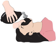 Bag-mask ventilation is a basic but critical airway management skill. It enables clinicians to provide adequate ventilation for patients requiring airway support and allows enough time to establish a more controlled approach to airway management, such as endotracheal (ET) intubation. Because the technique can be difficult to perform correctly, clinicians performing the procedure should continually practice and monitor their technique. Successful bag-mask ventilation depends on three things: Patent airway. Airway patency can be established using airway maneuvers and airway conduits described above. Adequate mask seal. In order to secure a good seal, the mask must be placed and held correctly. Disposable bag-mask units are often packaged with one large adult-sized mask. Even though facial anatomy differs for eachpatient, one sized face mask is usually sufficient for positive pressure ventilation with the bag mask. The nasal portion of the mask should be spread slightly and placed on the bridge of the patient's nose. The body of the mask is then placed onto the patient's face covering the nose and mouth. The provider's wrists or the mask cushion should not rest on the patient's eyes during bag-mask ventilation. There are two methods for holding the mask in place. The two-hand mask hold is most effective, however it requires a second clinician. Therefore, the clinician should be comfortable with both techniques. Proper ventilation (ie, proper volume, rate, and cadence). The three key errors to avoid when performing bag-mask ventilation are: Excessive tidal volumes: A volume just large enough to cause chest rise (no more than 8 to 10 cc/kg) should be used. During cardiopulmonary resuscitation (CPR), even smaller tidal volumes are adequate (5 to 6 cc/kg) due to the reduced cardiac output of such patients. Forcing air too quickly: The bag should not be squeezed explosively. It should be squeezed steadily over approximately one full second. Ventilating too rapidly. The ventilatory rate should not exceed 10 to 12 breaths per minute. BLS training is intended for certified or noncertified, licensed or nonlicensed, healthcare professionals, including: •physicians •nurses •paramedics •emergency medical technicians •respiratory, physical, and occupational therapists •physician's assistants •residents or fellows •medical or nursing students in training •aides, medical or nursing assistants, and other allied health personnel In addition, BLS training can be appropriate for first responders, such as police officers and firefighters, as well as for laypeople whose work brings them into contact with members of the public, such as school, fitness center, or other employees. Airway Is the airway open? Does the patient need an advanced airway? Breathing Is oxygenation and ventilation sufficient? If used, is the airway device properly placed and monitored? Are CO2 and O2 sats being monitored? Circulation What is the current cardiac rhythm? Is IV/IO access obtained? Does the patient need fluids or medications? Is a heart attack the same as cardiac arrest? No. What is a heart attack? A heart attack occurs when a blocked artery prevents oxygen-rich blood from reaching a section of the heart. If the blocked artery is not reopened quickly, the part of the heart normally nourished by that artery begins to die. The longer a person goes without treatment, the greater the damage. Symptoms of a heart attack may be immediate and intense. More often, though, symptoms start slowly and persist for hours, days or weeks before a heart attack. Unlike with sudden cardiac arrest, the heart usually does not stop beating during a heart attack. The heart attack symptoms in women can be different than men. What is cardiac arrest? Cardiac arrest is the abrupt loss of heart function in a person who may or may not have diagnosed heart disease. What to do: Sudden Cardiac Arrest Cardiac arrest is reversible in most victims if it's treated within a few minutes. First, call ________ for emergency medical services. Then get an automated external defibrillator if one is available and use it as soon as it arrives. Begin CPR immediately and CONTINUE until professional emergency medical services arrive. If two people are available to help, one should begin CPR immediately while the other calls ______ and finds an AED. Differential Diagnosis Why did arrest occur? Are there any other factors? Can we reverse the cause(s)? 7 H's and 5 T's: mnemonic for mechanisms •hypoxia •hypovolemia •hyperkalemia •hypokalemia •hypoglycemia •hypothermia •hydrogen ions (acidosis) •thrombosis (MI/heart attack) •tension pneumothorax •tamponade •toxins/therapeutics •thromboembolism •trauma Hypovolemia Hypovolemia or the loss of fluid volume in the circulatory system can be a major contributing cause to cardiac arrest. Looking for obvious blood loss in the patient with pusleless arrest is the first step in determining if the arrest is related to hypovolemia. After CPR, the most import intervention is obtaining intravenous access/IO access. A fluid challenge or fluid bolus may also help determine if the arrest is related to hypovolemia. Hypoxia Hypoxia or deprivation of adequate oxygen supply can be a significant contributing cause to cardiac arrest. You must ensure that the patient’s airway is open, and that the patient has chest rise and fall and bilateral breath sounds with ventilation. Also ensure that your oxygen source is connected properly. Hydrogen ion (acidosis) To determine if the patient is in respiratory acidosis, an arterial blood gas evaluation must be performed. Prevent respiratory acidosis by providing adequate ventilation. Prevent metabolic acidosis by giving the patient sodium bicarbonate. Hyper-/hypokalemia Both a high potassium level and a low potassium level can contribute to cardiac arrest. The major sign of hyperkalemia or high serum potassium is taller and peaked T-waves. Also, a widening of the QRS-wave may be seen. This can be treated in a number of ways which include sodium bicarbonate (IV), glucose+insulin, calcium chloride (IV), Kayexalate, dialysis, and possibly albuterol. All of these will help reduce serum potassium levels. The major signs of hypokalemia or low serum potassium are flattened T-waves, prominent U-waves, and possibly a widened QRS complex. Treatment of hypokalemia involves rapid but controlled infusion of potassium. Giving IV potassium has risks. Always follow the appropriate infusion standards. Never give undiluted intravenous potassium. Hypoglycemia Hypoglycemia or low serum blood glucose can have many negative effects on the body, and it can be associated with cardiac arrest. Treat hypoglycemia with IV dextrose to reverse a low blood glucose. Hypoglycemia was removed from the H’s but is still to be considered important during the assessment of any person in cardiac arrest. Hypothermia If a patient has been exposed to the cold, warming measures should be taken. The hypothermic patient may be unresponsive to drug therapy and electrical therapy (defibrillation or pacing). Core temperature should be raised above 86 F (30 C) as soon as possible. The T’s include: Toxins Accidental overdose of a number of different kinds of medications can cause pulseless arrest. Some of the most common include: tricyclics, digoxin, betablockers, and calcium channel blockers). Street drugs and other chemicals can precipitate pulseless arrest. Cocaine is the most common street drug that increases incidence of pulseless arrest. ECG signs of toxicity include prolongation of the QT interval. Physical signs include bradycardia, pupil symptoms, and other neurological changes. Support of circulation while an antidote or reversing agent is obtained is of primary importance. Poison control can be utilized to obtain information about toxins and reversing agents. Tamponade Cardiac tamponade is an emergency condition in which fluid accumulates in the pericardium (sac in which the heart is enclosed). The buildup of fluid results in ineffective pumping of the blood which can lead to pulseless arrest. ECG symptoms include narrow QRS complex and rapid heart rate. Physical signs include jugular vein distention (JVD), no pulse or difficulty palpating a pulse, and muffled heart sounds due to fluid inside the pericardium. The recommended treatment for cardiac tamponade is pericardiocentesis. Tension Pneumothorax Tension pneumothorax occurs when air is allowed to enter the plural space and is prevented from escaping naturally. This leads to a build up of tension that causes shifts in the intrathroacic structure that can rapidly lead to cardiovascular collapse and death. ECG signs include narrow QRS complexes and slow heart rate. Physical signs include JVD, tracheal deviation, unequal breath sounds, difficulty with ventilation, and no pulse felt with CPR. Treatment of tension pneumothorax is needle decompression. Thrombosis (heart: acute, massive MI) Coronary thrombosis is an occlusion or blockage of blood flow within a coronary artery caused by blood that has clotted within the vessel. The clotted blood causes an acute myocardial infarction which destroys heart muscle and can lead to sudden death depending on the location of the blockage. ECG signs during PEA indicating coronary thrombosis include ST-segment changes, T-wave inversions, and/or Q waves. Physical signs include: elevated cardiac markers on lab test. Treatments for coronary thrombosis before cardiac arrest include use of fibrinolytic therapy, or PCI (percutaneous coronary intervention). The most common PCI procedure is coronary angioplasty with or without stent placement. Thrombosis (lungs: massive pulmonary embolism) Pulmonary thrombus or pulmonary embolism (PE) is a blockage of the main artery of the lung which can rapidly lead to respiratory collapse and sudden death. ECG signs of PE include narrow QRS Complex and rapid heart rate. Physical signs include no pulse felt with CPR. distended neck veins, positive d-dimer test, prior positive test for DVT or PE. Treatment includes surgical intervention (pulmonary thrombectomy) and fibrinolytic therapy. Trauma The final differential diagnosis of the H’s and T’s is trauma. Trauma can be a cause of pulseless arrest, and a proper evaluation of the patients physical condition and history should reveal any traumatic injuries. Treat each traumatic injury as needed to correct any reversible cause or contributing factor to the pulseless arrest. Trauma was removed from the T’s but is still to be considered important during the assessment of any person in cardiac arrest. .. Respiratory Arrest In respiratory arrest, a pulse is present, though breathing is absent or ineffective. Perform BLS. Give one breath every 5-6 seconds, or 10-12 per minute. Check pulse (quickly) every two minutes. Stopping Recuscitation Each patient case is unique, and while clinical rules are developed, the decision to stop CPR is a complex one. Except in specific circumstances, such as hypothermia or drug overdose, BLS or ACLS are very unlikely to be successful after 20 minutes. Advanced Life Support (ALS) What is Advanced Life Support? Advanced cardiac life support (ACLS) describes the treatment of life-threatening cardiac rhythms. ACLS may only be provided by trained experts, as it includes advanced ECG diagnostics, procedural skills, and use of medications. It builds upon basic life support, or CPR, and the use of automated external defibrillators (AEDs). Basic Life Support Ambulance Basic Life Support care requires medical monitoring by a licensed EMT-Intermediate and may include monitoring vital signs, oxygen and IV therapy. The BLS Ambulance is equipped with state-of-the-art equipment including an automatic external defibrillator, blood pressure monitoring equipment, pulse oximetry and oxygen delivery devices. Advance Life Support Ambulance Advanced Life Support care requires medical monitoring and care by a licensed EMT-Paramedic and may include monitoring vital signs, advanced drug therapy, cardiac monitoring, oxygen and IV therapy. The ALS Ambulance is equipped with state-of-the-art heart and blood pressure monitoring equipment, pulse oximetry, IV pumps, oxygen delivery devices including a CPAP and advanced medications used to treat a variety of illnesses and provide pain relief. The Advanced Life Support team is composed of three members namely: the regular paramedic, the critical care paramedic and the emergency care practitioner. Here are further guidelines. Here are further guidelines. Pediatric Basic and Advanced Life Support Medical Diagnoses: Ineffective tissue perfusion (cardiac, renal, cerebral). Signs of inadequate tissue perfusion Depressed mental status Decreased urine output Metabolic acidosis Tachypnea Weak central pulses Deterioration in color such as mottling Hypotension Hypotension
|
Paramedic (EMT-P)
Critical Care Paramedic (CCEMTP)
Emergency Care Practitioner (ECP)
| Paramedics are licensed individuals who can perform tasks beyond that of an EMT. |
Human Vital Signs
|
What should you include in Human vital signs? What are vital signs? What is body temperature? What is the pulse rate? What is the respiration rate? What is blood pressure? What special equipment is needed to measure blood pressure? Is the patient conscious, oriented in time, space, and person? Conscious means able to see, hear, and talk. In pediatric patients younger than six months of age, the ability to make any verbal noise or cry is equivalent to talking. |
| Age-Appropriate Vital Signs | |||||||||||||||||||||||||||||||||||||||||||||||||||||||||||||||||||||||||||||||||
|
Ref: Pediatric Vital Signs
What is the normal respiratory rate for a newborn, infant, toddler, preschooler, school age child, and adolescent? What is the normal pulse rate for a newborn, infant, toddler, preschooler, school age child, and adolescent? What is the lower limit of normal systolic blood pressure in a newborn, infant, toddler, preschooler, school age child, and adolescent? | |||||||||||||||||||||||||||||||||||||||||||||||||||||||||||||||||||||||||||||||||
|
Vital Signs (Body Temperature, Pulse Rate, Respiration Rate, Blood Pressure) What are vital signs? Vital signs are measurements of the body's most basic functions. The four main vital signs routinely monitored by medical professionals and healthcare providers include the following: v * body temperature * pulse rate * respiration rate (rate of breathing) * blood pressure (Blood pressure is not considered a vital sign, but is often measured along with the vital signs.) Vital signs are useful in detecting or monitoring medical problems. Vital signs can be measured in a medical setting, at home, at the site of a medical emergency, or elsewhere. What is body temperature? The normal body temperature of a person varies depending on gender, recent activity, food and fluid consumption, time of day, and, in women, the stage of the menstrual cycle. Normal body temperature, according to the American Medical Association, can range from 97.8° F (or Fahrenheit, equivalent to 36.5° C, or Celsius) to 99° F (37.2° C). A person's body temperature can be taken in any of the following ways: What is fever? Fever (also called pyrexia) is defined as body temperature that is higher than normal for each individual. It generally indicates that there is an abnormal process going on within the body. The severity of a condition is not necessarily reflected by the degree of fever. For example, influenza may cause a fever of 104° F, while pneumonia may cause a very low-grade fever or no fever at all. Consult with your physician if you have any questions about whether a fever is significant. * orally Temperature can be taken by mouth using either the classic glass thermometer, or the more modern digital thermometers that use an electronic probe to measure body temperature. * rectally Temperatures taken rectally (using a glass or digital thermometer) tend to be 0.5 to 0.7° F higher than when taken by mouth. * axillary Temperatures can be taken under the arm using a glass or digital thermometer. Temperatures taken by this route tend to be 0.3 to 0.4° F lower than those temperatures taken by mouth. * by ear A special thermometer can quickly measure the temperature of the ear drum, which reflects the body's core temperature (the temperature of the internal organs). Body temperature may be abnormal due to fever (high temperature) or hypothermia (low temperature). A fever is indicated when body temperature rises above 98.6° F orally or 99.8° F rectally, according to the American Medical Association. Hypothermia is defined as a drop in body temperature below 95° F. About glass thermometers containing mercury: According to the Environmental Protection Agency (EPA), mercury is a toxic substance that poses a threat to the health of humans, as well as to the environment. Because of the risk of breaking, glass thermometers containing mercury should be removed from use and disposed of properly in accordance with local, state, and federal laws. Contact your local health department, waste disposal authority, or fire department for information on how to properly dispose of mercury thermometers. What is the pulse rate? The pulse rate is a measurement of the heart rate, or the number of times the heart beats per minute. As the heart pushes blood through the arteries, the arteries expand and contract with the flow of the blood. Taking a pulse not only measures the heart rate, but also can indicate the following: * heart rhythm * strength of the pulse The normal pulse for healthy adults ranges from 60 to 100 beats per minute. The pulse rate may fluctuate and increase with exercise, illness, injury, and emotions. Females ages 12 and older, in general, tend to have faster heart rates than do males. Athletes, such as runners, who do a lot of cardiovascular conditioning, may have heart rates near 40 beats per minute and experience no problems. Illustration demonstrating how to take a pulse Click Image to Enlarge How to check your pulse: As the heart forces blood through the arteries, you feel the beats by firmly pressing on the arteries, which are located close to the surface of the skin at certain points of the body. The pulse can be found on the side of the lower neck, on the inside of the elbow, or at the wrist. When taking your pulse: * Using the first and second fingertips, press firmly but gently on the arteries until you feel a pulse. * Begin counting the pulse when the clock's second hand is on the 12. * Count your pulse for 60 seconds (or for 15 seconds and then multiply by four to calculate beats per minute). * When counting, do not watch the clock continuously, but concentrate on the beats of the pulse. * If unsure about your results, ask another person to count for you. If your physician has ordered you to check your own pulse and you are having difficulty finding it, consult your physician or nurse for additional instruction. What is the respiration rate? The respiration rate is the number of breaths a person takes per minute. The rate is usually measured when a person is at rest and simply involves counting the number of breaths for one minute by counting how many times the chest rises. Respiration rates may increase with fever, illness, and with other medical conditions. When checking respiration, it is important to also note whether a person has any difficulty breathing. Normal respiration rates for an adult person at rest range from 15 to 20 breaths per minute. Respiration rates over 25 breaths per minute or under 12 breaths per minute (when at rest) may be considered abnormal. What is blood pressure? Blood pressure, measured with a blood pressure cuff and stethoscope by a nurse or other healthcare provider, is the force of the blood pushing against the artery walls. Each time the heart beats, it pumps blood into the arteries, resulting in the highest blood pressure as the heart contracts. One cannot take his/her own blood pressure unless an electronic blood pressure monitoring device is used. Electronic blood pressure monitors may also measure the heart rate, or pulse. Two numbers are recorded when measuring blood pressure. The higher number, or systolic pressure, refers to the pressure inside the artery when the heart contracts and pumps blood through the body. The lower number, or diastolic pressure, refers to the pressure inside the artery when the heart is at rest and is filling with blood. Both the systolic and diastolic pressures are recorded as "mm Hg" (millimeters of mercury). This recording represents how high the mercury column is raised by the pressure of the blood. High blood pressure, or hypertension, directly increases the risk of coronary heart disease (heart attack) and stroke (brain attack). With high blood pressure, the arteries may have an increased resistance against the flow of blood, causing the heart to pump harder to circulate the blood. According to the National Heart, Lung, and Blood Institute _______ of the National Institutes of Health (NIH), high blood pressure for adults is defined as: * 140 mm Hg or greater systolic pressure and * 90 mm Hg or greater diastolic pressure In an update of ______-guidelines for hypertension in 2003, a new blood pressure category was added called prehypertension: * 120 mm Hg – 139 mm Hg systolic pressure and * 80 mm Hg – 89 mm Hg diastolic pressure The new NHLBI guidelines now define normal blood pressure as follows: * Less than 120 mm Hg systolic pressure and * Less than 80 mm Hg diastolic pressure These numbers should be used as a guide only. A single elevated blood pressure measurement is not necessarily an indication of a problem. Your physician will want to see multiple blood pressure measurements over several days or weeks before making a diagnosis of hypertension (high blood pressure) and initiating treatment. A person who normally runs a lower-than-usual blood pressure may be considered hypertensive with lower blood pressure measurements than 140/90. Why should I monitor my blood pressure at home? For persons with hypertension, home monitoring allows your physician to monitor how much your blood pressure changes during the day, and from day to day. This may also help your physician determine how effectively your blood pressure medication is working. What special equipment is needed to measure blood pressure? Either an aneroid monitor, which has a dial gauge and is read by looking at a pointer, or a digital monitor, in which the blood pressure reading flashes on a small screen, can be used to measure blood pressure. About the aneroid monitor: The aneroid monitor is less expensive and easier to manage than the digital monitor. The cuff is inflated by hand by squeezing a rubber bulb. Some units even have a special feature to make it easier to put the cuff on with one hand. However, the unit can be easily damaged and become less accurate. Because the person using it must listen for heartbeats with the stethoscope, it may not be appropriate for the hearing-impaired. About the digital monitor: The digital monitor is automatic, with the measurements appearing on a small screen. Because the recordings are easy to read, this is the most popular blood pressure measuring device. It is also easier to use than the aneroid unit, and since there is no need to listen to heartbeats through the stethoscope, this is a good device for hearing-impaired patients. One disadvantage is that body movements or an irregular heart rate can change the accuracy. These units are also more expensive than the aneroid monitors. About finger/wrist blood pressure monitors: Tests have shown that finger and/or wrist blood pressure devices are not as accurate in measuring blood pressure as other types of monitors. In addition, they are more expensive than the other monitors. Before you measure your blood pressure: * Rest for three to five minutes without talking before taking a measurement. * Sit in a comfortable chair, with your back supported and your legs and ankles uncrossed. * Sit still and place your arm, raised level with your heart, on a table or hard surface. * Wrap the cuff smoothly and snugly around the upper part of your arm. The cuff should be sized to fit smoothly, while still allowing enough room for one fingertip to slip under it. * Be sure the bottom edge of the cuff is at least one inch above the crease in your elbow. It is also important, when taking blood pressure readings, that you record the date and time of day you are taking the reading, as well as the systolic and diastolic measurements. This will be important information for your physician to have. Ask your physician or another healthcare professional to teach you how to use your blood pressure monitor correctly. Have the monitor routinely checked for accuracy by taking it with you to your physician's office. It is also important to make sure the tubing is not twisted when you store it and keep it away from heat, to prevent cracks and leaks. Proper use of your blood pressure monitor will help you and your physician in monitoring your blood pressure. | |||||||||||||||||||||||||||||||||||||||||||||||||||||||||||||||||||||||||||||||||
Baseline Vital Signs
Vital Signs and Measurements
Normal Vital Signs
Detailed Vital Signs and SymptomsBreathing
Pulse
Skin
Pupils
Blood Pressure
Pulse Oximetry
Vital Sign ReassessmentThe SAMPLE HistoryMedical history obtained from the patient, family and bystanders
| |||||||||||||||||||||||||||||||||||||||||||||||||||||||||||||||||||||||||||||||||
A–Z alphabetical listing of human health emergency symptoms and signs.
Medical emergencies.
Examples of emergency symptoms, signs, and complaints
|
What are examples of emergency relevant to an individual?
Do you know what are emergency medical symptoms, signs, or complaints? What are various symptoms, signs, statements, questions, issues, and histories that should raise suspicion of a medical emergency? There are at least 159 such symptoms, signs, statements, questions, issues, histories, and scenarios.
What will happen if you do not diagnose and treat a medical emergency properly? It can lead to death. It can lead to disability. It can lead to other harms. It can lead to medical malpractice. It can lead to legal malpractice. |
Altered level of consciousness
|
What is an altered level of consciousness? Altered level of consciousness (ALOC) means that you are not as awake, alert, or able to understand or react as you are normally. What are the levels of impaired/altered human consciousness?
*Both stupor and coma are often further classified as mild, moderate or deep. The Glasgow Coma Scale (GCS) is the most widely accepted method for the evaluation and classification of coma, especially for head-injured patients. A GCS score of 8 or less is the generally accepted definition of coma. Those with a GCS of 8 or less are classified as severe, while those with a GCS score of 9 to 12 are categorized as moderate and those with a GCS score of 13 to 15 are mild. Coma grades 3 to 5 indicate potentially fatal damage, especially if accompanied by fixed pupils or absent oculovestibular responses. Conversely, scores of 9 and above correlate with good recovery. What causes an altered level of consciousness? Alcohol Dehydration Drugs Head injury Sedatives or other medicines Uncontrolled diabetes. An altered level of consciousness or coma have almost same causes. Coma is an extreme presentation of an altered level of consciousness. How should an emergency medical specialist go ahead in diagnosing and treating an altered level of consciousness? How do you diagnose and find out the underlying cause or causes of this medical complaint? What medical history should you ask relevant to this complaint? Syncope Signs and symptoms History and physical examination are the most specific and sensitive ways of evaluating syncope. A detailed account of the event must be obtained from the patient, including the following: Precipitant factors Activity the patient was involved in before the event Position the patient was in when the event occurred The following questions should be asked: Was loss of consciousness complete? Was loss of consciousness with rapid onset and short duration? Was recovery spontaneous, complete, and without sequelae? Was postural tone lost? Causes of altered level of consciousness · Structural: brain lesions that destroy tissue or occupy space that is normally occupied by the brain · Epilepsy · Tumors · Trauma · Cardiovascular: temporary or permanent interruption to the blood supply to the brain · Vasovagal response · CVA · TIA · Hypertensive encephalopathy · Shock · Dysrhythmias · Metabolic: abnormally high or low levels of circulating metabolites · Hypoxia · Hypoglycemia · Hyperglycemia · Renal failure (uremia) · Liver failure · Infection (sepsis) · Environmental: external factors that cause deterioration of central nervous system function · Overdose · Toxins · Behavioral: abnormal mental status that results from internal factors · Psychiatric disorders Mnemonic for Causes of Altered Level of Consciousness A - alcohol, acidosis, anoxia E - epilepsy, environment I - insulin (diabetes) O - overdose U - uremia (metabolic), underdose T - trauma, toxins, tumors I - infection (sepsis) P - psychiatric disorders S - stroke (CVA) Symptoms that may be associated with decreased consciousness are: •seizures •loss of bowel or bladder function •poor balance •falling •difficulty walking •fainting •lightheadedness •irregular heart beat •rapid pulse •low blood pressure •sweating •fever •weakness in the face, arms, or legs Common Underlying Causes of Decreased Consciousness Drugs, alcohol, substance abuse, drug overdose, certain medications, epilepsy, low blood sugar, stroke, and lack of oxygen to the brain are common causes of decreased consciousness. Other underlying causes of decreased consciousness include: •cerebral hemorrhage •dementia (Alzheimer’s) •head trauma •heart disease •heat stroke •liver disease •uremia (end stage kidney failure) •shock Sleep Sleep is a normal state of physical and mental inactivity from which the individual may be aroused to normal consciousness. A sleeping individual gives very little evidence of being aware of self or environment. Yet, he/she differs from the comatose patient in that he/she may still respond to unaccustomed stimuli and at times is capable of some form of mental activity in the form of dreams which leave their traces in memory. Altered Level Of Consciousness (Child) Level of consciousness is a measure of a person’s ability to interact with other people and to react to the surroundings. A child with an altered level of consciousness may be irritable for a number of hours. The child cannot be calmed by holding, rocking, or feeding. Some children may be hyperactive and can’t sleep. Others may be very sleepy and difficult to wake. Affected children may appear limp, with poor muscle tone. They may not respond to touch or voices. Their look may be vacant or blank. They may not make eye contact. Their cry may be weak and feeble. Some children may not move for a long time or show little interest in moving. They may be confused. There are many causes of altered consciousness. Generally, they are divided into medical reasons or injuries. The child may have low blood sugar, an infection, a metabolic abnormality, or a systemic disease. Other causes include drugs, abuse, or injuries such as a fall. A decreased level of consciousness is a medical emergency. Doctors must quickly determine the cause. This may include laboratory, radiology, and other types of testing. Initial treatment stabilizes breathing and heart rate. An intravenous (IV) line is used to give medications. Once the cause is determined, specific treatment for that cause is given. In almost all cases, the child will be admitted to the hospital for observation. History Perform a focused history and physical exam with particular attention to: A. When was the patient last completely well? B. Determine the onset, progression and duration of symptoms. C. What signs and symptoms were present before the change in level of consciousness (e.g., headaches, seizures, confusion, trouble breathing, fever/chills?) D. Obtain a past medical history, including alcohol abuse, diabetes, epilepsy, hypertension. E. What medications has the patient been, or is the patient supposed to be, taking (including over the counter medications)? F. Is there a history of head trauma? G. Has the patient noted any chest pain, dyspnea or irregular heartbeat? H. Has the patient been incontinent? Physical Examination A. Perform an initial assessment. B. Perform a focused history and physical exam with particular attention to: 1. Skin (a) Is the patient sweaty? (b) Is the skin hot or cold? (c) What is the skin color? 2. Assess patient from head to toe for injuries: (a) Is there any bruising? (b) Is there any evidence of head or neck trauma? C. Assess the level of consciousness. D. Assess the patient's neurological condition. 1. Check pupils for size, symmetry, reactivity. 2. Assess motor function. Is the patient moving all four extremities? Is there equal grip strength? Is there posturing? 3. Is sensation to touch intact in all four extremities? 4. What is the last thing the patient can recall? E. Are there signs of trauma which might have caused altered mental status/coma (e.g., head trauma, hematomas, Raccoon eyes, Battle's sign)? F. Are there other injuries, e.g., hip or wrist injury from a fall? G. Is there an unusual breath odor, e.g., alcohol, fruity/acetone)? H. Is there evidence of chemical use, e.g., needle tracks, runny nose? I. Inspect the surroundings 1. Check for pill bottles, syringes, etc. (bring them with the patient). 2. Note any odor in the house, unvented heaters, etc. (carbon monoxide is odorless). J. At the discretion of local medical direction, a specific prehospital stroke assessment may be made. The General Physical Exam Vital Signs The vital sign determinations may contribute important clues to the underlying process responsible for the altered level of consciousness. The temperature should be taken rectally. Fever suggests the presence of a sever systemic infection such as pneumonia, bacterial meningitis, or a brain lesion that has disturbed the temperature-regulating centers. An excessively high body temperature, ____ to _____, associated with dry skin should raise suspicion of heat stroke. Hypothermia is frequently observed in alcoholic or barbituate intoxication, extracellular fluid deficit, or in shock and peripheral circulatory failure. Slow respirations suggests barbituate, morphine, or other narcotic intoxication, whereas rapid deep breathing (Kussmaul respirations) suggests diabetic or uremic acidosis but may also occur in intracranial disease. Rapid breathing accompanied by an expiratory grunt and associated with fever is a common finding in lobar pneumonia. Diseases that elevate the intracranial pressure or cause damage to the brain, especially the brain stem, often cause slow, irregular or periodic (Cheynes-Stokes) breathing. Exhalation through pursed lips is suggestive of hypokalemia. Apneustic breathing is characterized by a pause of 2-3 seconds between inspiration and expiration and is attributed to a lesion in the pontine respiratory center. Ataxic respirations are manifested by an irregular pattern of inspiratory and expiratory respirations seen in association with lesions in the medullary respiratory center. If the pulse is exceptionally slow, it suggests a heart block and the possibility of Stokes-Adams syndrome, or if combined with hypertension and periodic breathing, increased intracranial pressure. A tachycardia of 150 or above suggests an arrhythmia with possible insufficiency of cerebral circulation. The presence of atrial fibrillation suggests the possibility of a cerebral embolism. Blood pressure observations are important. Marked hypertension occurs with cerebral hemorrhage and hypertensive encephalopathy and at times with increased intracranial pressure. Hypotension is seen in diabetic coma, alcohol or sedative drug intoxication or in loss of blood due to internal hemorrhage. The Ocular Fundi Careful observation of the optic fundi without the use of mydriatics that interfere with pupillary reflexes may produce information about hypertension, systemic arteriosclerosis, diabetes mellitus, hemorrhages, and the presence or absence of papilledema. If a lesion is suspected that is producing elevated intracranial pressure, examination of the ocular fundi for the presence or absence of papilledema should be considered an emergency procedure. Examination of the Skin Inspection of the skin may also yield valuable clinical information. Multiple bruises, especially in the scalp area, suggest cranial trauma. Bleeding from the nose, an ear or orbital hemorrhage also raises the possibility of trauma. Marked pallor suggests internal hemorrhage. Rashes are seen in a number of infectious diseases including meningococcemia, endocarditis, typhus, or Rocky Mountain fever. The dementia of pellagra is usually accompanied by typical skin lesions on the face and hands and diarrhea. The physician should examine the skin closely for evidence of needle marks suggestive of substance abuse or insulin usage. Odor of the Breath Often neglected, the odor of the breath may afford a valuable diagnostic clue. The odor of alcohol is easily recognizable, but may be a misleading clue that detracts from a more significant diagnosis. The “fruity” odor of diabetic acidosis, the uriniferous odor of uremia, and the musty, “mousy” odor of hepatic coma are sufficiently distinctive to provide valuable diagnostic clues. While checking breath odor, the physician has the opportunity to inspect the mouth for evidence of tongue-biting and gum hypertrophy, tell-tale evidence of epilepsy and anti-convulsant drug use. Cardiorespiratory and Abdominal Examinations The heart should be examined for the apical pulse rate and rhythm, the presence of murmurs and cardiac enlargement, as well as for evidence of congestive heart failure and carotid artery bruits and hemodynamics. The extremities should be examined for peripheral pulses, evidence of sclerosis, cyanosis, edema and clubbing of the fingers. The character of breathing should be noted and the lungs examined for evidence of percussive and auscultatory evidence of infiltration, consolidation, fluid or congestion. The abdominal examination should include evidence of hepatomegaly and the presence of masses. A rectal examination should include evaluation of the prostate and examination of the stool for occult or gross blood. The Neurological Examination Observation of the Patient Careful observation of the obtunded, stuporous or comatose patient may yield considerable information concerning the function or lack of function of various parts of the nervous system. One of the most helpful procedures is to sit at the patient’s bedside for five to ten minutes and observe what he/she does. The predominant postures of the body, the position of the head and eyes, the rate, depth and rhythm of respiration, and the pulse should be noted. The state of responsiveness should then be estimated by noting the patient’s reaction to verbal commands, his/her capacity to execute a simple response and the response to painful stimuli. The obtunded patient is usually capable of accurately localizing a painful stimulus and will fight forcefully against it accompanied by grimacing and vocalization. As unconsciousness progresses toward coma, these reactions may be replaced by stereotyped responses having specific anatomic or pathologic connotations. These “posturing” responses include the following: Decorticate posture or rigidity includes flexion of the arms and wrists with the legs extended with internal rotation and plantar flexion. This posture suggests severe bilateral damage in the hemispheres above the midbrain with involvement of the corticospinal tracts. Decerebrate posture or rigidity includes pronation and extension of the arms and extension of the legs with plantar flexion occasionally accompanied by opisthotonos and trismus. This posture suggests damage to the corticospinal tracts arising in the midbrain or caudal diencephalon. Abnormal posturing or motor activity is also seen under other circumstances. External rotation of a leg or lack of restless movements on one side suggest a hemiparesis. Multifocal myoclonus, characterized by sudden, generalized muscle contractions, are commonly seen in metabolic disorders especially uremia, anoxia or drug ingestion. Asterixis (palmar flipping) is commonly seen in metabolic encephalopathies associated with drowsiness and confusion. It is most commonly seen in hepatic encephalopathy and has been attributed to accompanying ammonia intoxication. Pupillary and Brainstem Reflexes Pupillary reflexes depend on the correct functioning of centers situated in the brainstem (diencephalon, pons and midbrain) and provide valuable information on the localization of lesions in the brainstem. Symmetrically reactive round pupils, 2.5 to 5 mm in diameter, usually exclude midbrain damage as a cause of coma. One enlarged (greater than 5 mm in diameter) unreactive or poorly reactive pupil usually results from either an intrinsic midbrain lesion on the same side or, far more commonly, is secondary to stretching or compression of the third nerve (oculomotor) by the secondary effects of a mass. Oval and slightly eccentric pupils accompany early midbrain-third nerve compression. Bilaterally dilated and unreactive pupils indicate severe midbrain damage usually by compression from transtentorial herniation or from anticholinergic drugs. Small, but not pinpoint, pupils (1 to 2.4 mm in diameter) that react are most commonly seen in metabolic encephalopathies. Bilateral pinpoint pupils that still react (but may require a magnifying glass to verify) are characteristic of narcotic or barbiturate overdose but may also occur as the result of a pontine hemorrhage. Skew deviation of the eyes (one up or down) is an indication of a brainstem lesion. Forced downward deviation of the eyes such as looking at one’s nose has been described in cases of thalamic hemorrhage and is usually accompanied by pinpoint nonreactive pupils. Eye Movements Eye movements are an additional valuable component of the neurological examination, contributing information concerning the location of the etiological mechanism causing an altered level of consciousness. The eyes are first observed by elevating the lids and noting the resting position and spontaneous movements of the globes. Unconscious patients, in whom the centers that mediate eye movements are intact, commonly have a slightly divergent straightforward gaze and slow, horizontal conjugate eye movements (roving eye movements). In these patients, turning the head briskly from side to side elicits conjugate eye movements directed opposite to the head rotation. This is the oculocephalic or doll’s head reflex which is absent in the normal alert individual. Cyclic vertical downward movements are seen in specific circumstances. “Ocular bobbing” describes a brisk downward and slow upward movement of the globes associated with loss of horizontal eye movements and is diagnostic of bilateral pontine damage. “Ocular dipping” is a slower arrhythmic downward movement followed by a faster upward movement in patients with normal reflex horizontal gaze and denotes diffuse anoxic damage of the cerebral cortex. The oculovestibular response is elicited by caloric stimulation of the semicircular canals by irrigating the external ear canal with cold water. This produces tonic bilateral eye deviation toward the stimulated side. In a normal awake individual, this type of stimulation causes nystagmus away from the stimulated side. Full horizontal excursion of the eyes to both sides is possible only when the nuclei and the internuclear connections between the third and sixth nerves are intact. A normal oculovestibular response indicates that the cause of unconsciousness is not a structural brainstem lesion. Unilateral lesions in the pontine gaze center adjacent to the sixth nerve nucleus result in a paralysis of ipsilateral gaze and contralateral deviation of the eyes and a failure of caloric stimulation to move the eyes beyond the midline. Conjugate horizontal ocular deviation at rest or incomplete conjugate eye movements with head turning indicate damage in the pons on the side of the gaze paresis or frontal lobe damage on the opposite side. This phenomenon may be summarized by the following aphorism: The eyes look toward a hemispheral lesion and away from a brainstem lesion. The pathway that connects the pontine gaze center to the midbrain oculomotor nuclei is called the medial longitudinal fasciculus (MLF). When its fibers are damaged, the ipsilateral medial rectus muscle fails to contract when required for voluntary or reflex conjugate horizontal gaze. This is called internuclear ophthalmoplegia and can be demonstrated in the unconscious patient by either oculocephalic or oculovestibular testing. Testing Office and Laboratory Blood chemistry tests are made routinely to investigate metabolic, toxic or drug-induced encephalopathies. The major areas of metabolic abnormalities encountered in clinical practice are those of electrolytes (sodium, potassium, chloride), calcium, blood urea nitrogen (BUN), serunm creatinine (Cr), glucose and hepatic dysfunction (ammonia). Toxicological analyses are of great value in any case of ALC where the diagnosis is not immediately apparent. However, the presence of exogenous drugs or toxins, especially alcohol, does not ensure that other factors, particularly head trauma, may not also contribute to the clinical state. The legal blood level above which alcohol is incriminated as a possible etiologic factor is 100 mg/dl in most states. Ethanol levels of 200 mg/dl in individuals who are non-habituated to alcohol generally cause confusion, and levels above 300 mg/dl are associated with stupor. The development of tolerance may allow the chronic alcoholic to maintain wakefulness above these levels. Radiologic Studies Skull roentgenograms, especially in cases of craniofacial trauma, are essential in detecting fractures, shifts of the pineal body from the midline, and abnormal calcifications. In the case of trauma and the possibility of vertebral damage, roentgenograms of the cervical and thoracic spine must be taken. Computerized tomography (CT) scanning and magnetic resonance imaging (MRI) yield information about structural damage in the supratentorial area and are well-suited for visualizing and diagnosing hemorrhages, tumors, hydrocephalus and brain edema and/or softening due to cerebral thrombosis or emboli. Patients for CT or MRI studies should be carefully selected because most cases of ALC are metabolic or toxic in origin. The notion that a normal CT scan excludes an anatomic lesion, however, is erroneous. Early bilateral hemisphere infarction, small brainstem lesions, encephalitis, meningitis, decreased or absent cerebral perfusion, superior saggital sinus thrombosis and even subdural hematomas that are isodense to adjacent brain may be overlooked. Even MRI may fail to demonstrate these processes early in their evolution. Nevertheless, in coma of unknown etiology, a CT or MRI scan should be obtained. In mass lesions, shifting of the pineal body correlates roughly with the level of consciousness. Three to 5 mm of horizontal displacement of the pineal body from the midline correlates with obtundation; 5 to 8 mm correlates with stupor; and greater than 8 mm displacement correlates with coma. The Electroencephalogram (EEG) The EEG tests neuronal physiology and therefore is important in the diagnosis and follow-up of metabolic or drug-induced encephalopathies. It is seldom diagnostic in cases of coma except to identify clinically unrecognized seizures. Specific EEG patterns have been described in metabolic coma due to hepatic failure and other mechanisms. A pattern described as “alpha coma” has been associated with either high pontine or diffuse cortical damage and is indicative of a poor prognosis. Cerebrospinal Fluid (CSF) Examination CSF examination is important when the differential diagnosis includes the possibility of meningitis, encephalitis or subarachnoid hemorrhage in which the CT is normal. In general, the CT has replaced the lumbar puncture as a diagnostic test in intracerebral hemorrhage. Spinal fluid pressure, cell count and differential, levels of protein and sugar, culture and viral studies are useful diagnostic contributions made by examination of the CSF. Differential Diagnosis In many instances, an altered state of consciousness spanning the spectrum of confusion through coma is part of an obvious medical problem such as known drug ingestion, hypoxia, stroke, trauma, or liver or kidney failure. Attention is then appropriately focused on the primary underlying illness. Some general rules are helpful in establishing the diagnosis if the underlying cause is unknown. Coma that appears subacutely or slowly is usually related to preceding medical or neurologic problems, including the secondary edema that surrounds a pre-existing lesion. Illnesses that cause sudden or acute coma are due to drug ingestion or to one of the catastrophic brain lesions such as hemorrhage, trauma, hypoxia or rarely, acute basilar artery thrombosis. The demonstration of focal brain disease or meningeal irritation with CSF abnormality and abnormalities in the CT, MRI or EEG, helps in the differential diagnosis of ALC. For purposes of differential diagnosis, the diseases that frequently result in ALC can be conveniently divided into three classes as shown in Table 3. Table 3 – The Differential Diagnosis of Altered States of Consciousness A. Disease that cause no focal or lateralizing neurologic signs or alterations of the cellular content of the CSF. 1. Intoxications (alcohol, barbiturates, narcotics) 2. Metabolic disturbances (diabetic acidosis, uremia, hepatic coma, hypoxia, hypoglycemia, Addisonian crisis 3. Severe systemic infections with our without septicemia 4. Circulatory shock from any cause 5. Hypertensive encepalopathy 6. Hypothermia or hyperthermia 7. Status epilepticus B. Diseases that cause meningeal irritation with either blood or an excess of white blood cells in the CSF, usually without focal or lateralizing signs. 1. Subarachnoid hemorrhage from ruptured aneurysm, occasionally trauma 2. Acute bacterial meningitis 3. Encephalitis C. Diseases that cause focal or lateralizing signs with our without changes in the CSF. (These may be subdivided into supratentorial and infratentorial lesions). CT and/or MRI are usually positive. 1. Brain hemorrhage 2. Cerebral thrombosis or embolism with secondary brain edema and/or softening 3. Brain abscess 4. Epidural and subdural hematoma with brain contusion and/or compression. 5. Brain tumor 6. Cerebral thrombosis Metabolic encephalopathies are diffuse disturbances of neuronal function that occur when the substrates required for neuronal metabolism are in short supply, when the internal environment of the cell is disturbed by external agents such as drugs or environmental poisons, or as a complication of the failure of another organ system such as the kidneys, liver or the endocrine, cardiovascular or respiratory systems. When the metabolic impairment is mild, the onset of symptoms can be insidious and nonspecific. Subtle changes in mentation such as mild drowsiness, dullness of affect, and decreased motor coordination precede the more ominous alteration in the level of consciousness such as confusion, delirium, stupor and eventually coma. Treatment Acute Care / Hospitalization It is apparent that in almost all instances, hospitalization will be required in all serious alterations in the level of consciousness. Until that can be safely accomplished, however, the role of the primary care physician, when confronted by a patient with an altered level of consciousness as a first priority is the institution of emergency treatment necessary to preserve and maintain vital signs and to stabilize the patient to prevent further damage to the nervous system and death. This includes the ABCs of basic life support – airway, breathing and circulation. An oropharyngeal airway is adequate to keep the pharynx open in patients who are breathing normally. Endotracheal intubation is indicated if there is apnea, upper airway obstruction or emesis, or if the patient is liable to aspirate. Mechanical ventilation will be required if there is hypoventilation. Intravenous access should be established as soon as possible and blood drawn for blood counts, blood chemistry determinations, toxic screen and blood gasses. Definitive Therapy Dextrose (50% if hypoglycemia is suspected) with added thiamine and naloxone should be administered if hypoglycemia and/or substance abuse are even remote possibilities. Thiamine is helpful in preventing Wernicke’s encephalopathy in malnourished alcoholic individuals. Ultimately, nasogastric intubation and bladder catheterization will probably be necessary. Overdistension of the bladder should be prevented. Only after initiation of urgent, potentially life-saving measures should a limited physical examination be done primarily to serve as a guide for the care of the patient in his/her transportation to the hospital. Evidence of head trauma should be sought and trauma to the neck and spine should be carefully considered at the time of first contact with the patient. The skull should be palpated for hematomas and the mastoid and periorbital tissues examined for ecchymoses. If neck trauma can be safely excluded, the patient should be placed in a semiprone position so that secretions and vomitus do not enter the endotracheal tree. The patient should be monitored closely during transportation to the hospital and most ambulances and emergency medical care vehicles are equipped to initiate emergency diagnostic and therapeutic measures. Therapeutic measures that can be instituted enroute to the hospital include treatment of shock and the administration of 100% oxygen by mask. Hypothermia can be helped with blankets and warming devices and hyperthermia by the use of alcohol sponging and the application of cooling solutions. If the patient is conscious, control of anxiety, restlessness and panic may be a problem since all but mild sedation should be avoided. The hospital should be alerted to the arrival of the patient so that appropriate neurological/neurosurgical consultation will be available for decisions concerning further diagnostic and therapeutic procedures. Complications The physician should be alert for the development of complications that may arise during a period of altered consciousness. Confused patients are prone to fall either in walking or getting out of bed. Many of the patients, especially postmenopausal women, will have accompanying osteoporosis and are at increased risk for serious vertebral and hip fractures. Households, hospital and convalescent, nursing and retirement facilities should provide and participate in providing instructions against falling as well as providing safety devices (handrails, etc.) for the prevention of falls. The consequences of hip fractures in this group of patients include long periods of disability and even death. Precautions should be taken to prevent the bedridden patient with prolonged coma from the development of decubitus ulcers as well as nococomial infections such as pneumonia and bladder infections secondary to an indwelling catheter. Many of the survivors of an altered state of consciousness will achieve complete or significant recovery of function even in the case of severe head trauma. However, as many as 30 to 40% will remain in prolonged states of severely reduced consciousness subsequent to achieving medical stability.23 The nature of the residual damage depends on the nature and location of the underlying causative agent and the severity of the process. Relatively few patients will remain in coma, that is, with eyes closed and no evidence of wakefulness for more than four weeks. Patients who show no signs of consciousness after their eyes open usually fit the criteria for the vegetative state (VS). Persistent vegetative state (PVS) is a prognostic term referring to a chronic condition in which basic arousal (i.e., wakefulness) and life-sustaining functions (e.g., respiration, blood pressure) are generally intact despite the absence of behavioral signs of meaningful interaction with the environment. The American Academy of Neurology (AAN) had recently adopted the position that the VS should be termed “persistent” at one month and considered “permanent” after three months following nontraumatic causes of unconsciousness and after twelve months following traumatic injury. However, exceptions to this have been cited. The locked-in syndrome (LIS) is another residual of the state of altered consciousness. It refers to a specific neurobehavioral diagnosis seen in patients who are alert, cognitively aware of their environment and capable of communication, but cannot move or speak. There are various subclassifications of LIS that relate to the extent of motor and verbal impairment ranging from complete to partial. Minimally responsive (Min-R) is a descriptive term that refers to patients who are no longer comatose or vegetative, but remain severely disabled. The term should be reserved for use with those patients whose responses are inconsistent but indicative of meaningful interaction with the environment. These patients will respond to a specific command or an environmental prompt (e.g., an attempt to shake an outstretched hand). Akinetic mutism (AM) is a neurobehavioral condition that is characterized by severely diminished neurologic drive or intention. Although movement and speech are markedly deficient, spontaneously visual tracking is always intact. AM is usually considered a subgroup of the minimally responsive state because meaningful responses are typically inconsistent but can usually be elicited after sensory or pharmacological stimulation. Most common are residual palsies and paralyses that are usually the residuals of brain damage or a focal lesion such as a brain tumor, hemorrhage, or a cerebrovascular thrombosis or embolism. These are often accompanied by defects in cognition and speech. Seizures may also occur secondary to a focal lesion. Headache, vertigo, light-headedness and hearing loss are also recognized as sequelae to lesions producing ALC, especially head trauma. Most of these bothersome but relatively mild conditions are amenable to palliative or remedial therapies such as physical medicine and rehabilitation, auditory and visual aids, behavioral therapy and biofeedback, and pharmacological agents such as anticonvulsants and antivertigo agents. Special Circumstances Vehicular accidents and substance abuse have resulted in an increase in incidence of altered levels of consciousness, especially in adolescents and young adults. The ready availability of motor vehicles in today’s society plus less stringent speed limitations has resulted in an increase in vehicular accidents and head trauma in the younger age groups. In many instances, this has been accompanied by the other burgeoning health problem of substance abuse. An adolescent or young adult seen in an altered level of consciousness in the absence of head trauma or other obvious cause id a definite candidate for drug overdosage and should be handled as such. Tell-tale needle tracks should be sought and nasogastric aspiration should be done to remove any residuals of an ingested agent. After withdrawal of a blood sample for appropriate analyses, the routine administration of naloxone is recommended in these cases. Airway patency should be established and treatment for shock initiated if present. When to Refer All ALC patients referred to a hospital should have the benefit of neurological and/or neurosurgical consultation. All such cases will certainly require further diagnostic and therapeutic procedures done by specialized personnel and facilities. In the patient presenting with delirium or catatonic stupor, psychiatric consultation should be obtained if a functional psychosis is suspected. When mild confusion is seen in an office or outpatient setting and the patient is in no danger of harming himself or others, the primary care physician can resort to appropriate outpatient consultation for diagnostic testing and therapeutic recommendations. The rapid rise in the frequency of occurrence of Alzheimer’s disease poses a problem of this type especially for the elderly. Prognosis The Glasgow Coma Scale (see Table 3) has predictive value especially in the case of head injury and traumatic brain injury (TBI). Major points include a 95% death rate in patients whose pupillary reactions or reflex eye movements are absent six hours after the onset of coma and there is a 91% death rate if the pupils are unreactive after 24 hours. Prognostication of nontraumatic alterations in consciousness is difficult because of the heterogeneity of the contributing disease. Metabolic coma has a generally more favorable prognosis than anoxic or traumatic coma. Statistics of this type from general municipal hospitals tend to be skewed by the preponderance (as high as 60%) of cases due to alcohol. Unfavorable signs in the first hours after admission of a comatose patient include the absence of any two of pupillary reactions, corneal reflex (blinking response to gentle stimulation of the cornea), or the oculovestibular response. The addition of absence of eye opening and muscle tone predict death, progressive disability or the vegetative state. The prognosis for regaining full mental faculties once the vegetative state is reached is almost nil and physicians are becoming less reluctant to withdraw life-support measures as prediction becomes more accurate. Diagnosis and treatment of decreased consciousness begins with a complete medical history and physical examination, which includes a detailed neurological evaluation. Medical personnel will want to know about any medical problems you have (such as diabetes, epilepsy, or depression), and any medications you are taking (such as insulin or anticonvulsants). They will also ask if you have a history of abusing illegal or prescription drugs or alcohol. In addition to your complete history and physical, the doctor may order the following tests: •CBC (complete blood count): a low hemoglobin level indicates anemia and an elevated white blood cell count indicates infections, such as meningitis or pneumonia •toxicology screen to detect the presence and levels of medications, illegal drugs and poisons in your system •electrolyte panel to measure levels of sodium, potassium, chloride, and bicarbonate •liver function tests to measure the health of your liver •EEG (electroencephalogram): a noninvasive test that uses scalp electrodes to evaluate brain activity •EKG (electrocardiogram): a non-invasive test that evaluates heart rate, rhythm, and health •chest X-ray to evaluates the heart and lungs •CAT scan of the head: a noninvasive test that uses X-rays to make high-resolution images of the brain •MRI of the head: a noninvasive test that uses nuclear magnetic resonance to make high-resolution images of the brain. Treatment Treatment for decreased consciousness depends upon its underlying cause. Your outlook worsens the longer you spend less than fully consciousness. {If acute arrhythmia/dysrhythmia, follow appropriate arrhythmia/dysrhythmia protocols.} {If shock is present, follow hypotension protocol.} {If trauma noted, follow appropriate protocol where indicated.} {If the patient has diabetes, follow diabetic emergencies protocol.} A. Establish an airway, maintain as indicated, suction as needed; assist ventilations as indicated. B. Administer high concentration oxygen. C. Transport the patient in the coma/recovery position (if trauma is suspected, transport supine with cervical collar and backboard). Intermediate D. If the patient is in respiratory arrest, perform advanced airway management. E. Secure IV access. Obtain blood specimen for glucose determination at the hospital if the receiving hospital desires it. F. Perform capillary blood glucose determination. G. If patient’s blood glucose level is <80 mg/dl, administer dextrose 50% 25 gm IV in a secure vein for an adult (standing order for paramedics) or 0.5 - 1 gm/kg for a child. H. Unless patient responded to dextrose administration, contact medical direction for an order to administer 2 mg of naloxone intravenously or intranasally to an adult (standing order for paramedics), 0.01 mg/kg for a child. I. ? Administer thiamine 100 mg IV if dextrose is to be administered. J. ? If IV access cannot be secured and the patient's blood glucose level is <80 mg/dl, administer 1 mg glucagon IM. Brain death What does it mean to be brain dead? Some of the signs of brain death include: The pupils don’t respond to light. The person shows no reaction to pain. The eyes don’t blink when the eye surface is touched (corneal reflex). The eyes don’t move when the head is moved (oculocephalic reflex). The eyes don’t move when ice water is poured into the ear (oculo-vestibular reflex). There is no gagging reflex when the back of the throat is touched. The person doesn’t breathe when the ventilator is switched off. An electroencephalogram test shows no brain activity at all. Brain death is not the same as coma What causes brain death? What causes brain death? What happens after a brain death diagnosis is made? Here are further guidelines. |
Airway obstruction
Upper airway obstruction
|
What is an airway obstruction? What causes an airway obstruction? What are the symptoms of partial and complete airway obstruction? When does a physician need to place an endotracheal tube for a person? What is endotracheal intubation? What kind of tube is used? How do they put the tube down into the trachea? What is the purpose of endotracheal intubation? What are the complications of endotracheal intubation? What should you know about managing airway obstruction? |
|
The airway can become narrowed or blocked due to various causes. Questions you need to answer. What is an airway obstruction? An airway obstruction is a blockage in the airway. It may partially or totally prevent air from getting into your lungs. Some airway obstructions are life-threatening emergencies. They require immediate medical attention to prevent death. What causes an airway obstruction? Anaphylaxis Chemical Burns Diphtheria Epiglottitis Fire or burns from breathing in smoke Infections of the upper airway area Injury to the upper airway area Laryngitis Peritonsillar Abscess Retropharyngeal Abscess Swallowed (or Inhaled) Foreign Object Tumor Tracheomalacia (weakness of the cartilage that supports the trachea) Vocal cord problems What are the symptoms of partial and complete airway obstruction? Symptoms of Partial Airway Obstruction Snoring Retraction of the sternum Rocking motion of the chest not in sync with respiratory effort Harsh, high-pitched sound upon inspiration (stridor) Hypoxemia Hypercarbia Symptoms of Complete Airway Obstruction Lack of any air movement perceived by feeling with the hand over the mouth or placing the ear over the mouth Lack of breath sounds while listening with stethoscope to lung fields Retraction of the sternum and rib cage Rocking motion of the chest not in sync with respiratory effort Hypoxemia Hypercarbia Symptoms vary depending on the cause. But some symptoms are common to all types of airway blockage. They include: •Agitation or fidgeting •Bluish color to the skin (cyanosis) •Changes in consciousness •Choking •Confusion •Difficulty breathing •Gasping for air •Panic •Unconsciousness •Wheezing, crowing, whistling, or other unusual breathing noises indicating breathing difficulty When does a physician need to place an endotracheal tube for a person? Endotracheal intubation is indicated in several clinical situations including acute hypoxemic or hypercapnic respiratory failure, or impending respiratory failure. This procedure is also used to protect the airway in conditions of upper airway obstruction, either mechanical or from airway pathology. Patients at risk for aspiration, most commonly from central nervous system derangements may benefit from elective intubation. In addition, elective EI is performed for many operative procedures; at times to facilitate certain diagnostic procedures (ex. COMPUTED tomographic scan); and to aid in respiratory hygiene. Another potential indication for EI includes the need to hyperventilate by mechanical ventilation, attempting to reduce intracranial pressure in patients with acute intracranial hypertension.6 What is endotracheal intubation? Endotracheal intubation is a procedure by which a tube is inserted through the mouth down into the trachea (the large airway from the mouth to the lungs). Before surgery, this is often done under deep sedation. In emergency situations, the patient is often unconscious at the time of this procedure. What kind of tube is used? The tube that is used today is usually a flexible plastic tube. It is called an endotracheal tube because it is slipped within the trachea. How do they put the tube down into the trachea? The doctor often inserts the tube with the help of a laryngoscope, an instrument that permits the doctor to see the upper portion of the trachea, just below the vocal cords. During the procedure the laryngoscope is used to hold the tongue aside while inserting the tube into the trachea. It is important that the head be positioned in the appropriate manner to allow for proper visualization. Pressure is often applied to the thyroid cartilage (Adam's apple) to help with visualization and prevent possible aspiration of stomach contents. What is the purpose of endotracheal intubation? The endotracheal tube serves as an open passage through the upper airway. The purpose of endotracheal intubation is to permit air to pass freely to and from the lungs in order to ventilate the lungs. Endotracheal tubes can be connected to ventilator machines to provide artificial respiration. This can help when a patient is unconscious and by maintaining a patent airway, especially during surgery. It is often used when patients are critically ill and cannot maintain adequate respiratory function to meet their needs. The endotracheal tube facilitates the use of a mechanical ventilator in these critical situations. What are the complications of endotracheal intubation? If the tube is inadvertently placed in the esophagus (right behind the trachea), adequate respirations will not occur. Brain damage, cardiac arrest, and death can occur. Aspiration of stomach contents can result in pneumonia and ARDS. Placement of the tube too deep can result in only one lung being ventilated and can result in a pneumothorax as well as inadequate ventilation. During endotracheal tube placement, damage can also occur to the teeth, the soft tissues in the back of the throat, as well as the vocal cords. This procedure should be performed by a physician with experience in intubation. In the vast majority of cases of intubation, no significant complications occur. Exams and Tests The health care provider will do a physical examination, which may show: •Decreased breath sounds in the lungs •Rapid, shallow, or slowed breathing Tests are usually not necessary, but may include: •Bronchoscopy •Laryngoscopy •X-rays Managing airway obstruction What should you know about managing airway obstruction? Treatment depends on the cause of the blockage. Simple maneuvers (manual maneuvers) Jaw thrust Chin lift Artificial airway devices Oropharyngeal airway (OPA). Nasopharyngeal airway (NPA). Bag-mask ventilation Laryngeal mask airway Endotracheal tube Objects stuck in the airway may be removed with special instruments. A tube may be inserted into the airway (endotracheal tube) to help with breathing. Sometimes, an opening is made through the neck into the airway (tracheostomy or cricothyrotomy). If the obstruction is caused by a foreign body, such as a piece of food that has been breathed in, doing abdominal thrusts can save the person's life. |
Burns
|
What are the classifications of burns? Rule of Nines for adult and child What causes burns? How long does it take for burns to heal? How are burns treated? What do I need to know about electrical and chemical burns? How are burns classified? What is the significance of the amount of body area burned? How important is the location of a burn? What about electrical burns? What about chemical burns? What are the potential complications of being severely burned? What is the prognosis of burn injury? |
|
Classification of Burns
What are the classifications of burns? Burns are classified as first-, second-, or third-degree, depending on how deep and severe they penetrate the skin's surface. First-degree (superficial) burns First-degree burns affect only the epidermis, or outer layer of skin. The burn site is red, painful, dry, and with no blisters. Mild sunburn is an example. Long-term tissue damage is rare and usually consists of an increase or decrease in the skin color. Second-degree (partial thickness) burns Second-degree burns involve the epidermis and part of the dermis layer of skin. The burn site appears red, blistered, and may be swollen and painful. Third-degree (full thickness) burns Third-degree burns destroy the epidermis and dermis and may go into the subcutaneous tissue. The burn site may appear white or charred Fourth degree burns. Fourth degree burns also damage the underlying bones, muscles, and tendons. There is no sensation in the area since the nerve endings are destroyed.. Here are further guidelines. www.qureshiuniversity.com/burns.html |
Tachycardia
|
What is tachycardia? What are the signs and symptoms of tachycardia? What are the risk factors for tachycardia? What are the causes of tachycardia? How is tachycardia diagnosed? What are the treatment options for tachycardia? What are the possible complications of tachycardia? What is tachycardia? Tachycardia refers to an abnormally fast resting heart rate - usually at least 100 beats per minute. The threshold of a normal heart rate (pulse) is generally based on the person's age. Tachycardia can be dangerous; depending on how hard the heart has to work. In general, the adult resting heart beats between 60 and 100 times per minute (some doctors place the healthy limit at 90, so some of them may diagnose tachycardia at slightly lower than 100 beats per minute). When an individual has tachycardia the upper or lower chambers of the heart beat significantly faster - sometimes this happens to both chambers. When the heart beats too rapidly, it pumps less efficiently and blood flow to the rest of the body, including the heart itself is reduced. The higher-than-normal heartbeat means there is an increase in demand for oxygen by the myocardium (heart muscle) - if this persists it can lead to myocardial infarction (heart attack), caused by the dying off of oxygen-starved myocardial cells. Some patients with tachycardia may have no symptoms or complications. Tachycardia significantly increases the risk of stroke, sudden cardiac arrest or death. Our heart rates are controlled by electrical signals which are sent across heart tissues. When the heart produces rapid electrical signals tachycardia occurs. What are the signs and symptoms of tachycardia? A symptom is something the patient feels and reports, while a sign is something other people, such as the doctor detect. For example, pain may be a symptom while a rash may be a sign. When the heart beats too rapidly blood may not be pumped to the rest of the body effectively; this may affect organs and tissues which are deprived of oxygen. The following signs and symptoms of tachycardia are possible: Accelerated heart rate (fast pulse) Chest pain (angina) - chest pain or discomfort that occurs when the heart muscle does not get enough blood. Angina is more likely if the heartbeat is very fast and the heart is being put under a lot of strain. Confusion Dizziness Hypotension (low blood pressure) Lightheadedness Palpitations - an uncomfortable racing feeling in the chest, sensation of irregular and/or forceful beating of the heart. Panting (shortness of breath) Sudden weakness yncope (fainting) It is not unusual for some patients with tachycardia to experience no symptoms at all. In such cases the condition is typically discovered when the individual comes in for a physical examination or a heart-monitoring test. What are the risk factors for tachycardia? A risk factor is something which increases the likelihood of developing a condition or disease. For example, obesity significantly raises the risk of developing diabetes type 2. Therefore, obesity is a risk factor for diabetes type 2. As you will see below, there is some overlap between risk factors and causes. Tachycardia risk is increased if the patient has a condition which either damages heart tissue and/or puts a strain on the heart. The following conditions are linked to a higher risk of tachycardia: Age - people over the age of 60 have a significantly higher risk of experiencing tachycardia, compared to younger individuals. Anxiety Consuming large quantities of alcohol regularly Consuming large quantities of caffeine Genetics - people who have close relatives (e.g. parents) with tachycardia or other heart rhythm disorders have a higher risk of developing the condition themselves, compared to other individuals. Heart disease Hypertension (high blood pressure) Mental stress Smoking Using recreational drugs What are the causes of tachycardia? Tachycardia is generally caused by a disruption in the normal electrical impulses that control our heart's pumping action rhythm - the rate at which our heart pumps. The following situations, conditions and illnesses are possible causes: A reaction to certain medications Congenital (present at birth) electrical pathway abnormalities in the heart Congenital abnormalities of the heart Consuming too much alcohol Consumption of cocaine and some other recreational drugs Electrolyte imbalance Heart disease which has resulted in poor blood supply and damage to heart tissues, including coronary artery disease (atherosclerosis), heart valve disease, heart failure, heart muscle disease (cardiomyopathy), tumors, or infections. Hypertension Hyperthyroidism (overactive thyroid gland) Smoking Certain lung diseases Sometimes the medical team may not identify the exact cause of the tachycardia. Atria, ventricles and the electrical circuitry of the heart The human heart consists of four chambers: Atria - the two upper chambers; a left atrium and a right atrium. Ventricles - the two lower chambers; a left ventricle and a right ventricle. The heart has a natural pacemaker called the sinus node; it is located in the right atrium. The sinus code produces electrical impulses; each one triggers an individual heartbeat. The electrical impulses leave the sinus mode and go across the atria, making the atria muscles contract. This atria muscle contraction pushes blood into the ventricles. The electrical impulses continue to the atrioventricular node (AV node), a cluster of cells. The AV node slows down the electrical signals, and then sends them on to the ventricles. By delaying the electrical signals the AV node is able to give the ventricles time to fill with blood first. When the ventricle muscles receive the electrical signals they contract, pumping blood either to the lungs or the rest of the body. When there is a problem with the electrical signals resulting in a faster-than-normal heartbeat, the patient has tachycardia. The most common types of tachycardia include: Atrial fibrillation - When the two upper chambers - the atria - contract at an excessively high rate, and in an irregular way, the patient has atrial fibrillation. During atrial fibrillation the contractions of the two upper chambers of the heart are not synchronized with the contractions of the two lower chambers, causing rapid and irregular heartbeats. Atrial fibrillation is caused by chaotic electrical impulses in the atria; the AV node is bombarded with chaotic signals. An atrial fibrillation episode may last from a few hours to several days. Sometimes the episode does not go away without treatment. Most atrial fibrillation patients have some heart abnormality related to the condition. Atrial flutter - the atria beats rapidly, but regularly. It is caused by a circuit problem within the atria. The contractions of the atria are weak because of the rapid heartbeat. There is a rapid and sometimes irregular ventricular rate, caused by rapid signals entering the AV node. An atrial flutter episode may last a few hours or some days. Sometimes it may not go away until treated. Atrial flutter is sometimes a complication of surgery, but it also can be caused by various forms of heart disease. Patients with atrial flutter commonly experience atrial fibrillation too. Supraventricular tachycardias (SVTs) - any tachycardic (accelerated) heart rhythm originating above the ventricular tissue. The abnormal circuitry in the heart if usually congenital (present at birth) and creates a loop of overlapping signals. An SVT episode may last from a few seconds to several hours. In one SVT form the AV node splits the electrical signal in two, with one signal going to the ventricles while the other goes back to the atria. There may also be an extra electrical pathway from the atria to the ventricles, effectively bypassing the AV node and resulting in a signal going down one pathway and up the other. Ventricular tachycardia - abnormal electrical signals in the ventricles result in a rapid heart rate. The speed of the heart beat does not allow the ventricles to fill and contract properly, resulting in poor blood supply to the body. This type of tachycardia is frequently a life threatening condition and is treated as a medical emergency. Ventricular tachycardia is linked to heart muscle damage from a previous heart attack or cardiomyopathy (disease of the heart muscle). Ventricular fibrillation - the ventricles quiver in an ineffective way, resulting in poor blood supply to the body. If normal heart rhythm is not restored rapidly, blood circulation will cease and the patient will die. Patients with an underlying heart condition, or those who have been struck by lightning causing serious trauma may experience ventricular fibrillation. How is tachycardia diagnosed? A good doctor can usually diagnose has tachycardia and what type it is by asking the patient some questions regarding symptoms, carrying out a physical exam, and ordering some tests. These may include: Blood tests - these help determine whether thyroid problems or other substances may be factors contributing to the patient's tachycardia. Blood tests can also reveal whether the individual has anemia, or problems with kidney function, which could complicate some tachycardias. Serum electrolytes may also be tested to determine sodium and potassium levels. Electrocardiogram (ECG) - electrodes are attached to the patient's skin to measure electrical impulses given off by the heart. The impulses are recorded as waves and displayed on a screen (or printed). This test will also show any previous heart disease that may have contributed to the tachycardia. The abnormality of the heart action is generally obvious right away. The doctor will look for patterns to determine what type of tachycardia the patient has. Holter monitor - the patient wears a portable device which records all their heartbeats. It is worn under the clothing and records information about the electrical activity of the heart while the person goes about his/her normal activities for one or two days. It has a button which can be pressed if symptoms are felt - then the doctor can see what heart rhythms were present at that moment. Event recorder - This device is similar to a Holter monitor, but it does not record all the heartbeats. There are two types: One uses a phone to transmit signals from the recorder while the patient is experiencing symptoms. The other is worn all the time for a long time; sometimes as long as a month (it must be taken off when showering or having a bath). This event recorder is good for diagnosing rhythm disturbances that happen at random moments. Electrophysiological testing (EP studies)- This is an invasive, relatively painless, non-surgical test and can help determine the type of arrhythmia, its origin, and potential response to treatment. The test is carried out in an EP lab by an electrophysiologist, and makes it possible to reproduce troubling arrhythmias in a controlled setting. During an EP study: The patient is given a local anesthetic. After an initial puncture an introducer sheath is inserted into a blood vessel. A catheter is inserted through the introducer sheath and is threaded up the blood vessel, through the body and into the right chambers of the heart. The electrophysiologist can see the catheter moving up the body on a monitor. When it is inside the heart the catheter stimulates the heart and records where abnormal impulses start, their speed, and which normal conduction pathways they bypass. Treatments can be given to find out whether they stop the arrhythmia. The catheter and introducer sheaths are then removed, and the insertion site is closed up either by applying pressure to the site or with stitches. Tilt-table test - if the patient experiences fainting spells, dizziness or lightheadedness, and neither the ECG nor the Holter revealed any arrhythmias, a tilt-table test may be performed. This monitors the patient's blood pressure, heart rhythm and heart rate while he/she is moved from a lying down to an upright position. A healthy patient's reflexes cause the heart rate and blood pressure to change when moved to an upright position - this is to make sure the brain gets an adequate supply of blood. If the reflexes are inadequate they could explain the fainting spells, etc. Chest X-ray - the X-ray images help the doctor check the state of the individual's heart and lungs. A chest X-ray may also help a doctor determine whether any congenital heart defects are present. Other conditions that may explain the signs and symptoms might also be detected. What are the treatment options for tachycardia? Treatment options vary, depending on what caused the condition, the patient's age and general health, and some other factors. The aim is to slow down an accelerated heartbeat when it occurs, prevent subsequent episodes of tachycardia and reduce risk complications. In some cases all that is required is to treat the cause, as may be the case with hyperthyroidism (an overactive thyroid gland). In some cases no underlying cause is found and the doctor may have to try out different therapies. Ways to slow down a fast heartbeat: Vagal maneuvers - this is a maneuver which affects the vagal nerve. The vagal nerve helps regulate our heartbeat. Maneuvers may include coughing, heaving (as if you were having a bowel movement), and placing an icepack on the patient's face. If this does not stop the rapid heartbeat the patient may need an anti-arrhythmic medication. Medication - an anti-arrhythmic injection is administered to restore a normal heartbeat. This is done in a hospital. The doctor might prescribe an oral anti-arrhythmic drug, such as flecainide (Tambocor) or propafenone (Rythmol). Controlling tachycardia can be approached in two ways: The normal heart rhythm can be restored. The rate at which the heart beats can be controlled. Available drugs can do one of three things: Restore normal heart rhythm. Control the heart rate. Both restore normal heart rhythm and control the heart rate. Which anti-rhythmic medication to use depends on: The type of tachycardia. Other medical conditions the patient might have. Side effects of the chosen drug. How well the patient's condition responds to treatment. Sometimes a patient will need to take more than one anti-arrhythmic drug. Cardioversion - paddles or patches are used to deliver an electric shock to the heart. This affects the electrical impulses in the heart and restores normal rhythm. This is carried out in a hospital. Doctors say that cardioversion has a success rate of over 90% in early-diagnosed patients. Cardioversion may be used when emergency care is needed, or when other therapies have not worked. Prevention of episodes of tachycardia Radiofrequency catheter ablation - this treatment is generally used when the tachycardia is caused by an extra electrical pathway. Catheters enter the heart via blood vessels. Electrodes at the ends of the catheter are heated to ablate (damage) the extra pathway, stopping it from sending electrical signals. Radiofrequency catheter ablation is especially effective for patients with supraventricular tachycardia. This procedure may also be used for atrial fibrillation and atrial flutter. Researchers from the Asklepios Klinik St Georg, Hamburg, Germany demonstrated that catheter ablation use before implanting ICD (implantable cardioverter-defibrillator) minimizes ventricular tachycardia recurrence risk at two years. They reported their findings in The Lancet, January 2010 issue. Medications - when taken regularly anti-arrhythmic medications may prevent tachycardia. Patients may be prescribed other medications which may be taken in combination with anti-arrhythmics, for example, channel blockers, such as diltiazem (Cardizem) and verapamil (Calan), or beta blockers, such as propranolol (Inderal) and esmolol (Brevibloc). ICD (implantable cardioverter defibrillator) - the device, which continuously monitors the patient's hearbeat, is surgically implanted into the chest. The ICD detects any heartbeat abnormality and delivers electric shocks to restore normal heart rhythm. Surgery - sometimes surgery is needed to destroy an extra electrical pathway. The surgeon may create a pattern or maze of scar tissue. Scar tissue is a bad conductor of electricity. This procedure is generally only used when other therapies have not been effective, or if the patient has another heart disorder. Warfarin - for patients with either high or moderate risk of developing stroke or heart attack. Warfarin makes it harder for the blood to clot. Although Warfarin increases the risk of bleeding, it is prescribed for patients whose risk of stroke or heart attack is greater than their risk of bleeding. Patients need to have regular blood tests - sometimes, depending on the results of the blood tests, the Warfarin dosages need to be altered. It is crucial that the patient takes the Warfarin as directed by the doctor. As Warfarin can interact harmfully with many medications, it is vital that the patient and doctor check for this every time a new medication is prescribed or bought over the counter. A trained pharmacist will know which medications interact with Warfarin. Even alcohol and cranberry juice can affect Warfarin. Pradaxa (Dabigatran) has similar bleeding rates to warfarin, the FDA informed in November 2, 2012. Pradaxa is gradually becoming the medication of choice for patients with non-valvular atrial fibrillation, because unlike warfarin, it does not require regular blood tests for international normalized ratio monitoring, and offers the same efficacy results. However, Pradaxa is much more expensive, and when bleeding starts, it is easier to stop with warfarin. What are the possible complications of tachycardia? The risk of complications depends on several factors, including: The severity The type The rate of tachycardia The duration of tachycardia Whether or not other heart conditions are present The most common complications include: Blood clots - these significantly increase the risk of heart attack or stroke. Heart failure - if the condition is not controlled the heart is likely to get weaker. This may lead to heart failure. Heart failure is when the heart does not pump blood around the body efficiently or properly. The patient's left side, right side, or even both sides of the body can be affected. Fainting spells Sudden death - generally only linked to ventricular tachycardia or ventricular fibrillation |
Mathematics in human health care
| Mathematics in human health care |
Inventory accountant in the state.
|
Questions you need to answer. How many road ambulances are there in the state? What types of ambulances are there in the state? What is the usually parking location of ambulances in the state? How many hospital emergency rooms are there in the state? What is the contact information of hospital emergency rooms in the state? How many hospital pharmacies are there in the state? How many emergency medical specialists (first responder) are there in the state? How many emergency medical specialists (emergency room) are there in the state? How many emergency technicians and paramedics are there in the state? How many hospital intensive care units are there in the state? How many other relevant staff are there in the state? How many computers with Internet relevant to emergency medical services are there in the state? What is the day, date, and address you are updating these facts in the state? |
What are examples of various emergency medical services in various states around the world?
North American States
|