Program Director: Doctor Asif Qureshi


Primary care physician
Primary care physician training program


Who has established these guidelines?
Doctor Asif Qureshi.
His biodata is available at this resource: http://www.qureshiuniversity.com/aboutthefounder.html

What is elaborated at this resource: http://www.qureshiuniversity.com/primarycarephysician.html?
Training programs for existing primary care physicians.
Training programs for aspiring primary care physicians.

Primary Care Physician
  1. How does Dr. Asif Qureshi elaborate on these issues?

  2. What best describes you? Answer

  3. Primary care physician: How many problems does this job resolve while on duty? Answer

  4. What problems does this job resolve while on duty? Answer

  5. What skills and knowledge are required for this job? Answer

  6. What are the duties and responsibilities of this job? Answer

  7. Why could existing directors of health departments in Illinois, New York, California, Texas, and similar entities not elaborate on these questions on or before November 1, 2023? Answer

  8. How do other primary care physicians in Illinois and around the United States answer these questions? Answer

  9. What do you have to do if you are outside the United States? Answer

  10. What types of blood tests may be required? Answer

  11. What is included in a complete blood count (CBC)? Answer

  12. What is included in a basic metabolic panel? Answer

  13. What is included in a comprehensive metabolic panel? Answer

  14. Why was there need for Dr. Asif Qureshi to elaborate on these issues via the internet? Answer

  15. What problems does a primary care physician manage? Answer

  16. How does a specific physician know what is a nonemergency medical problem and what is an emergency medical problem? Answer

  17. What has research shown relevant to primary care? Answer

  18. What does a primary care physician do? Answer

  19. What are the types of primary care physicians? Answer

  20. What is the curriculum for an aspiring primary care physician? Answer

  21. What should an existing primary care physician know? Answer

  22. What 10 problems relevant to adults, children, or women are most common for a primary care physician? Answer

  23. What are the most common issues for a primary care physician relevant to pediatrics? Answer

  24. How should a primary care physician record the consultation documentation? Answer

  25. At this point, what are the reasons for the consultation? Answer

  26. What organizations are relevant to a primary care physician? Answer

  27. How long did it take Dr. Asif Qureshi to author these resources and professional guidelines? Answer

What do you have to do if you are outside the United States?
Where is your profile?
When was your last complete blood count (CBC) done? Take a look at this.
When was your last basic metabolic panel done? Take a look at this.
Was this done fasting or random?
Where are the results of these blood tests?


If you are outside the United States, scan your blood test document and display it via the internet. Dr. Asif Qureshi will give you a diagnosis and outline treatment via the internet.

Primary Care Physician
What problems does a primary care physician manage?
A primary care physician manages the assigned nonemergency medical problems. A primary care physician provides nonemergency medical services.

How does a specific physician know what is a nonemergency medical problem and what is an emergency medical problem?
Here is a simple example. If you need an annual health assessment or have a new nonemergency medical problem, such as an adult's blood pressure going above 130/90 but less than 180/110, you will see a primary care physician. If at any point you have blood pressure that exceeds 180/110, this is a medical emergency and you have to see a medical emergency physician. There is a list of nonemergency medical problems and emergency medical problems that every specific physician must know.

Nonemergency Medical Problems
Here are further guidelines: https://qureshiuniversity.com/symptoms.html

Emergency Medical Problems
Here are further guidelines: https://www.qureshiuniversity.com/medicalemergency.html

What has research shown relevant to primary care?
Research shows that having access to competent primary care prolongs life and enhances the wellbeing of a person.

What does a primary care physician do?
1. Annual health assessment of an existing patient
2.
First-time consultation for this patient
3. New nonemergency medical problem
4. Follow-up
5. File a complaint
6. A case referred to this primary care provider
7. Provide referral to another specific healthcare provider
8. Report errors in medical records
9. I see something inaccurate in my chart
10. Non-urgent medical issue
11. Medication issue
12. Complaint against medical doctor license resource in America due to gross misconduct
13. Other specific issues to be elaborated upon

What are the types of primary care physicians?
Family practitioners
Internal medicine providers
Pediatricians
OB-GYN providers
Geriatricians

What 10 problems relevant to adults, children, or women are most common for a primary care physician?
1. Annual health assessment
2. High blood pressure in an adult
3. Diabetes in an adult
4. Stress in an adult
5. Relationship issue
6. Counseling issue
7. Missed milestone in a child
8. Juvenile justice issue
9. A woman who has been maliciously impregnated or as part of a criminal conspiracy
10. Abortion issue for a woman of childbearing age
11. Many more

What are the most common issues for a primary care physician relevant to pediatrics?
Missed milestones
A child with special needs
Juvenile justice issues

OB-GYN providers

Maliciously impregnated
Rape
Abortion
Similar issues

How should a primary care physician record the consultation documentation?
Record the exact reasons for the consultation, including the exact statement or question relevant to the consultation.

At this point, what are the reasons for the consultation?
Date: August 23, 2022
Time: 10:00 AM
Location: 5304 North Broadway, Chicago, Illinois 60640 Office: 773-784-2822 Fax: 773-784-3931
Name of the Primary Care Physician: Kimberly Therese Remski, MD
Specialty: Family Medicine
Reasons for consultation:
1. Routine checkup
2. Issue that needs research and deliberations with the current primary care physician. I would prefer for you to research and come up with answers relevant to the standard of healthcare. What did you understand? What do you suggest?

I can guide 72 types of physicians, including primary care physicians.
Take a look at this:
www.qureshiuniversity.com/physicians.html

Take a look at this: https://www.qureshiuniversity.com/primarycarephysician.html

What are the concerns relevant to this issue?
A person says one thing.
The primary care physician writes something else.
See the examples.

Why is there a need to elaborate on this issue?

What advice do you have for this situation?

If you give any diagnoses, make sure these questions are answered with the diagnoses.

How did you reach this diagnosis?

What are the symptoms?

What are the signs?

What is normal?

Is there any controversy in normal biochemistry values?

How do you plan to fix this?

What is your answer?


Billing
Medical billing

What do you have to place in the record on my behalf?
Insurance must cover the services you provide to me. It is your responsibility to verify reimbursement before providing the service. I will not be paying for anything with cash, a credit card or debit card, or any other source. This was revealed to you previously. I thought of forwarding a reminder.
What did you understand?

What organizations are relevant to a primary care physician?
American Academy of Family Physicians
https://www.aafp.org/about/policies/all/primary-care.html

How long did it take Dr. Asif Qureshi to author these resources and professional guidelines?
It has taken Dr. Asif Qureshi 22 years in Chicago, Illinois, United States to author these resources and professional guidelines. Take a look at this: https://www.qureshiuniversity.com/physicians.html

Questions you need to answer.

Why will you make these resources and professional guidelines part of your organization?
Why will you not make these resources and professional guidelines part of your organization?
What would you like to add that is not there?

What should an existing primary care physician know?
What should a primary care physician know?
  1. Attending physician issues.

  2. Annotation or definition of primary health care.

  3. Assessment of patients in a non-emergency situation by a primary care physician.

  4. Advice relevant to remaining healthy.

  5. Administrative Issues

  6. Annual health assessment.

  7. Appointment system for primary health care.

  8. Appointment reminders.

  9. Assessment of patient in nonemergency and emergency settings.

  10. Assessment/How to assess a patient.

  11. Assessment in medical emergency situation.

  12. Assessment in medical nonemergency situation.

  13. Branches of biology

  14. Community health center(s) in the state.

  15. Counseling services.

  16. Communication with state licensing board.

  17. Competence issues.

  18. Code of Conduct

  19. Common Medical Emergencies

  20. Dispatching for an emergency

  21. Diagnosis and treatment (nonemergency) in human healthcare.

  22. Diagnosis and treatment in the medical emergency room.

  23. Document for patient after seeing primary care physician.

  24. Document for referral patient.

  25. Emergency Diagnosis and Treatment

  26. Emergency Diagnosis Code

  27. Emergency call centers

  28. Emergency medical record/Emergency Department record

  29. Emergency medical responder

  30. Emergency medical record

  31. Emergency Medical Services

  32. Emergency and nonemergency human healthcare complaints.

  33. Emergency Medicine

  34. Everything about Community Health Center in the state and around the world.

  35. Emergency medicine specialist training programs and relevance to primary care physician.

  36. Follow-up.

  37. Facts every type of physician should know.

  38. Fitness for duty

  39. Health Care

  40. Grants, funding, and remuneration for these resources.

  41. Health assessment questions

  42. Human healthcare settings.

  43. Healthcare screening certificate.

  44. Human medical conditions

  45. Health Professionals & Allied Employees

  46. Human health complaint in the state.

  47. Hospital in the state.

  48. Human organ system functions

  49. Human Body Quiz

  50. Issues Primary Care Physician on Duty Has to Manage

  51. International classification of diseases

  52. Individualized consultation nonemergency patient

  53. Internet guidelines for patient.

  54. Internet human health care.

  55. Importance of nonemergency medical services (primary health care).

  56. Nonemergency medical services (primary health care).

  57. Location of work of primary health care.

  58. Low or high medical lab results

  59. List of medical emergencies

  60. Mathematics in human health care

  61. Mental health and role of primary care physician.

  62. Medico legal cases

  63. Medical Supplies and Equipment

  64. Medical biochemistry

  65. Mental Health

  66. Needs assessment.

  67. New patient relevant to primary health care (nonemergency).

  68. Normal lab values of human blood tests

  69. Nonemergency medical services (Primary care physician training program)

  70. Oral Health

  71. Primary Health Care Center

  72. Prescription

  73. Prescriptions and recommendations by a primary care physician differ.

  74. Primary care diseases and conditions with treatment.

  75. Program Director for this primary care physician training program.

  76. Physician types.

  77. Primary care physician or guide is not feeling well.

  78. Patient documents after seeing the primary care physician.

  79. Physician's specific types of abilities.

  80. Pharmacist

  81. What should every type of physician know?

  82. Procedure Classes for ICD-10-PCS

  83. Referral patient documents.

  84. Refer residents or patients to other physicians with relevant abilities.

  85. Referrals (essential departments in various states.)

  86. Reminder of health improvement-related programs or services.

  87. Role of guide for primary care physician.

  88. STDs (Sexually Transmitted Diseases)

  89. Statistics relevant to primary care physicians in various states.

  90. Symptoms and complaints relevant to human health.

  91. Seniority of specific job titles.

  92. Symptoms and signs (nonemergency) of a human.

  93. Symptoms and signs (emergency) of a human.

  94. State department of health administrative services.

  95. State department of public health.

  96. Signs and Symptoms of Common Health Issues at School

  97. Surgical Emergencies

  98. Symptoms & Signs A-Z List

  99. State Offices and Agencies of Emergency Management

  100. Stress counseling.

  101. Types of physicians.

  102. Treatment option recommendations.

  103. Training programs for various physicians and their subordinates.

  104. Use of the Internet in the physician patient relationship.

  105. What are various symptoms, signs, statements, questions, issues, and histories that should raise suspicion of a medical emergency?

  106. Value and benefits of these services to others.

Attending physician issues.
Take a look at this.
https://www.qureshiuniversity.org/physicians.html

Take a look at this.
https://qureshiuniversity.com/primarycarephysician.html
If you were an attending physician: What would you add to these resources?

How has the internet guidelines from Doctor Asif Qureshi (for example,
https://www.qureshiuniversity.org/physicians.html) changed the definition and role of attending physician?

In olden days, you would mention that you are an attending physician on your resume.
Mentioning that you are an attending physician on your resume, in person, or on the internet is not enough.
You must answer relevant questions at least through the Internet.

What was and actually should be the role of an attending physician?
Attending physicians are doctors who are responsible for supervising, teaching, and training interns, residents, fellows, and medical students.

Take a look at this.
https://www.qureshiuniversity.org/physicians.html
Existing attending physicians cannot display guidelines the way Doctor Asif Qureshi has displayed them.

Questions you need to answer at least through the Internet.

Do not recognize any person claiming to be attending physician on their resume or in person without answering these questions.

What types of physicians can you guide among the physicians listed at https://www.qureshiuniversity.org/physicians.html?

Where are the guidelines on the internet that you have authored?

Who has authored the guidelines that you are displaying?

How long did it take to author these guidelines?

Who assisted you when you authored these guidelines?

What professions are the guidelines you authored that are available through the Internet relevant to?


Careers in Medicine - Residency & Fellowship Program

What have been the various findings?
12 entities in Chicago, Illinois 60640 did not display these guidelines on or before August 10, 2019, the way Doctor Asif Qureshi has displayed through the Internet at https://www.qureshiuniversity.org/primarycarephysician.html resources.

Who all did not have a standard residency and fellowship program on or before August 10, 2019?
University of Illinois College of Medicine at Chicago Program Code 1201621488 Program Type ACGME
Swedish Covenant Hospital Program Code 1201600709
Swedish Covenant Hospital Program Code 1201631106
Presence Saints Mary and Elizabeth Medical Center Program Code 1201612693
Presence Saint Joseph Hospital (Chicago) Program Code 1201611103
Presence Resurrection Medical Center Program Code 1201611102
Norwegian American Hospital Program Code 1201600705
Mount Sinai Hospital Medical Center of Chicago Program Code 1201631618
McGaw Medical Center of Northwestern University Program Code 1201612701
Jackson Park Hospital Program Code 1201612363
Cook County Health and Hospitals System Program Code 1201611100
Advocate Health Care Program Code 1201621467

Who is responsible for reminding them to bring their residency and fellowship program up to existing requirements, including through internet the way Doctor Asif Qureshi has displayed (https://www.qureshiuniversity.org/primarycarephysician.html) on or before August 10, 2019?

Advocate residency fellowship program.

What is the mailing address?
Advocate Illinois Masonic Family Medicine Residency
836 W. Wellington Ave
Chicago, IL 60657
Phone: 773.296.8250
Fax: 773.296.3425
E-mail: ILMasonicFPR@advocatehealth.com

Who must receive these facts?
Advocate Health Care Program Code 1201621467
Catherine Plonka, MD
Program Director, Family Medicine Residency
Clinical Associate Professor, UIC College of Medicine
Catherine.Plonka@advocatehealth.com

Gina Schueneman, DO
Program Director, Osteopathic Family Medicine Residency
Gina.Schueneman@advocatehealth.com
https://www.advocatehealth.com/education/residency-opportunities/advocate-illinois-masonic-medical-center/residency/family-medicine/

On or before August 1, 2019, the existing curriculum of the Advocate Illinois Masonic Family Medicine Residency Curriculum cannot help specific primary care physicians in the real world.

On or before August 11, 2019, most of the Family Medicine Residency Curriculum was not relevant to the real world. ERAS and NRMP must circulate these facts to all.

Where are genuine guidelines for specific primary care physicians from Doctor Asif Qureshi?
Take a look at this.
https://www.qureshiuniversity.org/physicians.html

Take a look at this.
https://www.qureshiuniversity.org/primarycarephysician.html

Can you forward this to them?
Take a look at this.
https://www.qureshiuniversity.org/physicians.html

Take a look at this.
https://www.qureshiuniversity.org/primarycarephysician.html

Doctor Asif Qureshi’s guidelines for existing primary care physicians and aspiring primary care physicians.

Advocate Illinois Masonic Family Medicine Residency Curriculum has to be upgraded to follow the guidelines Doctor Asif Qureshi has provided. They must give recognition of this to the program director. Existing program directors do not need to worry. They will keep their jobs. I will be their guide.

Why do you need to forward this to them?
In 2005, Social Security and IDHS asked me to use my abilities and skills as a specific physician. For this, I was given some resources.

Once I complete them and display them via the internet, this will be reinforced and recognized. I have communicated with Social Security and IDHS regarding these issues. They have not responded. Someone has to reinforce and recognize the guidelines that I have researched for so many years and now display via the internet.
I conducted the research to develop these guidelines from June 1, 2005, to August 11, 2019, in Chicago, Illinois 60640.

Annotation or definition of a physician.
Primary health care: What is it?
What will happen if you ignore nonemergency medical services (primary health care)?
What is the role of guide to a primary care physician?
What does a primary care physician do?
Why are primary care physicians in high demand?
What's a Primary Care Physician (PCP)?
What are some benefits of being a primary care physician?
When is it appropriate to seek assistance from your PCP?
Is there a difference between a primary care physician and a primary care provider?
What is the difference between a primary care physician and a primary care provider?
What is a physician?
What is an example of a primary care physician?
What is the difference between a primary care physician training program and a primary care physician on duty at a location?
What are categories of human healthcare in the state and outside the state?
What are categories of medical emergencies?
What are the categories of nonemergency medical services (primary health care)?
What type of consultation is required for stable medical cases to be considered less serious than a medical emergency?
How does a walk-in consultation with a physician work at community health centers in the state?
What types of issues are handled at urgent care walk-in community health centers in the state?
What is a Primary Care Physician?
What is a Primary Care Doctor?
Which doctors qualify as Primary Care Physicians?
What is primary health care?
Can so many residents be screened for so many medical conditions in one day by a few medical doctors?
How many residents should be assigned to a medical doctor for primary health care?
What should a health care resource be able to provide in primary health care?
Should primary health care or hospitals organize health care discussions in communities?
What are advantages of organizing health care discussions?
What equipment and resources should ideally be in a primary health center?
What equipment and resources should ideally be in a hospital emergency facility?
Who nominates or recommends health care resources for the residents in the building and this area?
Who has duty to provide and supervise such health care services in this area from the state department of health?
Questions patient should answer
What happens during a primary health care annual visit?
What is an example of a primary care physician?
What is the difference between a primary care physician training program and a primary care physician on duty at a location?
What should a primary care physician know?
Primary health care: What is it?
Nonemergency medical services.

There are some other definitions of primary healthcare you should be aware of.

What will happen if you ignore nonemergency medical services (primary health care)?
This can become medical emergency.
If you completely ignore nonemergency medical services (primary health care), there can be many medical emergencies.

What is the role of guide to a primary care physician?
Guide from a distance.
Supervise from a distance.

What does a primary care physician do?
1. Primary care physicians (PCPs) are typically the first medical practitioners contacted by a patient.

Where should you go if you develop a cough, if you're feeling anxious and depressed, or if you are curious about a certain vaccine? The answer: your primary care doctor! Your primary care doctor focuses on holistic health care, meaning he or she is trained in treating a little bit of everything and coordinates patient health care in one central location. With a primary care doctor, you only need to make one appointment to discuss a variety of health issues you might be experiencing, whether they are physical, emotional or mental.

2. Better Manage Chronic Diseases

Chronic health conditions like diabetes, arthritis and lupus are often difficult to monitor by yourself. A primary care doctor can help you stay organized and aware of how your chronic disease is affecting your body. Scheduling regular visits and performing routine tests are two ways your doctor can help you better maintain your chronic disease.

3. Higher Level of Comfort

Most people don't love talking to strangers, especially about their health care needs and concerns. Choosing a primary care doctor you trust is the first step. The next step is scheduling regular appointments with your doctor so that you can discuss your concerns and overall health. In addition, researchers at the Journal of Health Affairs found that patients with primary care doctors reported higher levels of satisfaction than patients without primary care doctors. The more you visit a doctor you trust, the better your relationship with him or her and the better your care.

4. Transparency of Entire Health History

Routine visits with your primary care provider not only help with your comfort level, but they also build on your health history. Knowledge of your health history, as well as your family health history, is imperative for prevention of diseases and also helps your doctor catch early symptoms of serious conditions.

5. Lower Overall Health Costs

Attending regular appointments with your primary doctor might seem tedious and like a waste of money at times, but research from the Journal of Health Affairs suggests that routine appointments with a primary care doctor cut overall health costs for patients. Regular screenings, open communication between you and your doctor, and a record of your health history all contribute to an overall lower cost of health care.

6. Routine Screenings

Taking your blood pressure at every appointment might seem like a task, but screenings like these hold great importance when it comes to your health. Undergoing testing on a regular basis helps catch symptoms before they get worse or lead to debilitating health issues.

7. Catch Health Issues Early

While routine screenings may find certain symptoms of health issues before they become larger problems, your primary care doctor also knows you well enough to ask the right questions and detect other health issues in their early stages.

8. Referrals to Other Medical Specialists

Do you need a referral to see an ENT or a cardiologist? Start with an appointment with your primary care doctor. Chances are your doctor has helpful recommendations for specialists since he or she is familiar with how you prefer to participate in your health care.

9. Decrease in Hospital and ER Visits

Research shows that patients who regularly visit their primary care physicians have fewer hospitalizations and emergency visits than those who do not utilize primary care doctors. Waiting too long to see a doctor or ignoring symptoms can lead to a hospitalization or an ER visit, two of the most expensive and overwhelming outcomes for patients.

10. Better Patient-provider Communication

Communication is a two-way street, especially when it comes to your health. Communicating your health wants, needs and concerns are crucial for your doctor to address your health appropriately. Likewise, your primary care doctor should also feel comfortable holding honest conversations with you regarding their concerns and thoughts regarding your health.

Why are primary care physicians in high demand?
If you're weighing your options for medical specialties, you’d be wise to include primary care at the top of your list especially if employability is important to you.

What's a Primary Care Physician (PCP)?
A primary care physician is a physician who provides both the first contact for a person with an undiagnosed health concern as well as continuing care of varied medical conditions, not limited by cause, organ system, or diagnosis.

What are some benefits of being a primary care physician?
Studies show that having a primary care physician can help keep you healthy.

When is it appropriate to seek assistance from your PCP?
There are times when you should see your PCP and times when you should refer to other resources such as your state’s local Emergency Department or Urgent Care Center.

DO see your PCP for:
Non-Emergency needs
Follow-ups
Physicals
Screenings
Common illnesses (cold)
Immunizations

DO NOT see your PCP for:
Emergencies/life or death situations (heart attack, stroke, severe allergic reactions); go to the emergency department for immediate care. If you feel your health issue can't wait; go to urgent care

Is there a difference between a primary care physician and a primary care provider?
Yes.

What is the difference between a primary care physician and a primary care provider?
A primary care physician is a medical doctor who can provide a correct diagnosis and treatment of nonemergency healthcare problems.
A primary care provider can be a doctor, but could be a physician's assistant or a nurse practitioner.

What is a physician?
An individual who can diagnose and manage complex human medical problems in various healthcare setting.

In America, all medical practitioners are physicians.
In Australia and New Zealand, only specialists are referred to as physicians. This needs to be standardized.

Physician belongs to activity sectors or academic discipline of medicine, human healthcare.

What is an example of a primary care physician?
On September 10, 2015, Doctor Munawara Sultana worked as a primary care physician at Uptown Community Health Center, 4867 N. Broadway Ave., Chicago, Illinois 60640-3603. This is an example of a primary care physician.

What is the difference between a primary care physician training program and a primary care physician on duty at a location?
Establishing a training program for primary care physicians and working at a specific location 9-5 as a primary care physician are two different services.

What are categories of human healthcare in the state and outside the state?
  1. Emergency medical services

  2. Health associate professionals

  3. Internet Human Health Care Services

  4. Medical supplies and equipment

  5. Nonemergency medical services (primary care physician training program)

  6. Nurses

  7. Pharmacists

  8. State department of health administrative services

  9. State department of public health

  10. Training programs for various physicians and their subordinates

What are categories of medical emergencies?
1.Critical medical emergencies.
2.Medical emergencies that are not critical but are medical emergencies.

Take a look at this.
Assessment in medical emergency situation.
http://www.qureshiuniversity.com/emergencymedicalservices.html

What are the categories of nonemergency medical services (primary health care)?
Consultation required within 1 week.
Consultation can be sought after a week within 3 months.
Walk-in urgent care service at community health centers in the state.
Walk- in nonurgent care service at community health centers in the state.

Take a look at this.

Assessment in medical nonemergency situation.

What type of consultation is required for stable medical cases to be considered less serious than a medical emergency?
Walk-in consultation with a physician at community health centers in the state. In some regions, this is known as urgent care walk-in community health center service in the state.

Walk-in nonurgent care service at community health centers in the state is also possible if the caseload for the physician on duty at a community health center in the state allows it.

How does a walk-in consultation with a physician work at community health centers in the state?
A resident or patient walks into the community health center and approaches the receptionist or desk clerk and asks if specific physician consultation or any physician at community health center is available.

The receptionist or desk clerk takes date of birth, name, mailing address, reason for consultation and records on computer. A specific physician or available physician gets a message that the patient has come in on a walk-in basis with the profile of the patient.

The resident or patient has to wait until the physician completes consultations with other residents or patients who already had appointments. Walk-in waiting time at community health center can range from a few minutes to sometimes two or three hours.

Every specific geographical location in the state or outside the state serves specific residents of the state.

What types of issues are handled at urgent care walk-in community health centers in the state?
Treating minor medical problems or injuries that require a health care provider's attention but are not life-threatening, such as: allergies, burns, colds, coughs, cuts, earaches, fevers, flu, fractures, infections, sore throats and sprains.

In these situations, all vital signs are normal except there may be fever.
There are no open fractures or loss of alignment of fractures.
Burns are superficial only in epidermis of skin less than 1 percent without damage to any organ of human body.

If cases are life threading, consultation at a medical emergency room by an emergency medical specialist is required.

Which doctors qualify as Primary Care Physicians?
There are 24 specialties in medicine but typically only three qualify as PCPs. Although some women may think their Obstetrician/Gynecologist qualifies as a PCP, in most cases only doctors with the following three specialties are considered PCPs:
Family Practice or General Practice: A family practitioner is qualified to care for the entire family. A family practitioner can be board-certified and have training in a variety of subjects including Obstetrics and Gynecology, Pediatrics, Internal Medicine and Psychiatry. This specialty was more common in years past but can still be found to treat the entire family.

Internal Medicine: An internist can diagnose and treat disease with medicine. An internist is not a surgeon. There are several sub-specialties an internist can have, including: specializing in a particular organ, like the lungs or the kidneys, a particular disease, like diabetes, or a particular age group, like the elderly.

Pediatrics: A pediatrician specializes in the overall well-being of children. Most pediatricians treat children from birth until adolescence or about 14 years old. Pediatricians can have sub-specialties such as surgery or pediatric cardiology.

Primary Care

In defining primary care, it is necessary to describe the nature of services provided to patients, as well as to identify who are the primary care providers. The domain of primary care includes the primary care physician, other physicians who include some primary care services in their practices, and some non-physician providers. However, central to the concept of primary care is the patient. Therefore, such definitions are incomplete without including a description of the primary care practice.

The following five definitions relating to primary care should be taken together. They describe the care provided to the patient, the system of providing such care, the types of physicians whose role in the system is to provide primary care, and the role of other physicians, and non-physicians, in providing such care. Taken together they form a framework within which patients will have access to efficient and effective primary care services of the highest quality.

Definition #1 - Primary Care

Primary care is that care provided by physicians specifically trained for and skilled in comprehensive first contact and continuing care for persons with any undiagnosed sign, symptom, or health concern (the "undifferentiated" patient) not limited by problem origin (biological, behavioral, or social), organ system, or diagnosis.

Primary care includes health promotion, disease prevention, health maintenance, counseling, patient education, diagnosis and treatment of acute and chronic illnesses in a variety of health care settings (e.g., office, inpatient, critical care, long-term care, home care, day care, etc.). Primary care is performed and managed by a personal physician often collaborating with other health professionals, and utilizing consultation or referral as appropriate.

Primary care provides patient advocacy in the health care system to accomplish cost-effective care by coordination of health care services. Primary care promotes effective communication with patients and encourages the role of the patient as a partner in health care.

Definition #2 - Primary Care Practice

A primary care practice serves as the patient's first point of entry into the health care system and as the continuing focal point for all needed health care services. Primary care practices provide patients with ready access to their own personal physician, or to an established back-up physician when the primary physician is not available.

Primary care practices provide health promotion, disease prevention, health maintenance, counseling, patient education, diagnosis and treatment of acute and chronic illnesses in a variety of health care settings (e.g., office, inpatient, critical care, long-term care, home care, day care, etc.).

Primary care practices are organized to meet the needs of patients with undifferentiated problems, with the vast majority of patient concerns and needs being cared for in the primary care practice itself. Primary care practices are generally located in the community of the patients, thereby facilitating access to health care while maintaining a wide variety of specialty and institutional consultative and referral relationships for specific care needs. The structure of the primary care practice may include a team of physicians and non-physician health professionals.

Definition #3 - Primary Care Physician

A primary care physician is a generalist physician who provides definitive care to the undifferentiated patient at the point of first contact and takes continuing responsibility for providing the patient's care. Such a physician must be specifically trained to provide primary care services.

Primary care physicians devote the majority of their practice to providing primary care services to a defined population of patients. The style of primary care practice is such that the personal primary care physician serves as the entry point for substantially all of the patient's medical and health care needs - not limited by problem origin, organ system, or diagnosis. Primary care physicians are advocates for the patient in coordinating the use of the entire health care system to benefit the patient.

Definition #4 - Non-Primary Care Physicians Providing Primary Care Services

Physicians who are not trained in the primary care specialties of family medicine, general internal medicine, or general pediatrics may sometimes provide patient care services that are usually delivered by primary care physicians. These physicians may focus on specific patient care needs related to prevention, health maintenance, acute care, chronic care or rehabilitation. These physicians, however, do not offer these services within the context of comprehensive, first contact and continuing care.

The contributions of physicians who deliver some services usually found within the scope of primary care practice may be important to specific patient needs. However, the absence of a full scope of training in primary care requires that these individuals work in close consultation with fully-trained, primary care physicians. An effective system of primary care may utilize these physicians as members of the health care team with a primary care physician maintaining responsibility for the function of the health care team and the comprehensive, ongoing health care of the patient.

Definition #5 - Non-Physician Primary Care Providers There are providers of health care other than physicians who render some primary care services. Such providers may include nurse practitioners, physician assistants and some other health care providers.

These providers of primary care may meet the needs of specific patients. They should provide these services in collaborative teams in which the ultimate responsibility for the patient resides with the primary care physician.

Primary care physician

A primary care physician, or PCP, is a physician who provides both the first contact for a person with an undiagnosed health concern as well as continuing care of varied medical conditions, not limited by cause, organ system, or diagnosis.

All physicians first complete medical school (MD, MBBS, or DO). To become primary care physicians, medical school graduates then undertake postgraduate training in primary care programs, such as family medicine (also called family practice or general practice in some ______), pediatrics or internal medicine. Some HMOs consider gynecologists as PCPs for the care of women, and have allowed certain subspecialists to assume PCP responsibilities for selected patient types, such as allergists caring for people with asthma and nephrologists acting as PCPs for patients on kidney dialysis.

Emergency physicians are sometimes counted as primary care physicians. Emergency physicians see many primary care cases, but in contrast to family physicians, pediatricians and internists, are trained and organized to focus on episodic care, acute intervention, stabilization, and discharge or transfer or referral to definitive care, with less of a focus on chronic conditions and limited provision for continuing care.

What is a Primary Care Physician? What is a Primary Care Doctor?
A primary care physician is a specialist in Family Medicine, Internal Medicine or Pediatrics who provides definitive care to the undifferentiated patient at the point of first contact, and takes continuing responsibility for providing the patient's comprehensive care.

Trained in every major area of medicine, primary care doctors serve as the primary source of your health care throughout life.

Family Medicine

Family medicine doctors provide continuing comprehensive primary care to your whole family. They see patients of all ages.

Internal Medicine

Internal medicine doctors, internists, specialize in the study, diagnosis and treatment of non-surgical diseases in adult patients. An internist is trained to diagnose and treat complex illnesses, as well as manage the prevention of illness. Some internists, geriatricians, specialize in the health-care needs of the elderly.

Internal Medicine/Pediatrics (IM/Peds)

Doctors who specialize in internal medicine and pediatrics have completed a combined residency program in internal medicine and pediatrics. They are able to provide preventive care as well as care for the complex medical problems of adults and children.

Obstetrics and Gynecology

General obstetricians and gynecologists specialize in providing health care to women. Obstetricians care for the woman before, during and after a pregnancy, while gynecologists deal with disorders and diseases of the female reproductive tract.

Pediatrics

Pediatricians specialize in the treatment of newborns, infants, children and adolescents. It is their role to plan and carry out a medical care program for children – from birth through adolescence. They provide preventive health care as well as care for acute and chronic illness.

What is primary health care?
A health care resource advertises capabilities of screening hypertension and diabetes. That is not primary health care. That is quackery.

Such activities are planned on April 20, 2012, at Near North Center, Chicago, Illinois.
Heartland Alliance Chicago, Illinois, also is involved in similar quackery.

Asian Human Services has declared an annual health fair on April 25, 2012, in Chicago, Illinois, to screen specific medical conditions.

Can so many residents be screened for so many medical conditions in one day by a few medical doctors?
No, they cannot.

How many residents should be assigned to a medical doctor for primary health care?
One hundred patients per medical doctor.

What should a health care resource be able to provide in primary health care?
Annual health care assessment with screening of all medical conditions.
Answer all relevant questions via e-mail, fax, postal mail, and face-to- face.
Advise follow-up as per requirement.
Advice counseling under the supervision of a medical doctor.

Vision, hearing impairment, disability screening, blood cholesterol, blood glucose, blood pressure, bone density, body mass index, dental health, addiction screening, stress screening.

Health care services for individuals in all stages of life.
Maintain medical records of the residents.
Update state department of health regularly.
Assign 100 patients per medical doctor.

Establish or connect to nearby biochemistry laboratory in case lab services are required (blood chemistry).
Patient education.
Research/investigate in case of any medical emergency and measures to prevent such medical emergencies.
Update local medical emergency to place competent medical doctors with ability to reach correct diagnosis and treatment and answer all relevant questions.
Every individual needs annual health care assessment.

Should primary health care or hospitals organize health care discussions in communities?
Yes, they should.

What are advantages of organizing health care discussions?
Discussion relevant to stress decreases stress.
Patient education.

What should a medical emergency or primary health care provider answer in case of medical emergency?

Did the primary health care provider do a proper annual health care assessment?

Did the primary health care provider miss anything in the annual health care assessment?

Did the primary health care provider offer proper preventive health care advice?


Most of the time, there are wrong referrals to medical emergency.
Most of the time, any genuine medical emergency is due to improper primary health care.

Existing resident services can be educated to be medical doctors and provide primary health care.

What equipment and resources should ideally be in a primary health center?

1. Height measuring scale.
2. Weighing scale.
3. Snells chart for vision screening.
4. Blood biochemistry analyzer.
5. Computer with Internet connection.
6. Human thermometer.
7. Blood pressure equipment.
8. Competent medical doctors and staff.

What equipment and resources should ideally be in a hospital emergency facility?

1. All equipment required in primary health center plus equipment elaborated below.
2. Crash cart.
3. Hospital emergency bed.
4. Electronic monitor
5. Competent medical doctors and staff.

Who nominates or recommends health care resources for the residents in the building and this area?

Who has duty to provide and supervise such health care services in this area from the state department of health?

Those in primary health care should know you are in public service.
You should know about state economy and state budget.

Questions you need to answer

How many medical doctors are expected to attend?
Are they ready to answer relevant questions?
Is there going to be any discussion?
What is the time and duration of discussion?
Are medical doctors who are likely to see the residents able to answer relevant questions?
Where will residents go in case follow-up is required?
How will state department of health be updated?


Questions patient should answer

Do you have a primary health care provider?
Who is your primary health care provider?
When did you last see a medical doctor?
When did you last see your primary health care provider?
How long has he or she been your primary health care provider?
When did you first see him or her as primary health care provider?


What happens during a primary health care annual visit?

You present your identity card to the receptionist, who asks you a few questions.
The receptionist will ask you to fill out a form with a few questions if required.
You will be seated.
A nurse will come and take you to the lab, where a technician will take blood samples for blood chemistry. You will be taken to another room to test your vital signs.
A nurse will measure your height and weight for body mass index.

You will be taken to the medical doctor’s office for diagnosis and treatment.

If you get Internet health care, the routine is different.
Here are further guidelines.

Program Director: Doctor Asif Qureshi

Enhancing services of primary care physicians
What is the best method to enhance the quality of services of the primary care physician?
Ask the patient to put all problems, issues, complaints, and/or conditions in writing.
Verify the findings.
Scan the document.
Place the document in the patient’s medical records chart.
Experience in America has shown that the patient says one thing and the primary care physician writes something else.

The primary care physician has to do lots of research on his or her own to fix the issues.
Seek help from others to fix the issues. Fix the underlying cause.

Complaint about primary healthcare provider.

On or before May 21, 2019, the entity mentioned below was found to be a substandard healthcare provider.
The facility did not do anything to improve its healthcare services on or before May 21, 2019.

Near North Health Service Corporation
1276 N. Clybourn Ave. Chicago, IL 60610
Phone: 312-337-1073
Fax: 312-337-7616
Website: https://www.nearnorthhealth.org/locations

There primary care physicians were not competent either on or before May 21, 2019.

Swedish Covenant Hospital
Primary Care Physicians
https://swedishcovenant.org/find-a-doctor

Northwestern University
https://www.feinberg.northwestern.edu/centers/index.html

What describes the problems, issues, and/or complaints relevant to the quality of human healthcare?
The patient complained of the substandard quality of primary care physicians in America on or before May 21, 2019.
The primary care physician is not able to give a brief introduction of herself so that the patient has confidence in the primary care physician’s competence.
The primary care physician cannot understand the problems, issues, and/or complaints.
The primary care physician does not know the normal values of human biochemistry with proof presented from the FDA.
The primary care physician has substandard knowledge and, thus, is pushing for non-required referrals.
The primary care physician is only after a salary, with an utter disregard for the quality of human healthcare.
The primary care physician is not able to answer questions via an online portal, even after a reminder, proving the incompetency of the primary care physician.
The primary care physician is not able to answer relevant questions in person (face to face) or through the Internet relevant to the profession and/or services.
This is not in ICD 10.

Who has the answer?
Who is willing to answer?
Who has the duty and responsibility to answer?
How do you plan to resolve these issues?


What advice is there for existing in training and aspiring primary care physicians from Doctor Asif Qureshi?

No primary care physicians should depend on ICD 10 coding for evaluation and treatment due to the fact that ICD 10 was still evolving as of May 2019. Every year there is some kind of revision.

There are some serious medical issues that have not been included in ICD 10 as of May 22, 2019.

Most important governments, like the government of Illinois and the government of California, have not nominated specific physicians to supervise progress on ICD 10 as of May 22, 2019.

Primary care physicians must follow normal human biochemistry reference values.

Take a look at this.
https://qureshiuniversity.com/faculty.html#Debate Required

Grants, funding, and remuneration for these resources.
Who is responsible for providing grants, funding, and remuneration for these resources?

Assessment of patients in a non-emergency situation by a primary care physician.
Here are guidelines from Doctor Asif Qureshi for existing, in training, and aspiring primary care physicians.
Start with these questions.

Why are you seeking a consultation with a primary care physician at this point?
  1. Annual health assessment of an existing patient.

  2. First-time consultation for this patient.

  3. Follow-up.

  4. File a complaint.

  5. Referred to this primary care provider.

  6. Needs referral to another specific healthcare provider.

  7. Report errors in medical records

  8. I see something inaccurate in my chart.

  9. Non-urgent medical issue.

  10. Medication issue.

  11. Complaint against medical doctor license resource in America due to gross misconduct.

  12. Other specific issues to be elaborated upon.

A first-time consultation for this patient requires detailed profiling.

Where is the profile of the patient?

What is the profile of the patient?

What should a patient profile look like?

https://qureshiuniversity.com/patients.html

What describes the issue or issues?
Take a look at this.
  1. Issues

  2. Symptoms ICD 10
    Code Search

  3. Diagnosis
    Code Search

What describes the problems, issues, complaints, and/or conditions of the patient at this point among ICD 10?

What describes the symptoms, signs, issues, problems, and/or complaints among the list of issues?
Code Search

What describes the diagnoses or issues among the list of diagnoses and issues?

Code Search

There are some serious medical issues that have not been included in ICD 10 as of May 22, 2019.

Most important governments, like the government of Illinois and the government of California, have not nominated specific physicians to supervise progress on ICD 10 as of May 22, 2019.

What describes all problems, issues, complaints, and/or conditions of the patient at this point?

What is the diagnosis at this point?

What is the treatment at this point?

Document for patient after seeing primary care physician.
Providing Clinical Summaries to Patients after Each Office Visit
Who should provide this document to the patient whether within or outside the state?
The primary care physician provides this document to the patient during every clinical visit to the office.
One copy remains in the computer systems/office records and one copy is given to the patient.

Sample

What should be included in the Patient Clinical Summary during each office visit to a primary care physician?
Name of the patient: First and last name of patient
Date of birth of the patient: March 22, 1967
Consciousness: Able to answer relevant questions correctly
Pulse Rate: 78
Blood Pressure: 122/80
Temperature: 98.3°F oral temperature
Height: 70.60 inches
Weight: 176 lbs.
Body mass index: 24.99
Problem list: Upper respiratory infection, stress
Visit date: January 10, 2016
Visit location: 5042 N. Winthrop Ave. #237, Chicago, Illinois 60640
Visit provider: Asif Qrueshi, MD
At this point, Doctor Asif Qureshi does not see the patient in person/face to face.
Doctor Asif Qureshi guides existing physicians, medical students, and the public through the Internet guidelines.
Further guidelines can be found at http://www.qureshiuniversity.com/healthcareworld.html These types of guidelines are more effective.
Follow-up recommendations: Return to the clinic if symptoms persist.
Your care plan and instructions: Instructions were discussed during the visit.
You current allergy list: No known allergies
Test results from past 3 days: None
Orders from today’s visit: No current orders
You self-management goals: What best describes your self-management goals?
Self-management goals put in writing.
No current self-management goals.
Your current medication list: 600 mg of Ibuprofen, 1 by mouth every 6 hours as needed.
500 mg of Amoxicillin, 1 by mouth 3 times a day
Pharmacy: For example, Osco Pharmacy, 5343 North Broadway, Chicago, Illinois 60640 Telephone: (773) 784-1922

Document for referral patient.
Providing referral in a primary care setting
What should a referral within or outside the state look like?

Referral Order
Sample
The patient gets only this document. This is not a prescription.
This document has facts about the patient on this specific date and time, with prescription (if any).
If any prescription is issued, the prescription is transmitted to the pharmacy nearest the patient’s residence.

When were these resources last updated?
These resources were last updated on February 10, 2016.
Authorizing provider: Asif Qrueshi, MD
Signing provider: Asif Qrueshi, MD
Telephone: (773) 561-6102
Fax: (773) 337-9107
Service provider: King Fahed Hospital Medina al munawara counseling and psychiatry services
Service provider telephone:
Service provider fax:
Patient details
Address of patient: Sabha Masajid, Medina Al Munarawa, SA, Asia
Name of patient: Zakia
Date of birth of patient: Not known
Patient number: Not known
Age of patient: Approximately 35 to 45 years old
Any other relevant number:
Gender: Female
Preferred language: Arabic/English
Diagnosis of Zakia in Medina Al Munawara: Deprivation of rights under the color of law
Stress
Abuse
Women’s rights violation
Recommendations: Recommendations should be relevant to food, clothing, housing, healthcare, transportation, security, education, or any issues an individual is having that depend on the requirement or situation.

Doctor Asif Qureshi recommends counseling due to oppression. Should be slowly connected to Doctor Asif Qureshi.
One copy remains in the computer systems of the state healthcare organization.
One copy must be given to the patient.

When were these resources last updated?
These resources were last updated on February 10, 2016.

Importance of nonemergency medical services (primary health care)
What will happen if you ignore nonemergency medical services (primary health care)?
This can become medical emergency.
If you completely ignore nonemergency medical services (primary health care), there can be many medical emergencies.

Primary care physician
Primary care provider

Does the provider use email?
Is the office staff friendly and helpful?
Is the office good about returning calls?
Are the office hours convenient to your schedule?
How easy is it to reach the provider?
Do you prefer a provider whose communication style is friendly and warm, or more formal?
Do you prefer a provider focused on disease treatment, or wellness and prevention?
Does the provider have a conservative or aggressive approach to treatment?
Does the provider order a lot of tests?
Does the provider refer to other specialists frequently or infrequently?
What do colleagues and patients say about the provider?
Does the provider invite you to be involved in your care? Does the provider view your patient-doctor relationship as a true partnership?

Expressions a doctor uses about medicine and tests
http://www.qureshiuniversity.org/primaryhealthcareworld.html

http://www.qureshiuniversity.com/medicaldoctor.html

Human health complaint in the state.
What nonemergency problems/complaints do patients present to a primary care physician?
Take a look at chief complaints.
Patients can have one or combination of these problems/complaints.
I am here for a regular healthcare checkup.
I have a sore throat.
I have a cough that is troubling me.
I experience pain when swallowing.
Here are further facts.
Chief Complaint(s),
Depending on the situation.(Further questions)

What are the types of healthcare complaints?
Complaint about your health.
Complaint against health care professional.
Complaint against health care facility.
Complaint against others connected to health care.
Complaint against state department of health.
Complaint against drugs/medicines.

Complaints relevant to health care

Complaint about your health.

What seems to be the complaint?
The answer to this question depends on the type of complaint. Complaint solving is an essential skill.

What seems to be the complaint?
Is this complaint about your health or someone’s?
What best describes the complaint about your health?

How should you evaluate a medical complaint from a resident or patient to a physician?

This is relevant to a medical complaint from a resident/patient to a physician.

What seems to be the complaint?
Who has duty to resolve this compliant in the state or outside the state?
What type of complaint is this?
Do you think this is a medical or nonmedical complaint?
Is this one complaint or many complaints?
Is this complaint relevant to human healthcare?
If this complaint is relevant to human healthcare, here are further questions.
What seems to be the complaint(s)?
Is this an emergency or nonemergency medical complaint?
How did this start?
When did this start?
Where did this start?
Why do you think this happened?
How long has this been a complaint?
How severe is the complaint?
Mild, moderate, severe, incapacitating; if other, specify.
How frequent is the complaint?
Constant, daily, weekly, monthly, yearly, many years; if other, specify.
What are the aggravating and relieving factors?
What specific things do you think need to be done to fix the complaint(s)?
Did you ever have this complaint before?
Can you give more details?
What did you do up to now to fix this?
Is any physical examination or lab investigation required?
What type of physical examination or lab investigation is required?
What is the diagnosis?
What is the treatment?


Here are further guidelines.

Physician types
Traditional classifications of medical specialties that are not useful

What are the traditional classifications of medical specialties that are not useful?
Anatomy
Physiology
Biochemistry
Pathology
Microbiology
Pharmacology
Forensic Medicine
Community Medicine
General Medicine
Psychiatry
Dermatology, Venereology & Leprosy
General Surgery
Orthopedics
Otorhinolaryngology (ENT)
Ophthalmology
Obstetrics & Gynecology
Radiology
Pediatrics
Anesthesiology
Emergency Medicine

Take a look at this.
http://www.qureshiuniversity.com/medicalspecialty.html

What types of physicians are required in every state nowadays (as of 2015)?
1. Physician, primary care
2. Physician, intensive care unit
3. Physician, medical emergency room
4. Physician, medical emergency responder
5. Physician, surgeon medical emergency
6. Physician, surgeon elective surgery
7. Physician, anesthetist medical emergency
8. Physician, guide to physician(s)
9. Physician, hospitalist (takes care of hospital ward patients)
10. Physician, forensic psychiatry
11. Physician, forensic pathology
12. Physician, guide to other physicians through Internet

Competence issues.

What has experience revealed about primary care physicians?
Claims of being licensed to practice in the state, board certified, or board eligible in this specialty. They are not able to answer relevant questions.

Seniority of specific job titles.

Is there a difference between job title and seniority of a primary care physician?
Yes.

What can the job title and seniority of a primary care physician be?
Primary care physician, Head of the department.
Primary care physician, consultant.
Primary care physician, senior resident.
Primary care physician, junior resident.
Primary care physician, student (medical student).
Guide for primary care physicians through Internet.

What advice is there for primary care physicians?
Take every issue, problem, and/or complaint seriously.
Even if the issue, problem, and/or complaint seems minor, take it seriously.
Primary care physicians diagnose nonemergency medical problems.
If you do not take nonemergency medical problems seriously and address them, they can become medical emergencies or a medicolegal case.

Advice relevant to remaining healthy.
What advice is there for an individual to maintain good health?
Consume a balanced diet every day.
Do not take tea or coffee (weight loss occurs).
Do not consume over the counter medication even if freely available, for example ibuprofen.
Do not consume fish that has a bone that is difficult to remove.
Do not consume frozen meat like liver that has been there for months or years.
Do not smoke or consume alcohol.
Exercise every day.
Maintain a healthy relationship.
Maintain a healthy environment.
Stress can harm an individual. Decrease stress.

Tea

Do not consume tea every day.
Consuming tea everyday leads to weight loss.

In what situations tea should be consumed?
If you have a sore throat, tea helps.
Consume hot tea for sore throat with sugar or salt, Lipton or Namkeen tea, for at least an hour 2-4 cups slowly for 2-4 days during sore throat.

Consume tea if you have constipation.


Mental health and role of primary care physician.
What should a primary care physician know about mental health?
A primary care physician should know everything about mental health.
A primary care physician should rarely refer a case that is a diagnostic puzzle.

Questions you need to answer in the referral.

What complaints, issues, and problems did the individual present to you that need referral?

What complaints, issues, and problems do not need a referral?
If an individual feels stressed, this does not need referral from primary care physician to another physician of different abilities.

Here are further guidelines.
http://www.qureshiuniversity.com/psychiatryworld.html

Community health center
What staff is usually at a community health center?
Doctor
Nurse at community health center in the state.
Counselors
Receptionist or desk clerk at community health center.
Medical Director Community Health Center


It should be mandatory that an area have at least one doctor, one nurse, and one counselor for every 300 individuals at a community health center.

Community health care centers
(Community health center)


What are other names for a community health center?
Dispensary
Managed Care
Integrated Care Program (ICP)
Primary care providers (or PCPs)
Primary Care Case Management (PCCM)
Voluntary Managed Care (VMC)

What services are usually provided by a community health center?
Annual health assessment
Counseling services
Follow-up
Referrals
What is primary health care?
< A health care resource advertises capabilities of screening hypertension and diabetes. That is not primary health care. That is quackery.

Such activities are planned on April 20, 2012, at Near North Center, Chicago, Illinois.
Heartland Alliance Chicago, Illinois, also is involved in similar quackery.

Asian Human Services has declared an annual health fair on April 25, 2012, in Chicago, Illinois, to screen specific medical conditions.

Can so many residents be screened for so many medical conditions in one day by a few medical doctors?
No, they cannot.

How many residents should be assigned to a medical doctor for primary health care?
One handred patients per medical doctor.

What should a health care resource be able to provide in primary health care?

It should be mandatory that an area have at least one doctor, one nurse, and one counselor for every 300 individuals at a community health center.

What is the difference between a community health center and ambulatory health care?
Usually, lab for biochemical tests and X-rays are not associated with a community health center. If a biochemical lab and X-rays are associated with a community health center, this becomes ambulatory health care.

What should be ideal equipment, structures at community health centers in the state?
Computer with Internet.
Receptionist desk with computer, Internet, printing resource, telephone, and fax photocopy machine.
Nurse room to check blood pressure, temperature, weight, height, basic medical history.
Computer with Internet, weight machine, height machine, temperature equipment, blood pressure machine.

Here are examples of receptionist conversations.
Receptionist: Hello, this is Doctor Asif Qureshi Clinic. How can I help you?
Patient: I need an appointment. Can I get an appointment with Doctor Asif Qureshi within one week?
Doctor Asif Qureshi is not taking new patients.
You can see another doctor at this location.
Patient: What is the name of the available physician?
I would like to have an appointment on Monday at 2PM at your Chicago clinic.
Receptionist: What is your first and last name? What is your date of birth? What is your mailing address?
You appointment has been confirmed for Monday, April 13, 2015, at 2PM at our Chicago clinic.

Physicians room with computer, Internet, printing resource, telephone and fax photocopy machine, patient physical examination room, tongue depressors, strep swab, culture swab other equipment.

What are the Office Hours?
8AM to 4PM Monday through Friday.
On Saturday and Sunday, the community health center usually is closed.
At the same time, community health centers are encouraged to remain open Saturdays and Sundays also.
If a nonemergency ailment is deferred for two days, this can become an emergency.
Patients should seek treat as soon as possible.

Guidelines for primary care physicians are available 24 hours a day 7 days a week through the Internet.

A pharmacy sometimes is within a community health center. In some regions, the pharmacy is at a walkable distance and prescription gets transmitted through computer and Internet.

Receptionist or desk clerk at community health center.

What are the duties of a receptionist or desk clerk at a community health center?
A receptionist or desk clerk gets resident an appointment with a primary care physician on request. A receptionist or desk clerk answers resident questions relevant to availability of physician.

What are the Office hours?
8AM To 4PM Monday through Friday.

Nurse at community health center in the state.

What are duties of a nurse at a community health center?
Record vitals of patient.
Record weight, height.
Record basic medical history relevant to today’s medical consultation.
Keep detailed medical history for physician.

What are the Office hours?
8AM TO 4PM Monday through Friday.

On Saturday and Sunday, the community health center usually is closed.
At the same time, community health centers are encouraged to remain open Saturdays and Sundays also.
If a nonemergency ailment is deferred for two days, this can become an emergency.

Guidelines for primary care physicians are available 24 hours a day 7 days a week through the Internet.

Program Director
Program Director for this primary care physician training program.
What is a program director?
A program director researches, plans, develops, implements, and circulates one or more of the professional training programs or professional services.

What types of training programs can a program director offer?
Here are various examples.
I have training programs for 611 professions.
This includes training programs for physicians, teachers, lawyers, and engineers.
I have guidelines for 43 departments in the state and outside the state.

Who is the program director of this primary care physician training program?
Doctor Asif Qureshi.

What other programs does Doctor Asif Qureshi guide?
I have training programs for 611 professions.
This includes training programs for physicians, teachers, lawyers, and engineers.
I have guidelines for 43 government departments in the state and outside the state.
This all is available up to various extents publicly.
Improvements of this program.

Primary care physician training program.

What suggestions do you have to improve this program?

STDs (Sexually Transmitted Diseases)
What are sexually transmitted diseases (STDs)?
Chlamydia
Chancroid
Ectoparasitic Infections
Genital herpes
Gonorrhea
Hepatitis B
Hepatitis C
Human papilloma virus/Genital warts
Syphilis

How Can I Protect Myself From STDs?
Here are some basic steps that you can take to protect yourself from STDs:
•Consider that not having sex or sexual relations (abstinence) is the only sure way to prevent STDs. •Use a latex condom every time you have sex. (If you use a lubricant, make sure it is water-based.) •Limit your number of sexual partners. The more partners you have, the more likely you are to catch an STD.
•Practice monogamy. This means having sex with only one person. That person must also have sex with only you to reduce your risk.
•Choose your sex partners with care. Don't have sex with someone whom you suspect may have an STD. And keep in mind that you can't always tell by looking if your partner has an STD.
•Get checked for STDs. Don't risk giving the infection to someone else.
•Don't use alcohol or drugs before you have sex. You may be less likely to use a condom if you are drunk or high.
•Know the signs and symptoms of STDs. Look for them in yourself and your sex partners.
•Learn about STDs. The more you know, the better you can protect yourself.
Here are further guidelines.

Symptoms and complaints relevant to human health.
What best describes symptoms or complaints relevant to human health?
Nonemergency medical symptoms or complaints.

Emergency medical symptoms or complaints.

Why should a physician know everything about symptoms or complaints relevant to human health?
Correct diagnosis and treatment is based on proper analysis of symptoms or complaints relevant to human health in various healthcare setting.

What can be symptoms or complaints relevant to the human brain and central nervous system?
What can be symptoms or complaints relevant to the human circulatory system?
What can be symptoms or complaints relevant to the human digestive system?
What can be symptoms or complaints relevant to the human endocrine system?
What can be symptoms or complaints relevant to the human integumentary system?
What can be symptoms or complaints relevant to the human lymphatic system?
What can be symptoms or complaints relevant to the human muscular system?
What can be symptoms or complaints relevant to the human reproductive system?
What can be symptoms or complaints relevant to the human respiratory system?
What can be symptoms or complaints relevant to the human skeletal system?
What can be symptoms or complaints relevant to the human urinary system?
What can be symptoms or complaints relevant to human environmental harms issues?

Survival needs issues
Abuse issues
Trauma issues
Other harms.

Fever or pain that is recurrent or new can be symptoms or complaints relevant to any human body system.

Here are further facts.

Nonemergency medical symptoms or complaints.
Codes
  1. •R00-R09 Symptoms and signs involving the circulatory and respiratory systems

  2. •R10-R19 Symptoms and signs involving the digestive system and abdomen

  3. •R20-R23 Symptoms and signs involving the skin and subcutaneous tissue

  4. •R25-R29 Symptoms and signs involving the nervous and musculoskeletal systems

  5. •R30-R39 Symptoms and signs involving the genitourinary system

  6. •R40-R46 Symptoms and signs involving cognition, perception, emotional state and behavior

  7. •R47-R49 Symptoms and signs involving speech and voice

  8. •R50-R69 General symptoms and signs

  9. •R70-R79 Abnormal findings on examination of blood, without diagnosis

  10. •R80-R82 Abnormal findings on examination of urine, without diagnosis

  11. •R83-R89 Abnormal findings on examination of other body fluids, substances and tissues, without diagnosis

  12. •R90-R94 Abnormal findings on diagnostic imaging and in function studies, without diagnosis

  13. •R97-R97 Abnormal tumor markers

  14. •R99-R99 Ill-defined and unknown cause of mortality
Symptoms ICD 10
Code Search
Diagnosis
Code Search
Brain & Nervous System

  1. A headache that changes or is different

  2. Altered Alertness.

  3. Babbling speech

  4. Back pain which radiates to the feet, toes, or other parts of the body

  5. Dizziness

  6. Eye Problems, Noninjury.

  7. Head Injury, Age 3 and Younger.

  8. Head Injury, Age 4 and Older

  9. Headaches

  10. Impaired mental ability

  11. Lack of coordination

  12. Lightheadedness

  13. Loss of balance

  14. Loss of feeling or tingling

  15. Memory Loss

  16. Muscle rigidity

  17. Muscle wasting and slurred speech

  18. Persistent or sudden onset of a headache

  19. Seizures.

  20. Sudden loss of sight or double vision

  21. Tremors and seizures

  22. Vertigo

  23. Weakness and Fatigue.

  24. Weakness or loss of muscle strength

  25. Whirling sensation

    Circulation Symptoms

  26. Abnormal pulse

  27. Chest pain or pressure

  28. Cyanosis

  29. Dyspnea (Shortness of Breath)

  30. Difficulty breathing

  31. Sudden weakness or numbness on one side of the body

  32. Slurred speech

  33. Loss of vision

  34. Paralysis or inability to move a body part

  35. Poor wound healing

  36. Sudden, severe headache; or confusion or loss of consciousness, even for a brief moment

    Endocrinology

    Diabetes Symptoms


  37. Urinating often

  38. Feeling very thirsty

  39. Feeling very hungry - even though you are eating

  40. Extreme fatigue

  41. Blurry vision

  42. Cuts/bruises that are slow to heal

  43. Weight loss - even though you are eating more (type 1)

  44. Tingling, pain, or numbness in the hands/feet (type 2)

    Symptoms of Hypothyroidism

  45. Abnormal menstrual cycles

  46. Coarse, dry hair and dry skin

  47. Constipation

  48. Decreased libido

  49. Depression

  50. Fatigue

  51. Hair loss

  52. Increase in tongue size (severe cases)

  53. Irritability

  54. Jaundice (severe cases)

  55. Memory loss

  56. Muscle cramps and aches

  57. Sensitivity to cold

  58. Slowed speech (severe cases)

  59. Weakness

  60. Weight gain or difficulty losing weight (despite reduced food intake)

    Gastrointestinal Symptoms:


  61. Abdominal Bloating

  62. Abdominal Pain

  63. Abdominal pain and discomfort

  64. Black Stools

  65. Bleeding in the digestive tract

  66. Bloody Stools

  67. Change in Bowels

  68. Constipation

  69. Constipation and diarrhea

  70. Diarrhea

  71. Difficulty swallowing

  72. Excess Belching

  73. Excess Flatus

  74. Heartburn

  75. Heartburn and gastroesophageal reflux disease (GERD)

  76. Hemorrhoids

  77. Hiatal hernias

  78. Jaundice

  79. Nausea

  80. Painful Swallowing

  81. Poor Appetitie

  82. Regurgitation

  83. Stomach upset, nausea, vomiting

  84. Unexplained weight loss

  85. Vomiting

  86. Weight Loss

    Muscle Symptoms


  87. Muscle Cramps

  88. Muscle pain Muscle weakness


  89. Duchenne muscular dystrophy
    Signs and symptoms typically appear between the ages of 2 and 3, and may include: Frequent falls
  90. Difficulty getting up from a lying or sitting position

  91. Trouble running and jumping

  92. Waddling gait

  93. Walking on the toes

  94. Large calf muscles

  95. Muscle pain and stiffness

  96. Learning disabilities

    ALS
    Initial Symptoms of the Disease

  97. Muscle weakness in one or more of the following: hands, arms, legs or the muscles of speech, swallowing or breathing

  98. Twitching (fasciculation) and cramping of muscles, especially those in the hands and feet Impairment of the use of the arms and legs

  99. "Thick speech" and difficulty in projecting the voice

  100. In more advanced stages, shortness of breath, difficulty in breathing and swallowing

  101. Symptoms involving Skin and Other Integumentary Tissue

  102. Disturbance of skin sensation 782.0
    including anesthesia, hypoesthesia, numbness, paresthesia, tingling, burning or prickling sensation, dysesthesia, formication, pain, hyperesthesia, hypersensitivity, tenderness

  103. Rash and other nonspecific skin eruption 782.1
    including exanthem, eruption

  104. Localized superficial swelling, mass, or lump 782.2
    including subcutaneous nodules

  105. Edema 782.3 including anasarca, dropsy, swelling, pitting, localized edema NOS
    Jaundice, unspecified 782.4
    including cholemia, icterus, hyperbilirubinemia
    newborn jaundice

  106. Cyanosis 782.5

  107. Pallor 782.61 including livedo reticularis, cutis marmorata
    Flushing / blushing 782.62 including plethora

  108. Spontaneous ecchymoses / petechiae 782.7

  109. Changes in skin texture 782.8
    including induration, thickening of skin, crusts, scabs, scales

  110. Other symptoms involving skin and integumentary tissues 782.9
    including abnormal fat distribution


    Respiratory Symptoms
    Respiratory symptoms could mean you have a mild cold, or a life threatening condition.
  111. Abnormal respiration

  112. Asphyxia

  113. Breath-holding spell

  114. Bronchophony

  115. Cheyne–Stokes respiration

  116. Choking

  117. Cold air-provoked respiratory symptoms: the mechanisms and management

  118. Cough

  119. Crackles

  120. Dyspnea (Shortness of Breath)

  121. Egophony

  122. Hamman's sign

  123. Hemoptysis

  124. Hiccup

  125. Honeymoon rhinitis

  126. Hyperpnea

  127. Hyperventilation

  128. Hypocapnia

  129. Hypoventilation

  130. Merciful anosmia

  131. Mouth breathing

  132. Mucus

  133. Nasal congestion

  134. Nosebleed

  135. Orthopnea

  136. Pectoriloquy

  137. Phlegm

  138. Pleural friction rub

  139. Pleurisy

  140. Post-viral cough

  141. Respiratory arrest

  142. Respiratory distress

  143. Rhinorrhea

  144. Rhonchi

  145. Shortness of Breath (Dyspnea)

  146. Sneeze

  147. Sneeze

  148. Snoring

  149. Sore throat

  150. Sputum

  151. Stridor

  152. Tachypnea

  153. Trepopnea

  154. Velopharyngeal inadequacy

  155. Wheeze

  156. Whispered pectoriloquy

    What are the symptoms of upper respiratory infection?
    Cough (from laryngeal swelling and post nasal drip)
    Fever (more common in children).
    Malaise
    Nasal breathing
    Nasal congestion
    Nasal discharge (may change from clear to white to green)
    Painful swallowing (odynophagia)
    Runny nose (rhinorrhea)
    Sneezing
    Sore or scratchy throat


    Pregnancy Symptoms
    Early signs of pregnancy


  157. Food aversions

  158. Mood swings

  159. Abdominal bloating

  160. Frequent urination

  161. Fatigue

  162. Tender, swollen breasts

  163. Nausea (Morning Sickness)

  164. Missed Period

  165. Your basal body temperature stays high

  166. The proof: A positive home pregnancy test

  167. Headaches and back pain

  168. Dizziness

  169. Constipation

  170. Spotting and Cramping (implantation bleeding)

    A pregnant woman could have all of these symptoms, or maybe have only one or two.

    Human Pregnancy
    Here are further guidelines.


    Human Pregnancy Emergencies

    Danger signs during pregnancy

    Call your physician if you experience any of the following symptoms during your pregnancy:
    Abdominal or epigastric pain Dizziness, blurred or double vision and spots before your eyes Fever over 101° and chills Hard, rigid abdomen with severe pain Noticeable decline in fetal movement Painful, difficult or scanty urination Persistent vomiting Sudden gush of fluid from the vagina Severe headaches Swelling around the eyes with accompanying swelling of the hands (some swelling in the legs and feet can be normal) Seizure(Convulsion) Vaginal bleeding

    Here are further guidelines.

    Women's health
    Here are further guidelines.
    Skeletal symptoms


  171. Aching joints

  172. Fracture Swollen Joints
    Here are further facts.

    Urinary system

    Lower urinary tract symptoms
    Filling or irritative symptoms

  173. Increased frequency of urination

  174. Increased urgency of urination

  175. Painful urination

  176. Excessive passage of urine at night

    Voiding or obstructive symptoms


  177. Poor stream

  178. Hesitancy

  179. Terminal dribbling

  180. Incomplete voiding

  181. Overflow incontinence (occurs in chronic retention) Symptoms of Chronic Kidney Disease
    Most people do not have any severe symptoms until their kidney disease gets worse. However you may:
  182. Have muscle cramps at night

  183. Have swollen feet and ankles

  184. Have puffiness around your eyes, especially in the morning

  185. Feel more tired

  186. Have less energy

  187. Have trouble thinking clearly

  188. Have a poor appetite

  189. Have trouble sleeping

  190. Have dry, itchy skin

  191. Need to urinate more often, especially at night.
Emergency medical symptoms or complaints.

Here are further guidelines.
http://www.qureshiuniversity.com/emergencymedicalservices.html

Importance of nonemergency medical services (primary health care)
What will happen if you ignore nonemergency medical services (primary health care)?
This can become medical emergency.
If you completely ignore nonemergency medical services (primary health care), there can be many medical emergencies.

Branches of biology
What are the different branches of biology?
Biology, the scientific study of life, includes several relevant branches. Below is a list of major branches of biology with a brief description for each.

  1. Agriculture - science and practice of producing crops and livestock from the natural resources of the earth.

  2. Anatomy - study of the animal form, particularly human body

  3. Astrobiology - branch of biology concerned with the effects of outer space on living organisms and the search for extraterrestrial life.

  4. Biochemistry - the study of the structure and function of cellular components, such as proteins, carbohydrates, lipids, nucleic acids, and other biomolecules, and of their functions and transformations during life processes

  5. Bioclimatology - a science concerned with the influence of climates on organisms, for instance the effects of climate on the development and distribution of plants, animals, and humans

  6. Bioengineering - or biological engineering, is a broad-based engineering discipline that deals with bio-molecular and molecular processes, product design, sustainability and analysis of biological systems.

  7. Biogeography - a science that attempts to describe the changing distributions and geographic patterns of living and fossil species of plants and animals

  8. Bioinformatics - information technology as applied to the life sciences, especially the technology used for the collection, storage, and retrieval of genomic data

  9. Biomathematics - mathematical biology or biomathematics is an interdisciplinary field of academic study which aims at modelling natural, biological processes using mathematical techniques and tools. It has both practical and theoretical applications in biological research.

  10. Biophysics - or biological physics is an interdisciplinary science that applies the theories and methods of physical sciences to questions of biology

  11. Biotechnology - applied science that is concerned with biological systems, living organisms, or derivatives thereof, to make or modify products or processes for specific use

  12. Botany - the scientific study of plants

  13. Cell biology - the study of cells at the microscopic or at the molecular level. It includes studying the cells’ physiological properties, structures, organelles, interactions with their environment, life cycle, division and apoptosis

  14. Chronobiology - a science that studies time-related phenomena in living organisms

  15. Conservation Biology - concerned with the studies and schemes of habitat preservation and species protection for the purpose of alleviating extinction crisis and conserving biodiversity

  16. Cryobiology - the study of the effects of low temperatures on living organisms

  17. Developmental Biology - the study of the processes by which an organism develops from a zygote to its full structure

  18. Ecology - the scientific study of the relationships between plants, animals, and their environment

  19. Ethnobiology - a study of the past and present human interactions with the environment, for instance the use of diverse flora and fauna by indigenous societies

  20. Evolutionary Biology - a subfield concerned with the origin and descent of species, as well as their change over time, i.e. their evolution

  21. Freshwater Biology - a science concerned with the life and ecosystems of freshwater habitats

  22. Genetics - a science that deals with heredity, especially the mechanisms of hereditary transmission and the variation of inherited characteristics among similar or related organisms

  23. Geobiology - a science that combines geology and biology to study the interactions of organisms with their environment

  24. Immunobiology - a study of the structure and function of the immune system, innate and acquired immunity, the bodily distinction of self from nonself, and laboratory techniques involving the interaction of antigens with specific antibodies

  25. Marine Biology - study of ocean plants and animals and their ecological relationships

  26. Medicine - the science which relates to the prevention, cure, or alleviation of disease

  27. Microbiology - the branch of biology that deals with microorganisms and their effects on other living organisms

  28. Molecular Biology - the branch of biology that deals with the formation, structure, and function of macromolecules essential to life, such as nucleic acids and proteins, and especially with their role in cell replication and the transmission of genetic information

  29. Mycology - the study of fungi

  30. Neurobiology - the branch of biology that deals with the anatomy and physiology and pathology of the nervous system

  31. Paleobiology - the study of the forms of life existing in prehistoric or geologic times, as represented by the fossils of plants, animals, and other organisms

  32. Parasitology - the study of parasites and parasitism

  33. Pathology - the study of the nature of disease and its causes, processes, development, and consequences

  34. Pharmacology - the study of preparation and use of drugs and synthetic medicines

  35. Physiology - the biological study of the functions of living organisms and their parts

  36. Protistology - the study of protists

  37. Psychobiology - the study of mental functioning and behavior in relation to other biological processes

  38. Toxicology - the study of how natural or man-made poisons cause undesirable effects in living organisms

  39. Virology - study of viruses

  40. Zoology - The branch of biology that deals with animals and animal life, including the study of the structure, physiology, development, and classification of animals

  41. Ethology - the study of animal behavior

  42. Entomology - the scientific study of insects

  43. Ichthyology - the study of fishes

  44. Herpetology - the study of reptiles and amphibians

  45. Ornithology - the study of birds

  46. Mammalogy - the study of mammals

  47. Primatology - the science that deals with primates

Communication with state medical licensing board.
What should happen if the state medical licensing board does not reply?
If you have communicated, you have fulfilled your responsibilities.

What type of state medical licensing board should you communicate with?
Make sure you communicate with at least one state licensing board on this planet.
If you are enhancing Internet human health care and guidelines are displayed publicly, communicating with state medical licensing board is not required. Everything is known publicly.

Location of work of primary health care.
What is the location of work of a primary care physician?
Community health center.
A primary care physician also can work in a hospital.
Guide for primary care physician can update guidelines on Internet from home office from a distance.

What should a so-called medical specialist be reminded of?
You must participate in emergency medical services.
You must participate in nonemergency medical services (primary health care).
You must participate in state department of health administrative services.
You must participate in services of the state department of public health.
If you participate in these services, then you can be recognized as a medical specialist.

How many criminal offenses are there that also are medical conditions?
255

Assessment of patient in nonemergency and emergency settings.
Assessment in medical emergency situation.
http://www.qureshiuniversity.com/emergencymedicalservices.html

Assessment in medical nonemergency situation.
Search by Symptom
What best describes your issues?
Brain and central nervous system Issues
Circulatory Issues
Digestive Issues
Endocrine Issues
Integumentary Issues
Lymphatic Issues
Muscular Issues
Reproductive Issues
Respiratory Issues
Skeletal Issues
Urinary Issues
Environmental harms Issues
    Survival needs Issues
    Abuse Issues
    Trauma Issues
Fever Issues
Pain recurrent or new Issues


Do you have any of these symptoms?
What best describes the problem, symptom, complaint, or situation in the list?
Brain & Nervous System

  1. Anxiety

  2. A headache that changes or is different

  3. Altered Alertness.

  4. Babbling speech

  5. Back pain which radiates to the feet, toes, or other parts of the body

  6. Dizziness

  7. Eye Problems, Noninjury.

  8. Fits/faints/loss of consciousness

  9. Head Injury, Age 3 and Younger.

  10. Head Injury, Age 4 and Older

  11. Headaches

  12. Impaired mental ability

  13. Lack of coordination

  14. Lightheadedness

  15. Loss of balance

  16. Loss of feeling or tingling

  17. Loss of memory/personality change

  18. Memory Loss

  19. Muscle rigidity

  20. Muscle wasting and slurred speech

  21. Numbness/tingling

  22. Persistent or sudden onset of a headache

  23. Seizures.

  24. Sudden loss of sight or double vision

  25. Tremors and seizures

  26. Vertigo

  27. Weakness and Fatigue.

  28. Weakness or loss of muscle strength

  29. Whirling sensation

    Circulation Symptoms

  30. Abnormal pulse

  31. Ankle swelling

  32. Chest pain/angina

  33. Chest pain or pressure

  34. Cyanosis

  35. Dyspnea (Shortness of Breath)

  36. Difficulty breathing

  37. Orthopnoea

  38. Paroxysmal nocturnal dyspnoea

  39. Palpitations

  40. Shortness of breath (including on exercise)

  41. Sudden weakness or numbness on one side of the body

  42. Slurred speech

  43. Loss of vision

  44. Paralysis or inability to move a body part

  45. Poor wound healing

  46. Sudden, severe headache; or confusion or loss of consciousness, even for a brief moment

    Endocrinology

  47. Menstrual abnormalities

  48. Hirsutism/alopecia

  49. Abnormal secondary sexual features

  50. Polyuria/polydipsia

  51. Amount of sweating

  52. Quality of hair

    Diabetes Symptoms


  53. Urinating often

  54. Feeling very thirsty

  55. Feeling very hungry - even though you are eating

  56. Extreme fatigue

  57. Blurry vision

  58. Cuts/bruises that are slow to heal

  59. Weight loss - even though you are eating more (type 1)

  60. Tingling, pain, or numbness in the hands/feet (type 2)

    Symptoms of Hypothyroidism

  61. Abnormal menstrual cycles

  62. Coarse, dry hair and dry skin

  63. Constipation

  64. Decreased libido

  65. Depression

  66. Fatigue

  67. Hair loss

  68. Increase in tongue size (severe cases)

  69. Irritability

  70. Jaundice (severe cases)

  71. Memory loss

  72. Muscle cramps and aches

  73. Sensitivity to cold

  74. Slowed speech (severe cases)

  75. Weakness

  76. Weight gain or difficulty losing weight (despite reduced food intake)

    Gastrointestinal Symptoms:

    Mouth Symptoms

  77. Bad breath

  78. Bleeding

  79. Burning or scalding sensation

  80. Deformity of the mouth or its structures

  81. Difficulty chewing or swallowing

  82. Excessive salivation or drooling Itching

  83. Pain and soreness including dental pain due to dental disease and problems

  84. Patches or spots that can be red or white, smooth or raised

  85. Rash, blisters, ulcers or sores

  86. Swelling or enlargement of the gums, tonsils, lips or tongue
    www.qureshiuniversity.com/oralhealth.html
    Other

  87. Appetite/weight loss

  88. Abdominal Bloating

  89. Abdominal Pain

  90. Abdominal pain and discomfort

  91. Black Stools

  92. Bleeding in the digestive tract

  93. Bloody Stools

  94. Change in Bowels

  95. Constipation

  96. Constipation and diarrhea

  97. Diarrhea

  98. Difficulty swallowing

  99. Dysphagia

  100. Excess Belching

  101. Excess Flatus

  102. Heartburn

  103. Heartburn and gastroesophageal reflux disease (GERD)

  104. Hemorrhoids

  105. Hiatal hernias

  106. Jaundice

  107. Nausea

  108. Nausea/vomiting/haematemesis

  109. Indigestion/heart burn

  110. Painful Swallowing

  111. Poor Appetitie

  112. Regurgitation

  113. Stomach upset, nausea, vomiting

  114. Unexplained weight loss

  115. Vomiting

  116. Weight Loss

    Muscle Symptoms


  117. Muscle Cramps

  118. Muscle pain

  119. Muscle weakness

  120. Pain/swelling/stiffness – muscles/joints/ back

  121. Restriction of movement or function

    Duchenne muscular dystrophy
    Signs and symptoms typically appear between the ages of 2 and 3, and may include: Frequent falls

  122. Difficulty getting up from a lying or sitting position

  123. Trouble running and jumping

  124. Waddling gait

  125. Walking on the toes

  126. Large calf muscles

  127. Muscle pain and stiffness

  128. Learning disabilities

    ALS
    Initial Symptoms of the Disease

  129. Muscle weakness in one or more of the following: hands, arms, legs or the muscles of speech, swallowing or breathing

  130. Twitching (fasciculation) and cramping of muscles, especially those in the hands and feet Impairment of the use of the arms and legs

  131. "Thick speech" and difficulty in projecting the voice

  132. In more advanced stages, shortness of breath, difficulty in breathing and swallowing

  133. Symptoms involving Skin and Other Integumentary Tissue

  134. Disturbance of skin sensation 782.0
    including anesthesia, hypoesthesia, numbness, paresthesia, tingling, burning or prickling sensation, dysesthesia, formication, pain, hyperesthesia, hypersensitivity, tenderness

  135. Rash and other nonspecific skin eruption 782.1
    including exanthem, eruption

  136. Localized superficial swelling, mass, or lump 782.2
    including subcutaneous nodules

  137. Edema 782.3 including anasarca, dropsy, swelling, pitting, localized edema NOS
    Jaundice, unspecified 782.4
    including cholemia, icterus, hyperbilirubinemia
    newborn jaundice

  138. Cyanosis 782.5

  139. Pallor 782.61 including livedo reticularis, cutis marmorata
    Flushing / blushing 782.62 including plethora

  140. Spontaneous ecchymoses / petechiae 782.7

  141. Changes in skin texture 782.8
    including induration, thickening of skin, crusts, scabs, scales

  142. Other symptoms involving skin and integumentary tissues 782.9
    including abnormal fat distribution


    Respiratory Symptoms
    Respiratory symptoms could mean you have a mild cold, or a life threatening condition.
  143. Abnormal respiration

  144. Asphyxia

  145. Breath-holding spell

  146. Bronchophony

  147. Cheyne–Stokes respiration

  148. Choking

  149. Cold air-provoked respiratory symptoms: the mechanisms and management

  150. Cough

  151. Crackles

  152. Cough/sputum/haemoptysis

  153. Dyspnea (Shortness of Breath)

  154. Egophony

  155. Hamman's sign

  156. Hemoptysis

  157. Hiccup

  158. Honeymoon rhinitis

  159. Hyperpnea

  160. Hyperventilation

  161. Hypocapnia

  162. Hypoventilation

  163. Merciful anosmia

  164. Mouth breathing

  165. Mucus

  166. Nasal congestion

  167. Nosebleed

  168. Orthopnea

  169. Pectoriloquy

  170. Phlegm

  171. Pleural friction rub

  172. Pleurisy

  173. Post-viral cough

  174. Respiratory arrest

  175. Respiratory distress

  176. Rhinorrhea

  177. Rhonchi

  178. Shortness of Breath (Dyspnea)

  179. Sneeze

  180. Sneeze

  181. Snoring

  182. Sore throat

  183. Sputum

  184. Stridor

  185. Tachypnea

  186. Trepopnea

  187. Velopharyngeal inadequacy

  188. Wheeze

  189. Whispered pectoriloquy


  190. What are the symptoms of upper respiratory infection?
    Cough (from laryngeal swelling and post nasal drip)
    Fever (more common in children).
    Malaise
    Nasal breathing
    Nasal congestion
    Nasal discharge (may change from clear to white to green)
    Painful swallowing (odynophagia)
    Runny nose (rhinorrhea)
    Sneezing
    Sore or scratchy throat

    Pregnancy Symptoms
    Early signs of pregnancy
  191. Food aversions

  192. Mood swings

  193. Abdominal bloating

  194. Frequent urination

  195. Fatigue

  196. Tender, swollen breasts

  197. Nausea (Morning Sickness)

  198. Missed Period

  199. Your basal body temperature stays high

  200. The proof: A positive home pregnancy test

  201. Headaches and back pain

  202. Dizziness

  203. Constipation

  204. Spotting and Cramping (implantation bleeding)


  205. A pregnant woman could have all of these symptoms, or maybe have only one or two.

    Human Pregnancy
    Here are further guidelines.


    Human Pregnancy Emergencies

    Danger signs during pregnancy

    Call your physician if you experience any of the following symptoms during your pregnancy:
    1. Abdominal or epigastric pain

    2. Dizziness, blurred or double vision and spots before your eyes

    3. Fever over 101° and chills

    4. Hard, rigid abdomen with severe pain

    5. Noticeable decline in fetal movement

    6. Painful, difficult or scanty urination

    7. Persistent vomiting

    8. Sudden gush of fluid from the vagina

    9. Severe headaches

    10. Swelling around the eyes with accompanying swelling of the hands (some swelling in the legs and feet can be normal)

    11. Seizure(Convulsion)

    12. Vaginal bleeding


    Here are further guidelines.

    Women's health
    Here are further guidelines.
    Skeletal symptoms


  206. Aching joints

  207. Fracture Swollen Joints

  208. Here are further facts.

    Urinary system
  209. Frequency/dysuria/nocturia/polyuria/oliguria

  210. Haematuria

  211. Incontinence/urgency

  212. Prostatic symptoms

  213. Impotence

  214. Menstruation (if appropriate):
      menarche (age at onset)
      duration of bleeding, periodicity
      menorrhagia (blood loss)
      dysmenorrhoea, dyspareunia
      menopause, post-menopausal bleeding
    Lower urinary tract symptoms
    Filling or irritative symptoms

  215. Increased frequency of urination

  216. Increased urgency of urination

  217. Painful urination

  218. Excessive passage of urine at night

    Voiding or obstructive symptoms


  219. Poor stream

  220. Hesitancy

  221. Terminal dribbling

  222. Incomplete voiding

  223. Overflow incontinence (occurs in chronic retention) Symptoms of Chronic Kidney Disease
    Most people do not have any severe symptoms until their kidney disease gets worse. However you may:
  224. Have muscle cramps at night

  225. Have swollen feet and ankles

  226. Have puffiness around your eyes, especially in the morning

  227. Feel more tired

  228. Have less energy

  229. Have trouble thinking clearly

  230. Have a poor appetite

  231. Have trouble sleeping

  232. Have dry, itchy skin

  233. Need to urinate more often, especially at night.
Symptoms
Listed in alphabetical order

  1. Abdominal Pain, Long-term

  2. Abdominal Pain, Short-term

  3. Abuse

  4. Ankle Problems

  5. Bad Breath

  6. Breathing difficulties.

  7. Breast Problems in Men

  8. Breast Problems in Women

  9. Chest Pain in Infants and Children

  10. Chest Pain, Acute

  11. Chest Pain, Chronic

  12. Cold and Flu

  13. Cough

  14. Diarrhea

  15. Ear Problems

  16. Elimination Problems

  17. Elimination Problems in Infants and Children

  18. Eye Problems

  19. Facial Swelling

  20. Feeding Problems in Infants and Children

  21. Fever

  22. Fever in Infants and Children

  23. Foot Problems

  24. Genital Problems in Infants

  25. Genital Problems in Men

  26. Genital Problems in Women

  27. Hair Loss

  28. Hand/Wrist/Arm Problems

  29. Headaches

  30. Hearing Problems

  31. Hip Problems

  32. Knee Problems

  33. Leg Problems

  34. Lower Back Pain

  35. Menstrual Cycle Problems

  36. Mouth Problems

  37. Mouth Problems in Infants and Children

  38. Nausea and Vomiting

  39. Nausea and Vomiting in Infants and Children

  40. Neck Pain

  41. Neck Swelling

  42. Shortness of Breath

  43. Shortness of Breath in Infants and Children

  44. Shoulder Problems

  45. Skin Rashes and Other Changes

  46. Throat Problems

  47. Tooth Problems

  48. Urination Problems
Chief Complaint(s),
Depending on the situation.(Further questions)

History of the present illness


________________________

Symptoms analysis

What should you elaborate about a symptom or sign?

When did it start?
How did it start?
Where did it start?
What is the most likely diagnosis?
What do you think causes it?
Why do you think this happened?

Harms Evaluation/Medico-legal

When did it happen?
How did it happen?
Where did it happen?
Who all are involved?
What are their profiles?
Which, whose profile is required?
Why did they do this?
What should be done?

What human organ system or systems is this symptom relevant to?
Are any environmental harms involved?

Is it a medical emergency or nonemergency?

Assessment in medical emergency situation.

Do you know what are emergency medical symptoms, signs, or complaints?
What are emergency medical symptoms, signs or complaints?
Does this need on-the-spot diagnosis and treatment?
What questions should you ask in case of a medical emergency?
What questions should you ask the patient?
What questions should you ask person with or reporting for the patient?

Are you the person reporting a medical emergency for yourself?
http://www.qureshiuniversity.com/emergencymedicalservices.html

Assessment in medical nonemergency situation.

Do you have any existing medical condition?
Did you have any previous medical condition that does not exist now?
Do you take any medications now?
Did you take any medications in the past that you are not taking now?
Do you think any health care details need to be added?

What is your Email address?

________________________

What is the name of the individual who needs doctor consultation?

________________________

What is the date of birth of the individual who needs doctor consultation?

________________________

Address

What is your mailing address? <

________________________

What was your mailing address from birth until now?

________________________

Where is the patient now?

________________________

Where do you live now?

How long have you lived at this address?

________________________

How long do you plan to live at this address?

________________________

What is your contact information including current mailing address, telephone, e-mail, and any other details, and person to contact in case of emergency?

________________________

What is the gender of the patient?

What best describes the patient?: Child Adolescent girl Adolescent boy Woman Man

________________________

In general, how is your physical and mental health? Excellent Good Fair Poor

________________________

What is your telephone number?

________________________

Have you been in the hospital in the last month?
Yes No

________________________

Do you have health problems that you need help with right away?
Yes No

________________________

Do you have any appointments scheduled with doctors or other specialists?
Yes No

________________________

Screening for survival needs

Do you have enough of these resources from the state?
Food
Clothing
Housing
Health care
Transportation
Security
Education
Consumer goods
Communication

Do you need any of these resources to be enhanced?

________________________

What are the issues?

________________________

Do you need extra help to access services, such as a wheelchair ramp, a computer screen reader or large print materials?
Yes No

________________________

What is the number on your medical card?
A medical card number is usually a nine digit number.

________________________

What state or entity has issued this medical card?

________________________

What is troubling you?

________________________

How old is the patient?

________________________

What languages can you understand?

________________________

What are the sources of medical history?

________________________

Where are you located now?

________________________

Is your complete medical history ready?

________________________

Who is writing answers to these questions?

________________________

The patient.
Someone else on behalf of patient.

If someone else is answering these questions on behalf of the patient, how are you related to the patient?
Sister
Cousin
Brother
Mother
Father
Case manager
Relative Primary care physician
Nurse
If other, specify.
I have read and agree to the Terms & Conditions.

These are basic questions.
There are many more.

Once the above listed relevant questions about comprehensive patient assessment are answered and received, you will get another questions list relevant to age, gender, location, problems, or issues, if any.

This will be followed by specific recommendations.

http://www.qureshiuniversity.com/emergencymedicalservices.html

Human organ system functions
What should you know about human organ systems’ functions?
Human organ systems functions exist from birth.

How is a human body organized?
Cells are the basic unit of life.
Tissues are clusters of cells that perform a similar function.
Organs are made of tissues that perform one specific function.
Organ systems are groups of organs that perform a specific purpose in the human body.

How many organ systems does the human body have?
The human body has 11 organ systems.

What are the organ systems of the human body?
Take a look at this.
Organ Systems of the Body
  1. Brain & central nervous system (nervous system)

  2. Circulatory System

  3. Digestive System

  4. Endocrine System

  5. Integumentary system

  6. Lymphatic (immune) system

  7. Muscular system

  8. Reproductive System

  9. Respiratory System

  10. Skeletal System

  11. Urinary system
Here are further guidelines.

Organs/Structures
The purpose of the 11 organ systems is for the human body to maintain homeostasis.

Organs and Functions

What are the human organs and their function?
Adrenal glands - Fight or flight emergency explosive action and mental clarity.
Appendix - No longer in direct use, theorized to help Immune system.
Bladder - Temporally collects liquids from food waste.
Brain - A neural network of interdependent systems to send signals to muscles.
Epidermis - protect against pathogens, oxidant stress (UV light) and chemicals.
Esophagus - Muscular tube through which food travels to the stomach.
Eyes - Conversion of photons into a data stream for the optic nerve.
Gall bladder - Fat conversion/digestion with high powered chemicals.
Heart - Pump to move blood around the body.
Kidney - Regulate acidity, blood pressure, salt/water balance, signal hormones.
Large intestine - Absorb water and last remaining nutrients from waste.
Liver - Filter out the blood of impurities and toxins.
Lungs - Absorption of Oxygen and release of Carbon Dioxide.
Mouth - Temporary storage area for food while it is evaluated and crushed.
Ovaries - In Females, secrete estrogen, progesterone and create ovums.
Pancreas - Break down the carbohydrates, proteins, and lipids in food.
Parathyroid - Control the amount of calcium in the blood and within the bones.
Pleura - Lubricant and structure to convert muscle movements to inhale/exhale.
Prostate gland - In Males, Assist in the preparation of semen.
Rectum - About 12cm of temporary storage site for feces.
Small intestine - Primary absorption of nutrients and minerals in food.
Spine - Bendable support structure for upper body, protects wires from brain to lower body.
Spleen - Secondary backup systems to regulate blood and immune system.
Stomach - Dissolve and churn eaten foods with acids.
Testes - In Males, create sperm containing the DNA code to build another human.
Thyroid gland - Configuration for energy storage, dial in sensitivity to hormones.
Tongue - Evaluate and reposition food in the mouth and push down esophagus.
Uterus - Hold and supply nutrients to the ovum while it grows into a human.
Human
Adrenal glands
Appendix
Bladder
Blood
Blood vessels
Bones
Brain
Bronchi
Bronchial tubes
Cartilage
Diaphragm
Ears
Epidermis
Esophagus
Eyes
Gall bladder
Gonads
Hair
Heart
Intestines
Joints
kidneys
Large intestine
Ligaments
Liver
Lungs
Lymph
Lymph nodes
Lymph vessels
Mammary glands
Mouth
Muscles
Nails
Nasal passages
Nerves
Nose
Ovaries
Pancreas
Parathyroid
Penis
Pharynx
Pineal gland
Pituitary gland
Pleura
Prostate gland
Rectum
Scrotum
Skin
Pituitary gland
Small intestine
Spinal cord
Spine
Spleen
Stomach
Sweat glands
Teeth
Tendons
Testes
Thymus
Thyroid gland
Tongue
Tonsils
Trachea
Ureters
Urethra
Urinary bladder
Uterus
Vagina
Vas deferens
Here are further guidelines.

Osteology
Here are further guidelines.

Brain & Nervous System
What is the nervous system?
The nervous system is a complex, sophisticated system that regulates and coordinates body activities. It is made up of two major divisions, including the following:
•Central nervous system. This consists of the brain and spinal cord.
•Peripheral nervous system. This consists of all other neural elements.

In addition to the brain and spinal cord, principal organs of the nervous system include the following:
•Eyes
•Ears
•Sensory organs of taste
•Sensory organs of smell
•Sensory receptors located in the skin, joints, muscles, and other parts of the body

What are some disorders of the nervous system?
The nervous system is vulnerable to various disorders. It can be damaged by the following:
•Trauma
•Infections
•Degeneration
•Structural defects
•Tumors
•Blood flow disruption
•Autoimmune disorders

Disorders of the nervous system

Disorders of the nervous system may involve the following:

•Vascular disorders, such as stroke, transient ischemic attack (TIA), subarachnoid hemorrhage, subdural hemorrhage and hematoma, and extradural hemorrhage

•Infections, such as meningitis, encephalitis, polio, and epidural abscess

•Structural disorders, such as brain or spinal cord injury, Bell's palsy, cervical spondylosis, carpal tunnel syndrome, brain or spinal cord tumors, peripheral neuropathy, and Guillain-Barré syndrome

•Functional disorders, such as headache, epilepsy, dizziness, and neuralgia

•Degeneration, such as Parkinson's disease, multiple sclerosis, amyotrophic lateral sclerosis (ALS), Huntington's chorea, and Alzheimer's disease

Any environmental change due to which any organism shows reaction is known as stimulus. The reaction shown by the organism is called response. The organism gets many stimuli from its environment, these are called external stimuli. The organism gets these stimuli from the other organisms or any other living things or heat, water, temperature, light and wind. Some stimuli come from the inside of the body of an organism, these are called internal stimuli. Nerve Impulse is an electrochemical change occurring in the membrane of a nerve fiber produced by a stimulus.

What is Nervous System?

The human body has quite a few systems. These systems work together but perform separate particular processes. All the systems are closely related to each other. So, it is necessary to keep co-ordination among all of them and among the organs of each system. This coordination is done by a system, called nervous system (Fig. 6.1), which is a network of various nerves. A nerve consists of a bundle of nerve fibre, a number of neurons make a nerve fibre, there are about 30,000 million nerves in human body. The speed of nerve impulse is 100 meters per second.

Nervous System

Need of Nervous System:

Nervous system is required in our body due to some factors:

1. Nervous system informs us about the outside world through the sense organs.

2. Nervous system helps us to think, to remember.

3. This system regulates involuntary activities like heart beat and breathing.

4. It controls and keep co-ordination among various system of the body.

Structure of Neuron:

A neuron is a structural and functional unit of nervous system. A neuron consists of the following parts (Fig. 6.2).

Generalized Structure of a Nerve Cell

(a) Cyton:

The main part of neuron is a cell body, it is called cyton. Cyton contain nucleus and cytoplasm.

(b) Dendrite:

These are highly branched structures of cyton. Dendrite has specialized structured to receive message.

(c) Axon:

Axon is long specialized process aries from the cyton. It may be from few mm to up to more than one metre in length. Axon is surrounded by a sheath called myelin sheath. Places, where one neuron communicates with another are called synapses. There are three kinds of neurons.

1. Sensory neurons:

These neurons carry impulses from the sense organs up to the brain.

2. Motor neurons:

These are made up of motor nerve fibre and carry impulses from central nervous system to various organs.

3. Association neurons:

These are located in the brain and spinal cord, which connect sensory and motor centres.

There are three main divisions of the nervous system:

1. Central nervous system (CNS):

Central nervous system includes the brain and the spinal cord.

The Brain:
Human brain diagram


Brain Research
Can our experiences change our brain?
Scientists now know that the brain is remarkably “plastic:” it continues to change throughout life in accordance with our experiences. It is also clear that our surroundings influence our experiences, to a large degree driving our behavior and thinking as we adapt to our environment. Our brain, in turn, reflects our behavior, since behaviors are the sum total of patterns of neural activation. In essence, then, brain, behavior, and environment are all intricately linked in an interactive loop: changes in the environment lead to changes in behavior, which lead to changes in the brain.

How can “basic” neuroscience research help to find cures for neurological disorders?
A better understanding of the brain at every level – molecules, cells, and neural systems – is critical to finding better treatments and, perhaps more important, ways to prevent disease altogether.

What capacity does the brain have to repair itself after injury or disease?
Is memory loss an early sign of Alzheimer’s disease?
How have brain-imaging methods such as PET and MRI affected neuroscience research and clinical care?
Can stroke be prevented?
How might stem cells and other tools of “regenerative medicine” be used to treat brain diseases?
Do we know what causes mental illnesses, or how best to treat them?
Is there anything I can do to keep my brain healthy in older age?
What do alcohol and illicit drugs do to the brain?
How can brain research improve the treatment of pain?
How is the brain involved in the immune system?
What are neurodegenerative diseases and how might they be treated?
How can we prevent the misuse of discoveries about the brain, such as those that suggest how learning and cognition might be enhanced?
What purpose does sleep serve for the brain?
How can I become involved in supporting brain research?


Brain is the most vital and delicate part of human body. It is enclosed inside the skull. Brain is protected by three membranous coverings called meninges. The dura mater is the outermost tough covering, arachnoid is the middle one contains blood vessels and the tender. Innermost layer is pia mater. The membranes contain a fluid between them is called cerebrospinal fluid. It nourishes the brain and absorb shocks. Brain has three main parts (Fig. 6.3).

1. The cerebrum

2. The cerebellum

3. Medulla oblongata.

Human Brain

The Cerebrum:

This is the largest part of the brain it is divided into right and left halves called cerebral hemisphere. This covers all the parts of the brain. The outer part of this is lightly convoluted with ridges and grooves, these increases the surface area of the brain.

The two hemisphere are held together by a structure called corpus callosum. Surface of cerebrum is made up of grey matter and inner side is made up of white matter. It controls mental activities, like thinking and reasoning, it is seat of intelligence and centre for memory. It perceives the impulses such as pain, touch, smell, taste, hearing and light.

The Cerebellum:

This is smaller part of the brain, present just at the base and under the large cerebrum and above the medulla oblongata. It consists of two large lobes called cerebellar hemispheres. It has an inner core of white matter which is surrounded by grey matter. Its main function is to maintain body ‘balance’ and co­ordinate muscular activities.

Medulla oblongata:

This is triangular in shape and is the lowest part of the brain located at the base of the skull. It controls involuntary activities such as heart beat, respiratory system, coughing and sneezing.

The Spinal Cord:

The spinal cord (Fig. 6.4) is a long, un-segmented structure extends from the medulla oblongata. It runs down almost the whole length of the backbone to the end at the second lumbar vertebra. It is covered by three covering called meninges. Cerebrospinal fluid is filled between the meninges. Spinal cord is hollow from inside containing a cavity called central canal. Spinal cord is concerned with three functions, reflexes below the neck, carrying sensory impulses from the skin and muscles to the brain, conducting motor responses from the brain to the muscles of limbs and trunk.

Cross-section of Spinal Cord

2. Peripheral nervous system: (PNS):

Includes the nerves that emerge from the brain and spinal cord. Cranial nerves emerge from the brain, there are twelve (12) pairs of cranial nerves. There are thirty one (31) pairs of spinal nerves emerge from the spinal cord. There are 8 pairs present in neck region, 12 pairs on the thorax region, 5 pairs in the lumbar region, 5 pairs are present in sacral and 1 pair in the coccygeal region.

3. Autonomic nervous system (ANS):

The autonomic nervous system consists of a pair of chains of nerves and ganglia on either side of the back bone. It controls all the involuntary activities of various body viscera (means internal body organs, especially those in the abdomen) (Fig. 6.5).

Automatic Nervous System

Reflex Action:

Reflex action is involuntary, automatic action without the involvement of the brain. The path that an impulse takes in a reflex action is called a reflex arc (Fig. 6.6). All the reflexes taking place are of two types, unconditional reflexes and conditional reflexes. Pavlov is father of conditional reflexes. Closure of eyes on seeing very bright light, knee jerk, withdrawal of hand on touching fire, sneezing are the examples of unconditional reflexes.
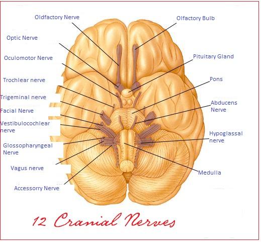
Reflex arc

The Visual System


1. What is vision? Why is vision important for life on earth?
Vision is the ability of some living beings to perceive, to distinguish and to interpret luminous stimuli.

Vision is important on earth mainly in the terrestrial and in the superficial aquatic habitats because our planet is intensely exposed to sunlight and thus light and colors become distinguishing factors of objects present in the environment, even at distance. This distinction provided new survival strategies for the organisms, new protection mechanisms against external dangers, new ways to find food and to communicate with other individuals, new types of courting and reproduction behaviors, etc. That is, it created new possibilities of interaction with the surrounds and increased capacity to explore new ecological niches.

2. How does photosensitivity in cnidarians, annelids and worms differ from insects, cephalopods and vertebrates?
In the first mentioned group of animals there are photoreceptor cells organized in ocelli or diffusely dispersed in the body. These animals do not form images.

In the animals of the second group the photoreceptor cells are part of more sophisticated structures, the eyes, able to form images and to send them to the nervous system.

3. What are the structures that compose the human vision apparatus?
The organs of the human visual apparatus are the eyes, the optical nerves and the visual areas of the brain (located in the occipital lobes of both hemispheres).

4. What are the main structures of the human eye?
The main structures of the human eye are the cornea, the iris, the pupil, the ciliary muscles, the crystalline lens and the retina (the space between the crystalline lens and the retina within the eyeball is filled with vitreous humor).

5. What is the function of the iris and of the pupil?
The iris works like the diaphragm of a photographic camera since it has muscles that contract or relax varying the pupil diameter. When the luminous intensity heightens the parasympathetic nervous system commands the contraction of the pupil; when there is shortage of light the sympathetic nervous system stimulates the dilation of the pupils. These movements depend upon the muscles of the iris.

6. Which is the part of the human visual system where the receptors that sense light, i.e., the photoreceptor cells, are located? How do those cells work?
The photoreceptor cells form the retina, a lamina that covers the internal posterior region of the eyeball. The photosensitive cells of the retina are divided into two types: the cone cells and the rod cells. These cells have pigments that sense specific light wave ranges (frequencies) and trigger action potentials conducted by the optical nerves to the visual area of the brain.

7. Since the visual images are projected in an inverted manner on the retina why don't we see things upside down?
Since the crystalline lens is a convex spherical lens it forms inverted images on the retina (every converging lens forms inverted images). The inverted information follows through the optical nerves until the occipital cerebral cortex that contains the visual area of the brain. In the brain the interpretation of the image takes place and the inverted information is reverted.

8. What type of structure is the crystalline lens? What is its function?
The crystalline is a converging spherical lens. This natural lens has the function to project images of objects onto the retina.

9. What is visual accommodation?
Visual accommodation is the phenomenon of varying the curvature of the crystalline lens to make possible the variation of its refractivity to adjust the images of objects exactly onto the retina. The visual accommodation is accomplished by the action of the ciliary muscles.

The nitid vision depends on the visual accommodation since, if the images are not projected onto the retina but in front or behind it, they will appear blurred. The closer an object is more the ciliary muscles must compress the crystalline lens (increasing its curvature); the more distant an object is more the ciliary muscles must relax.

10. What are the near point and the far point of the vision?
The near point is the closest distance between an object and the eye that makes possible the formed image to be focused, i.e., it is the point in which the ciliary muscles are in their maximum contraction. The far point is the most distant point from the eye in which an object can be placed and its image is still focused, i.e., it is the situation of maximum relaxation of the ciliary muscles. The zone between the near point and the far point is called the accommodation zone.

11. How can the visual deficiencies known as myopia and hypermetropia be optically explained?
Myopia is the visual condition in which the images are formed before (in front of) the retina. Hypermetropia is the visual condition in which the point of image formation is beyond (behind) the retina. Actually myopia is due to an increase in the distance between the retina and the crystalline lens, mainly caused by a slight flattening of the eyeball. In hypermetropia the retina is too close to the crystalline lens due to slight shortening of the eyeball.

In myopia the near point and the far point of vision come closer (the refractivity of the crystalline lens that corresponds to the maximum distension capacity of the ciliary muscles is not enough to provide visual accommodation). In hypermetropia the ciliary muscles are not able to contract more to compensate the inadequate position of the retina, i.e., the near point becomes more distant.

12. What are presbyopia and astigmatism?
Presbyopia is the visual impairment in which there is loss of the cililary muscle strength thus reducing the capability of the crystalline lens to adjust images of near objects onto the retina. In presbyopia the near point of vision becomes more distant. The disease generally occurs in old people.

Astigmatism is caused by irregular shape of the refractive structures, mainly the cornea. In astigmatism a single object-point may produce more than one image onto the retina and so the vision becomes distorted.

The Hearing System


1. What are the structures that participate in the human auditory sensitivity?
The structures of the human auditory sensitivity are the ears (external, middle and internal), the vestibulocochlear nerves and the auditory areas of the brain (located in the temporal lobes of both hemispheres).

2. What are the main parts of the human ear?
The human ear is divided into three mains parts: the external ear, the middle ear and the internal ear.

3. What are the structures that form the external ear? What is its function?
The internal ear comprises the pinna, or auricle, and the auditory canal. Its function is to conduct the sound waves to the tympanum.

4. What are the elements that form the middle ear? What are the names of the three middle ear ossicles that participate in the phonosensitivity?
The middle ear is formed by the tympanum, the ossicular chain and the oval window. The functional ossicles of the middle ear are the hammer (malleus), the incus and the stapes.

5. What is the tympanum? In which part of the ear is it located and what is its function?
The tympanum (or ear drum) is a membrane located in the middle ear just after the auditory canal and so it separates the middle ear from the external ear. The function of the tympanum is to vibrate with the same frequency of the sound waves that reach it.

6. How is the sound vibration captured by the tympanum transmitted through the ossicular chain of the middle ear?
The acoustic transmission from the external to the middle ear (and to the internal ear too) is entirely mechanical. The vibration of the tympanic membrane triggers the vibration of the hammer that then causes the incus to vibrate. The incus then causes the stapes to vibrate.

7. What are the elements that constitute the internal ear? What are the functions of those structures?
In the internal ear there are the cochlea and the semicircular canals. The fluid that fills the cochlea receives vibration from the ossicular chain of the middle ear and transmits the pressure to the semicircular canals. Within the semicircular canals the pressure variation of the filling fluid moves cilia of the hair cells of these structures. The hair cells then generate action potentials that are transmitted to the brain through the auditory nerves.

8. Why is there a sense of pressure change inside the ear when someone goes down a mountain?
The pressure inside the middle ear is maintained equal to the external ear (so to the exterior too) due to a communicating duct between the middle ear and the pharynx called the auditory tube, or Eustachian tube. When someone goes down a mountain the air pressure upon the middle ear increases and it is necessary to do some exercises like fake swallowing to force the opening of the pharyngeal orifice of the auditory tube to equalize the pressure again.

9. What is the vestibular system? How does it operate?
The vestibular system is the part of the ear that participates in the control and regulation of the equilibrium of the body (balance).

The semicircular canals of the inner (internal) ear are perpendicularly placed and detect changes in the gravitational position of the head (this is another sensorial function of the inner ear, besides auditory perception). When the head rotates the pressure of the fluid within the canals upon the cilia of specific receptor cells varies and these cells generate action potentials transmitted by the vestibulocochlear nerve. The neural impulse is then interpreted by the brain as information about the gravitational position of the head.

The Nervous System - Questions and Answers

1. What are the physiological systems known as integrative systems? Why is this designation justified?
The integrative systems are the nervous system and the endocrine system. The designation is justified since both systems control and regulate biological functions and act at distance receiving information from organs and tissues and sending effector commands (nervous impulses or hormones) to organs and tissues thus integrating the body.

2. Which are the structures that are part of the nervous system?
The structures that form the nervous system can be divided into the central nervous system (CNS) and the peripheral nervous system (PNS).

The organs of the CNS are the brain (cerebrum, brainstem and cerebellum) and spinal cord. The PNS is made of nerves and neural ganglia. Besides these organs the meninges (dura-mater, arachnoid and pia-mater) are part of the nervous system too since they cover and protect the encephalon and the spinal cord.

3. Which are the main cells of the nervous system?
The main cells of the nervous system are the neurons. Besides the neurons the nervous system is also constituted of glial cells.

4. What are the functional differences between neurons and glial cells?
Glial cells and neurons are the cells that form the nervous system. Neurons are cells that have the function of receiving and transmitting the neural impulses and glial cells (astrocytes, microgliacytes, ependymal cells and oligodendrocytes) are the cells that support, feed and insulate (electrically) the neurons. The Schwann cells that produce the myelin sheath of the peripheral nervous system can also be considered glial cells.

5. What are the three main parts into which a neuron can be divided? What are their respective functions?
The three mains parts into which a neuron can be didactically divided are: dendrites, cell body and axon.

Dendrites are projections of the plasma membrane that receive the neural impulse from other neurons. The cell body is where the nucleus and the main cellular organelles are located. Axon is the long membrane projection that transmits the neural impulse at distance to other neurons, to muscle cells and to other effector cells.

6. What is the name of the terminal portion of the axon?
The terminal portion of the axon is called presynaptic membrane. Through this membrane neurotransmitters are released into the synaptic junction.

7. What are synapses?
Synapses are the structures that transmit the neural impulse between two neurons.

When the electric impulse arrives the presynaptic membrane of the axon releases neurotransmitters that bind to postsynaptic receptors of the dendrites of the next cell. The activated state of these receptors alters the permeability of the dendritic membrane and the electric depolarization propagates along the neuron plasma membrane to its axon.

8. What is an example of a situation in which the neuron cell body is located in a part of the body and its axonal terminal portion is in another distant part of the body? Why does this happen?
Most of the neurons are situated within the brain and the spinal cord (central nervous system) in places known as neural nuclei. Neural ganglia, or simply ganglia, are structures of the peripheral nervous system located beside the spinal column or near some organs where neuron cell bodies are also located.

Neurons situated at specific points can present distant axonal terminations and they also can receive impulses from axons of distant neurons. The inferior motor neurons situated in the spinal cord are examples since their axons can transmit information to the extremities of the inferior limbs triggering contractions of the foot.

9. According to the function of the transmitted neural impulse which are the types of neurons? How different are the concepts of afference and efference of the neural impulse transmission?
There are three types of neurons: afferent neurons, efferent neurons and interneurons. Afferent neurons are those that only transmit sensory information from the tissues to neural nuclei and ganglia (where they make connection with interneurons or effector neurons). Efferent neurons are those that transmit commands to tasks performed in several parts of the body. Interneurons, also known as association neurons or relay neurons, serve as connection between two other neurons.

Afference is the conduction of sensory impulses and efference is the conduction of effector impulses (impulses that command some body action).

10. What are nerves?
Axons extend throughout the body inside nerves. Nerves are axon-containing structures presenting many axons and covered by connective tissue. The nerves connect neural nuclei and ganglia with the tissues.

Nerves may contain only sensory axons (sensory nerves), only motor axons (motor neurons) or both types of axons (mixed nerves).

11. What are ganglia?
Ganglia (singular ganglion), or neural ganglia, are structures located outside the central nervous system (for example, beside the spinal column or near viscera) made of concentration of neuron bodies.

Examples of neural ganglia are the ganglia that concentrate cell bodies of sensory neurons in the dorsal roots of the spinal cord and the ganglia of the myenteric plexus responsible for the peristaltic movements of the digestive tube.

In the central nervous system (CNS) the concentrations of neuron bodies are called nuclei and not ganglia.

12. What is meant by the peripheral nervous system (PNS)?
The peripheral nervous system comprehends the nerves and ganglia of the body.

13. What is the function of the myelin sheath? Do all axons present a myelin sheath?
The function of the myelin sheath is to improve the safety and speed of the neural impulse transmission along the axon. The myelin sheath serves as an electrical insulator preventing the dispersion of the impulse to other adjacent structures. Since the myelin sheath has gaps called Ranviers’ nodes in its length, the neural impulse “jumps” from one node to another thus increasing the speed of the neural transmission.

Not all neurons have a myelin sheath. There are myelinated axonal fibers and unmyelinated ones.

14. What are the cells that produce the myelin sheath? Of which substance is the myelin sheath formed?
In the central nervous system (CNS) the myelin sheath is made by apposition of oligodendrocyte membranes. Each oligodendrocyte can cover portions of axons of several different neurons. In the peripheral nervous system (PNS) the myelin sheath is made by consecutive Schwann cell membranes covering segments of a single axon. The Ranviers’ nodes appear in the intercellular space between these cells.

The myelin sheath is rich in lipids but it also contains proteins.

15. What are some diseases characterized by progressive loss of the axonal myelin sheath?
Multiple sclerosis is a severe disease caused by progressive destruction of the myelin sheath in the central nervous system. The Guillain-Barré disease is due to destruction of the myelin sheath in the peripheral nervous system caused by autoimmunity (attack by the own immune system). The genetic deficiency in the formation or preservation of the myelin sheath is an X-linked inheritance called adrenoleukodystrophy. The movie “Lorenzo’s Oil” featured a boy with this disease and his father's dramatic search for treatment.

16. What are meninges and cerebrospinal fluid?
Meninges are the membranes that enclose and protect the central nervous system (CNS). Cerebrospinal fluid is the fluid that separates the three layers that form the meninges and it has the functions of nutrient transport, defense and mechanical protection for the CNS.

The cerebrospinal fluid fills and protects cavities of the brain and the spinal cord.

17. What is the difference between brain and cerebrum? What are the main parts of these structures?
The concept of brain, or encephalon, comprehends the cerebrum (mostly referred to as the hemispheres, but actually the concept also includes the thalamus and the hypothalamus), the brainstem (midbrain, pons and medulla) and the cerebellum. Brain and spinal cord form the central nervous system (CNS).

18. How is the cerebrum anatomically divided?
The cerebrum is divided into two cerebral hemispheres, the right and the left. Each hemisphere is made of four cerebral lobes: frontal lobe, parietal lobe, temporal lobe and occipital lobe.

Each cerebral lobe contains the gray matter and the white matter. The gray matter is the outer portion and it is made of neuron bodies; the gray matter is also known as the cerebral cortex. The white matter is the inner portion and it is white because it is in the region where axons of the cortical neurons pass.

19. Which is the brain region responsible for the coordination and equilibrium of the body?
In the central nervous system the cerebellum is the main controller of the motor coordination and equilibrium of the body. (Do not confuse this with muscle command, performed by the cerebral hemispheres).

20. Why is the cerebellum more developed in mammals that jump or fly?
The cerebellum is the main brain structure that coordinates the movement and the equilibrium of the body. For this reason it appears more developed in mammals that jump or fly (like bats). The cerebellum is also very important for the flight of birds.

21. Which is the brain region responsible for the regulation of breathing and blood pressure?
The neural regulation of breathing, blood pressure and other physiological parameters like heartbeat, digestive secretions, peristaltic movements and transpiration is performed by the medulla.

The medulla, together with the pons and the midbrain, is part of the brainstem.

22. Which is the brain region that receives conscious sensory information? Which is the brain region that triggers the voluntary motor activity?
In the brain conscious sensory information is received by the neurons situated in a special region called postcentral gyrus (or sensory gyrus). Gyri are the convolutions of the cerebrum. Each of the two postcentral gyri are located in one of the parietal lobes of the cerebrum.

The voluntary motor activity (voluntary muscle movement) is commanded by neurons situated in the precentral gyrus (or motor gyrus). Each of the two precentral gyri are located in one of the frontal lobes of the cerebrum.

The names post- and pre-central refer to the fact that the motor and sensory gyri are spaced apart in each cerebral hemisphere by the sulcus centralis, a fissure that separates the parietal and frontal lobes.

23. What is the spinal cord? Of which elements is the spinal cord constituted?
The spinal cord is the dorsal neural cord of vertebrates. It is the part of the central nervous system that continues in the trunk to facilitate the nervous integration of the whole body.

The spinal cord is made of groups of neurons situated in its central portion forming the gray matter and of axon fibers in its exterior portion forming the white matter. Neural bundles connect to both lateral sides of the spinal cord segments to form the dorsal and ventral spinal roots that join to form the spinal nerves. The dorsal spinal roots present a ganglion with neurons that receive sensory information; the ventral spinal roots contain motor fibers. Therefore the dorsal roots are sensory roots and the ventral roots are motor roots.

24. Which are the brain regions associated with memory?
According to researchers some of the main regions of the nervous system associated with the memory phenomenon are the hippocampus, situated in the interior portion of the temporal lobes, and the frontal lobe cortex, both part of the cerebral hemispheres.

25. How is it structurally explained that the motor activity of the left side of the body is controlled by the right cerebral hemisphere and the motor activity of the right side of the body is controlled by the left cerebral hemisphere?
In the cerebral hemispheres there are neurons that centrally command and control muscle movements. These neurons are called superior motor neurons and they are located in a special gyrus of both frontal lobes known as motor gyrus (or precentral gyrus). The superior motor neurons send axons that transmit impulses to the inferior motor neurons of the spinal cord (for neck, trunk and limb movements) and to the motor nuclei of the cranial nerves (for face, eyes and mouth movements).

The fibers cross to the other side in specific areas of those axon paths. About 2/3 of the fibers that go down the spinal cord cross at the medullar level forming a structure known as pyramidal decussation. The other (1/3) of fibers descend in the same side of their original cerebral hemisphere and cross only within the spinal cord at the level where their associated motor spinal root exit. The fibers that command the inferior motor neurons of the cranial nerves cross to the other side just before the connection with the nuclei of these nerves.

The motor fibers that descend from the superior motor neurons to the inferior motor neurons of the spinal cord form the pyramidal tract. Injuries in this tract, for example, caused by spinal sections or by central or spinal tumors may lead to paraplegia and tetraplegia.

26. What is meant by the arch reflex?
In some situations the movement of the skeletal striated muscles does not depend upon commands of the superior motor neurons, i.e., it is not triggered by volition.

Involuntary movements of those muscles may happen when sensory fibers that make direct or indirect connection with inferior motor neurons are unexpectedly stimulated in situations that suggest danger to the body. This happens, for example, in the patellar reflex, or knee jerk reflex, when a sudden percussion on the knee patella (kneecap) triggers an involuntary contraction of the quadriceps (the extension muscle of the thigh). Another example of the arch reflex occurs when someone steps on a sharp object: one leg retracts and the other, by the arch reflex, distends to maintain the equilibrium of the body.

27. Which are the types of neurons that participate in the spinal arch reflex? Where are their cell bodies situated?
In the arch reflex first a sensory neuron located in the ganglion of a dorsal spinal root collects the stimulus information from the tissues. This sensory neuron makes direct or indirect (through interneurons) connection with inferior motor neurons of the spinal cord. These motor neurons then command the reflex reaction. So sensory neurons, interneurons and inferior motor neurons participate in the arch reflex.

28. What are the respective constituents of the gray matter and of the white matter of the spinal cord?
The gray matter, or gray substance, of the spinal cord contains predominantly neuron bodies (inferior motor neurons, secondary sensory neurons and interneurons). The white matter is mainly made of axons that connect neurons of the brain with spinal neurons.

29. Is the neural impulse generated by the stimulus that triggers the arch reflex restricted within the neurons of this circuit?
The sensory fiber that first conducts the arch reflex connects with neurons of the arch reflex but it also connects with secondary sensory neurons of the spinal cord that transmit information upwards to other neurons of the brain. This is obvious since the person that received the initial stimulus (e.g., the percussion on his/her kneecap) perceives it (meaning that the brain became conscious of the fact).

30. How is it explained that a person with the spinal cord sectioned at the cervical level is still able to perform the patellar reflex?
The arch reflex depends only on the integrity of the fibers at a single spinal level. In the arch reflex the motor response to the stimulus is automatic and involuntary and does not depend upon the passage of information to the brain. So it happens even if the spinal cord is damaged at other levels.

31. How does poliomyelitis affect the neural transmission in the spinal cord?
The poliovirus parasites and destroys spinal motor neurons causing paralysis of the muscles that depend on these neurons.

32. Concerning volition of the individual how can the reactions of the nervous system be classified?
The efferences (reactions) of the nervous system can be classified into voluntary, when controlled by the will, and involuntary, those not consciously controlled. Examples of reactions triggered by volition are the movements of the limb, tongue and respiratory muscles. Examples of involuntary efferences are those that command the peristaltic movements, the heartbeat and the arterial wall muscles. The skeletal striated muscles are voluntarily contracted; the cardiac striated and the smooth muscles are involuntarily contracted.

33. What are the functional divisions of the nervous system?
Functionally the nervous system can be divided into the somatic nervous system and visceral nervous system.

The somatic nervous system includes the central and peripheral structures that make voluntary control of efferences. Central and peripheral structures that participate in the control of the vegetative (unconscious) functions of the body are included in the concept of visceral nervous system.

The efferent portion of the visceral nervous system is called the autonomic nervous system.

34. What are the two divisions of the autonomic nervous system?

The autonomic nervous system is divided into the sympathetic nervous system and the parasympathetic nervous system.

The sympathetic nervous system comprehends the nerves that come out from the ganglia of the neural chains lateral to the spinal column (near the spinal cord) and thus are distant from the tissues they innervate. The central and peripheral neurons associated to those neurons are also part of the sympathetic.

The parasympathetic nervous system is made of nerves and central or peripheral neurons related to the visceral ganglia, neural ganglia situated near the tissues they innervate.

35. What is the antagonism between the sympathetic and the parasympathetic neural actions?
In general the actions of the sympathetic and the parasympathetic are antagonistic, i.e., while one stimulates something the other inhibits and vice versa. The organs, with few exceptions, get efferences from these two systems and the antagonism between them serves to modulate their effects. For example, the parasympathetic stimulates salivation while the sympathetic inhibits it; the parasympathetic constricts pupils while the sympathetic dilates it; the parasympathetic contracts the bronchi while the sympathetic relaxes them; the parasympathetic excites the genital organs while the parasympathetic inhibits the excitation.

36. Using examples of invertebrate nervous systems how can the process of evolutionary cephalization be described?
Considering the example of invertebrates it is observed that evolution makes the increasing of the complexity of the organisms to be accompanied by convergence of nervous cells to special structures for controlling and commanding: the ganglia and the brain. In simple invertebrates, like cnidarians, the nervous cells are not concentrated but they are found dispersed in the body. In platyhelminthes a beginning of cephalization with the anterior ganglion concentrating neurons is already verified. In annelids and arthropods the existence of a cerebral ganglion is evident. In cephalopod molluscs the cephalization is even greater and the brain commands the nervous system.

37. What are some main differences of the vertebrate nervous systems comparing to invertebrates?
In vertebrates the nervous system is well-characterized, having the brain and dorsal neural cord protected by rigid skeletal structures. In most invertebrates the nervous system is predominantly ganglial, with ventral neural cords.

38. What are the protective structures of the central nervous system present in vertebrates?
In vertebrates the brain and the spinal cord are protected by membranes, the meninges, and by osseous structures, respectively the skull and the vertebral column. These protections are fundamental for the integrity of those important organs that command the functioning of the body.

39. What is the nature of the stimulus received and transmitted by the neurons?
Neurons receive and transmit chemical stimuli through neurotransmitters released in the synapses. Along the neuron body however the impulse transmission is electrical. So neurons conduct electric and chemical stimuli.

40. What are the two main ions that participate in the electrical impulse transmission in neurons?
The two main ions that participate in the electrical impulse transmission in neurons are the sodium cation (Na+) and the potassium cation (K+).

41. Which is the normal sign of the electric charge between the two sides of the neuron plasma membrane? What is the potential difference (voltage) generated between these two sides? What is that voltage called?
As in most cells the region just outside the surface of the neuron plasma membrane presents a positive electrical charge in relation to the region just inside that thus is negative.

The normal (at rest) potential difference across the neuron membrane is about –70 mV (millivolts). This voltage is called the resting potential of the neuron.

42. How do the sodium and potassium ions maintain the resting potential of the neuron?
The plasma membrane of the neuron when at rest maintains an electric potential difference between its external and internal surfaces. This voltage is called resting potential. The resting potential about –70 mV indicates that the interior is more negative than the exterior (negative polarization). This condition is maintained by transport of sodium and potassium ions across the plasma membrane.

The membrane is permeable to potassium ions but not to sodium ions. At rest the positive potassium ions exit the cell in favor of the concentration gradient since within the cell the potassium concentration is higher than in the extracellular space. The positive sodium ions cannot however go into the cell. As positive potassium ions exit the cell with not enough compensation of positive ions entering the cell, the intracellular space becomes more negative and the cell stays polarized.

43. How is the depolarization of the neuronal plasma membrane generated? How does the cell return to its original rest?
When the neuron receives a stimulus by the binding of neurotransmitters to specific receptors sodium channels open and the permeability of the plasma membrane in the postsynaptic region is altered. Sodium ions then go into the cell causing lowering (less negative) of the membrane potential. If this reduction of the membrane potential reaches a level called the excitation threshold, or threshold potential, about –50 mV, the action potential is generated, i.e., the depolarization intensifies until reaching its maximum level and the depolarization current is transmitted along the remaining length of the neuronal membrane.

If the excitation threshold is reached voltage-dependent sodium channels in the membrane open allowing more sodium ions to enter the cell in favor of the concentration gradient and an approximate –35 mV level of positive polarization of the membrane is achieved. The voltage-dependent sodium channels then close and more voltage-dependent potassium channels open. Potassium ions then exit the cell in favor of the concentration gradient and the potential difference of the membrane decreases, a process called repolarization.

The action potential triggers the same electrical phenomenon in neighboring regions of the plasma membrane and the impulse is thus transmitted from the dendrites to the terminal region of the axon.

44. What is the excitation threshold of a neuron? How does this threshold relate to the “all-or-nothing” rule of the neural transmission?
The excitation threshold of a neuron is the depolarization level that must be caused by a stimulus to be transmitted as a neural impulse. This value is about –50 mV.

The transmission of the neural impulse along the neuronal membrane obeys an all-or-nothing rule: or it happens with maximum intensity or nothing happens. Always and only when the excitation threshold is reached the depolarization continues and the membrane reaches its maximum possible positive polarization, about +35 mV. If the excitation threshold is not reached nothing happens.

45. How does the depolarization of the neuronal membrane start?
The primary cause of the neuronal depolarization is the binding of neurotransmitters released in the synapse (by the axon of the neuron that sent the signal) to specific receptors in the membrane of the neuron that is receiving the stimulus. The binding of neurotransmitters to those receptors is a reversible phenomenon that alters the membrane permeability of the region since the binding causes sodium channels to open. When positive sodium ions enter the cell in favor of their concentration gradient, the membrane voltage increases, thus lessening the negative polarization. If this depolarization reaches the excitation threshold (about –50 mV) the depolarization continues, the action potential is reached and the impulse is transmitted along the cell membrane.

46. How different are the concepts of action potential, resting potential and excitation threshold concerning neurons?
Action potential is the maximum positive voltage level achieved by the neuron in the process of neuronal activation, around + 35 mV. The action potential triggers the depolarization of the neighboring regions of the plasma membrane and thus the propagation of the impulse along the neuron.

Resting potential is the membrane voltage when the cell is not excited, about –70 mV.

Excitation threshold is the voltage level, about –50 mV, that the initial depolarization must reach for the action potential to be attained.

47. In chemical terms how is the neuronal repolarization achieved?
Repolarization is the return of the membrane potential from the action potential (+35 mV) to the resting potential (-70 mV).

When the membrane reaches its action potential voltage-gated sodium channels close and voltage-gated potassium channels open. So sodium stops entering into the cell and potassium starts to exit. Therefore the repolarization is due to exiting of potassium cations from the cell.

The repolarization causes the potential difference temporarily to increase under –70 mV, below the resting potential, a phenomenon known as hyperpolarization.

48. What is the mechanism by which the neural impulse is transmitted along the axon?
The neural impulse is transmitted along the neuronal membrane through depolarization of consecutive neighboring regions. When a region in the internal surface of the membrane is depolarized it becomes more positive in relation to the neighboring internal region. So positive electrical charges (ions) move towards this more negative region and voltage-gated sodium channels are activated and open. The action potential then linearly propagates along the membrane until near the presynaptic region of the axon.

49. What is the structure through which the neural impulse is transmitted from one cell to another? What are its parts?
The structure through which the neural impulse passes from one cell to another is the synapse. The synapse is composed by the presynaptic membrane in the terminal portion of the axon of the transmitter cell, the synaptic cleft (or synaptic space) and the postsynaptic membrane in the dendrite of the receptor cell.

50. How does synaptic transmission between neurons take place?
The propagation of the action potential along the axon reaches the region immediately anterior to the presynaptic membrane causing its permeability to calcium ions to change and these ions to enter the cell. In the presynaptic area of the axon there are many neurotransmitter-repleted vesicles that by means of exocytosis activated by the calcium influx release the neurotransmitters into the synaptic cleft. The neurotransmitters then bind to specific receptors of the postsynaptic membrane. (The binding of neurotransmitters to their receptors is reversible, i.e., the neurotransmitters are not consumed after the process.) With the binding of neurotransmitters to the postsynaptic receptors the permeability of the postsynaptic membrane is altered and the depolarization that will lead to the first action potential of the postsynaptic cell begins.

51. What are some important neurotransmitters?
The following are some neurotransmitters: adrenaline (epinephrine), noradrenaline (norepinephrine), acetylcholine, dopamine, serotonin, histamine, gaba (gamma aminobutyric acid), glycine, aspartate, nitric oxide.

52. Since neurotransmitters are not consumed in the synaptic process, what are the mechanisms to reduce their concentrations in the synaptic cleft after they have been used?
Since the binding of neurotransmitters to the postsynaptic receptors is reversible, after these neurochemicals perform their role they must be eliminated from the synaptic cleft. Neurotransmitters can then bind to specific proteins that carry them back to the axon they came from in a process called neurotransmitter re-uptake. They can also be destroyed by specific enzymes, like acetylcholinesterase, an enzyme that destroys acetylcholine. Or they can simply diffuse out of the synaptic cleft.

53. Fluoxetine is an antidepressant drug that presents an action mechanism related to the synaptic transmission. What is that mechanism?
Fluoxetine is a substance that inhibits the re-uptake of serotonin, a neurotransmitter that acts mainly in the central nervous system. By inhibiting the re-uptake of the neurotransmitter the drug increases its availability in the synaptic cleft thus improving the neuronal transmission.

54. What is the neuromuscular synapse?
Neuromuscular synapse is the structure through which the neural impulse passes from the axon of a motor neuron to the muscle cell. This structure is also known as neuromuscular junction, or motor end plate.

As in the nervous synapse, the axonal terminal membrane releases the neurotransmitter acetylcholine in the cleft between the two cells. Acetylcholine binds to specific receptors of the muscle membrane, dependent sodium channels then open and the depolarization of the muscle membrane begins. The impulse is then transmitted to the sarcoplasmic reticulum that releases calcium ions into the sarcomeres of the myofibrils thus triggering contraction.

55. How does the nervous system get information about the external environment, the organs and the tissues?
Information about the conditions of the external and internal environments, like temperature, pressure, touch, spatial position, pH, metabolite levels (oxygen, carbon dioxide, etc.), light, sounds, etc., are collected by specific neural structures (each for each type of information) called sensory receptors. Sensory receptors are distributed throughout the tissues according to their specific roles. The receptors get that information and transmit them through their own axons or through dendrites of neurons that connect to them. The information reaches the central nervous system that interprets and uses it to control and regulate the body.

56. What are sensory receptors?
Sensory receptors are structures specialized in the acquiring of information, like temperature, mechanical pressure, pH, chemical environment and luminosity, transmitting them to the central nervous system. Sensory receptors may be specialized cells, e.g., the photoreceptors of the retina, or specialized interstitial structures, for example the vibration receptors of the skin. In this last case they transmit information to dendrites of sensory neurons connected to them. There are also sensory receptors that are specialized terminations of neuronal dendrites (e.g., the olfactory receptors).

57. According to the stimuli they collect how are the sensory receptors classified?
The sensory receptors are classified according to the stimuli they get: mechanoreceptors are stimulated by pressure (e.g., touch or sound); chemoreceptors respond to chemical stimuli (olfactory, taste, pH, metabolite concentration, etc.); thermoreceptors are sensitive to temperature changes; photoreceptors are stimulated by light; nocireceptors send pain information; proprioceptors are sensitive to the spatial position of muscles and joints (they generate information for the equilibrium of the body).

Signs and symptoms of nervous system disorders

The following are the most common general signs and symptoms of a nervous system disorder. However, each individual may experience symptoms differently. Symptoms may include:

A headache that changes or is different
Altered Alertness.
Babbling speech
Back pain which radiates to the feet, toes, or other parts of the body
Dizziness
Eye Problems, Noninjury.
Head Injury, Age 3 and Younger.
Head Injury, Age 4 and Older
Headaches
Impaired mental ability
Lack of coordination
Lightheadedness
Loss of balance
Loss of feeling or tingling
Memory Loss
Memory loss
Muscle rigidity
Muscle wasting and slurred speech Persistent or sudden onset of a headache
Seizures.
Sudden loss of sight or double vision
Tremors and seizures
Vertigo
Weakness and Fatigue.
Weakness or loss of muscle strength
whirling sensation

The symptoms of a nervous system disorder may resemble other medical conditions or problems. Always consult your doctor for a diagnosis.

Doctors who treat nervous system disorders

Doctors who treat nervous system disorders may have to spend a lot of time working with the patient before making a probable diagnosis of the specific condition. Many times, this involves performing numerous tests to eliminate other conditions, so that the probable diagnosis can be made.

Neurology. The branch of medicine that manages nervous system disorders is called neurology. The medical doctors who treat nervous system disorders are called neurologists.

Neurological surgery. The branch of medicine that provides surgical intervention for nervous system disorders is called neurosurgery, or neurological surgery. Surgeons who operate as a treatment team for nervous system disorders are called neurological surgeons or neurosurgeons.

Rehabilitation for neurological disorders. The branch of medicine that provides rehabilitative care for patients with nervous system disorders is called physical medicine and rehabilitation. Doctors who work with patients in the rehabilitation process are called physiatrists.

Functions and placements of 12 cranial nerves

Cranial Nerves


1. Olfactory

This is a type of sensory nerve that contributes in the sense of smell in human being. These basically provide the specific cells that are termed as olfactory epithelium. It carries the information from nasal epithelium to the olfactory center in brain.

2. Optic nerve

This again is a type of sensory nerve that transforms information about vision to the brain. To be specific this supplies information to the retina in the form of ganglion cells.

3. Oculomoter nerve

This is a form of motor nerve that supplies to different centers along midbrain. Its functions include superiorly uplifting eyelid, superiorly rotating eyeball, construction of pupil on the exposure to light and operating several eye muscles.

4. Trochlear

This motor nerve also supplies to the midbrain and performs the function of handling the eye muscles and turning the eye.

5. Trigeminal

This is a type of largest cranial nerve in all and performs many sensory functions related to nose, eyes, tongue and teeth. It basically is further divided in three branches that are ophthalmic, maxillary and mandibular nerve. This is a type of mixed nerve that performs sensory and motor functions in brain.

6. Abducent

This is again a type of motor nerve that supplies to the pons and perform function of turning eye laterally.

7. Facial

This motor nerve is responsible for different types of facial expressions. This also performs some functions of sensory nerve by supplying information about touch on face and senses of tongue in mouth. It is basically present over brain stem.

8. Vestibulocochlear

This motor nerve is basically functional in providing information related to balance of head and sense of sound or hearing. It carries vestibular as well as cochlear information to the brain and is placed near inner ear.

9. Glossopharyngeal

This is a sensory nerve which carries sensory information from pharynx (initial portion of throat) and some portion of tongue and palate. The information sent is about temperature, pressure and other related facts.

It also covers some portion of taste buds and salivary glands. The nerve also carries some motor functions such as helping in swallowing food.

10. Vagus

This is also a type of mixed nerve that carries both motor and sensory functions. This basically deals with the area of pharynx, larynx, esophagus, trachea, bronchi, some portion of heart and palate. It works by constricting muscles of the above areas. In sensory part, it contributes in the tasting ability of the human being.

11. Spinal accessory nerve

As the name intimates this motor nerve supplies information about spinal cord, trapezius and other surrounding muscles. It also provides muscle movement of the shoulders and surrounding neck.

12. Hypoglossal nerve

This is a typical motor nerve that deals with the muscles of tongue.

Cranial Nerves
Spinal Nerves
Nerves

History

Have you had a temporary loss of function?
Yes

Do you have a persistent nervous system problem, such as a tremor or lack of coordination?
Yes

Do you think your nervous system problem may be caused by a medicine?
Yes

Alzheimer's Disease
Autism
Bell’s Palsy
Coma
Concussion
Creutzfeldt-Jakob Disease
Deep Brain Stimulation Dementia
Dizziness
Encephalitis
Epilepsy
Essential Tremor
Guillain-Barré Syndrome
Hydrocephalus
Lou Gehrig's Disease (ALS)
Mad Cow Disease
Meniere's Disease
Multiple Sclerosis
Myasthenia Gravis
Nerve Pain
Parkinson's Disease
Peripheral Neuropathy
Preventing Falls
Reflex Sympathetic Dystrophy Syndrome
Restless Legs Syndrome
Seizure Disorders
Speech and Language Disorders
Stroke
Tourette's
Tremors
Trigeminal Neuralgia
Vertigo

Circulation Symptoms
Circulatory System
Abnormal pulse
Chest pain or pressure
Cyanosis
Difficulty breathing
Sudden weakness or numbness on one side of the body
Slurred speech
Loss of vision
Paralysis or inability to move a body part
Poor wound healing
Sudden, severe headache; or confusion or loss of consciousness, even for a brief moment

What is circulatory system?
On average, your body has about 5 liters of blood continually traveling through it by way of the circulatory system. The heart, the lungs, and the blood vessels work together to form the circle part of the circulatory system. The pumping of the heart forces the blood on its journey. How does the blood circulatory system work?
The body's circulatory system really has three distinct parts: pulmonary circulation, coronary circulation, and systemic circulation. Or, the lungs (pulmonary), the heart (coronary), and the rest of the system (systemic). Each part must be working independently in order for them to all work together.

Circulatory system: The system that moves blood throughout the body. The circulatory system is composed of the heart, arteries, capillaries, and veins. This remarkable system transports oxygenated blood from the lungs and heart throughout the body via the arteries. The blood goes through the capillaries which are situated between the arteries and veins. And the blood that has been depleted of oxygen by the body is then returned to the lungs and heart via the veins.

What are the signs of circulation problems?
Circulation symptoms occur when blood flow to a part of your body is reduced, usually as a result of a narrowing of the arteries, the blood vessels that carry blood rich in oxygen to all parts of the body. Typically this occurs due to the plaque buildup in the artery walls, a condition known as atherosclerosis. Risk factors for atherosclerosis include smoking, diabetes, obesity, and elevated blood lipid (fat) levels. It results in a narrowing of the arteries, and this narrowing prevents adequate blood flow.

Circulation symptoms can also arise in the veins (blood vessels that carry blood back to the heart). Blood clots in the veins may cause local inflammation and swelling (thrombophlebitis), or the blood clots may break off and travel to other sites in the body, in a process known as embolization. Pulmonary embolism is a life-threatening condition caused by a blockage of an artery in the lung due to a blood clot that has traveled to this location.

Circulation symptoms can occur in the head, arms, kidneys, stomach, and other organs. When blood flow to an organ is interrupted, especially over time, organ failure can result. Interruption of blood flow to the brain (stroke) or heart (heart attack) are serious causes of circulation symptoms. The legs are another common site for circulation symptoms. As the flow of blood is blocked or slowed significantly due to peripheral artery disease in the legs, you may feel pain, heaviness and numbness in your legs, especially during and after walking or climbing stairs, when your muscles need more blood and are not able to get enough.

You may prevent or improve circulation symptoms by making important lifestyle changes, particularly by giving up smoking and limiting fats, cholesterol, and refined sugars in your diet, as well as asking your physician to recommend a good exercise program for you.

The Circulatory System

1. What is circulation?
Circulation is the movement of substances like nutrients and gases within blood vessels and cavities throughout the organism.

2. Do all animals have a circulatory system?
Not all animals have a circulatory system.

Poriferans, cnidarians, platyhelminthes and nematodes (in these there are the pseudocoelom fluid but no vessels) are avascular animals. Echinoderms do not have true circulatory systems either.

3. What is the alternative means for transport of substances in animals without a circulatory system? Why is blood important for larger animals?
In animals that do not present the circulatory system the transport of substances occurs by cell to cell diffusion.

The blood is a fundamental means of substance transport for larger animals since in these animals there are tissues distant from each other and from the environment thus making diffusion impossible.

4. What are the two types of circulatory systems?
The circulatory systems can be classified into open circulatory system and closed circulatory system.

5. What is an open circulatory system?
Open circulatory system is the one in which blood does not circulate only inside blood vessels but it also falls in cavities that irrigate tissues. In the open circulatory system the blood pressure is low and generally the blood (called hemolymph) has low cellularity.

Arthropods, molluscs (the cephalopods are exception) and protochordates have open circulatory system.

6. What is a closed circulatory system?
A closed circulatory system is one in which blood circulates only inside blood vessels. For this reason the blood pressure is higher in animals with closed circulatory system. The cellularity of the blood is also higher with many specific blood cells.

The closed circulatory system is a feature of annelids, cephalopod molluscs and vertebrates.

7. What are the advantages of the closed circulatory system over the open circulatory system?
The closed circulatory system is more efficient. Since blood circulates only inside blood vessels it can do it with more pressure reaching farther distances between the organs where hematosis happens and the peripheral tissues. In addition the circulatory speed also heightens making possible more oxygen supply to great consuming tissues, like the muscle tissues that then can perform faster movements. Animals with an open circulatory system (with the exception of insects that do gas exchange independently from the circulation) are generally slower and have a low metabolic rate.

8. What is the difference between octopuses and mussels regarding their circulatory systems? How does that difference influence the mobility of these animals?
Cephalopod molluscs, like octopuses and squids, have a closed circulatory system with blood pumped under pressure flowing within vessels. Bivalve molluscs, like mussels and oysters, have an open circulatory system (also known as lacunar circulatory system) where blood flows under low pressure since it falls in cavities of the body and does not only circulate within blood vessels. Molluscs with closed circulatory systems are larger, agile and can actively move; molluscs with open circulatory systems are smaller, slow and some are practically sessile.

9. Why, even thoough they have an open circulatory system, can flying insects like flies beat their wings with great speed?
In insects the circulatory system is open but this system does not participate in the gas exchange process and in oxygen supply to the tissues. Gases go in and out through the independent tracheal system that allows direct contact of cells with the ambient air. Therefore an insect can supply the great oxygen demand of its fast-beating wing muscles even having open circulatory system.

10. What are the typical components of a closed circulatory system?
The typical components of the closed circulatory system are the blood vessels within which blood circulates (arteries, veins and capillaries), a pumping organ (heart) and the blood or bloodlike fluid.

11. How does the heart impel the blood?
The heart is a muscular organ that contains chambers (right atrium and right ventricle and left atrium and right ventricle) through which blood passes. The blood enters the heart in the atria, goes to the ventricles and then leaves the organ.

The blood is pumped out of the heart by the contraction of the muscle fibers that form the ventricular walls. The contraction reduces the ventricle volume thus increasing the internal pressure and the blood then flows to the exit vessels (pulmonary artery for the right ventricle and aorta for the left ventricle). When ventricular muscle fibers distend the ventricles regain their original size and receive new blood flow coming from the atria.

12. What is the difference between systole and diastole
Systole and diastole are the two stages into which the cardiac cycle is divided. Systole is the stage when the contraction of ventricular muscle fibers occurs and the ventricles are emptied. Diastole is the stage of the cardiac cycle when the ventricular muscle fibers distend and the ventricles are filled with blood.

13. What are arterial vessels, arteries and arterioles?
Arterial vessels are every blood vessel that carries blood from the heart to the tissues. Arteries and arterioles are arterial vessels. Arterioles are thin arteries that end in capillaries.

Not all arteries however contain arterial blood (highly oxygenated blood). The pulmonary artery and its ramifications, arteries that carry blood from the right heart ventricle to the lungs, contain venous blood.

14. What are venous vessels, veins and venules?
Venous vessels are every blood vessel that carries blood from the tissues to the heart. Veins and venules are venous vessels. Venules are thin veins that are continuous to capillaries.

In general venous vessels carry venous blood. The pulmonary veins that carry blood from the lungs to the left atrium of the heart however contain arterial blood.

15. What are the capillaries of the vascular system?
Capillaries are small blood vessels that perform exchange of substances between the blood and the body tissues. Capillaries are neither arteries nor are they veins since they have distinct features. In capillaries the wall is made of a single layer of endothelial cells through which substances are exchanged. These vessels receive blood from the arterioles and drain to the venules.

16. What is the part of the vascular system that performs exchange of gases and other substances with the tissues?
Only capillaries perform exchange of gases and other substances with the tissues.

17. Are the arteries or the veins constituted of more muscle tissue? How different are the walls of these two types of blood vessels?
The arterial system has thicker muscle walls since within arteries the blood circulates under higher pressure. The veins are more flaccid than arteries.

From the lumen to the external layer both types of vessels are made of endothelium, muscle tissue and connective tissue. In both the endothelium is a single layer of cells. In arteries the muscle tissue portion is thicker than in veins and in these vessels the external connective tissue is thicker than in arteries.

Arteries are the pulsating blood vessels. The arterial pulse can be felt in a medical examination, for example, by the palpation of the radial artery in the internal and lateral face of the wrist near the base of the thumb.

18. What are the valves of the venous system? What is their function?
The valves of the venous system are structures inside the veins that permit blood to flow only in the normal way (from the tissues to the heart) and forbid it to return in the reverse way in favor of gravity. The valves close when the pressure of the fluid column above (after, regarding normal flux) is higher than the fluid pressure before them. Valves are thus fundamental for the returning of blood to the heart.

19. How do the muscles of the legs and of the feet contribute to the venous return?
The muscles of the legs, mainly the muscles of the calves, contract and compress the deep veins of the legs impelling the blood to the heart.

The plantar portion of the feet retains blood and when it is compressed against the ground it impels its blood volume and aids venous return.

20. What are varices? Why are they more common in the inferior limbs?
Varix means abnormal enlargement of veins. Varices occur when excessive pressure against the normal blood flux creates enlargement of the vein and thus insufficient functioning of its valves (venous insufficiency).

Varices are more common in the veins of the inferior limbs since the fluid column above these vessels is higher. This is the reason why people that spend much time standing (e.g., surgeons) are more susceptible to varices.

In general varices are not the apparent superficial veins that appear in the leg of varix patients. These apparent vessels are the consequences of internal varices (venous insufficiency) in the deep internal veins of the legs and they appear because the blood flux is diverted to superficial veins. (Popularly however apparent superficial veins are called varices.)

21. What is the lymphatic system?
The lymphatic system is a network of specialized valved vessels that drain interstitial fluid (lymph). The lymphatic system is also responsible for the transport of chylomicrons (vesicles that contain lipids) made after the absorption of fats by the intestinal epithelium.

In the way of the lymphatic vessels there are ganglial-like structures called lymph nodes that contain many cells of the immune system. These cells filter impurities and destroy microorganisms and cellular wastes. The lymphatic vessels drain to two major lymphatic vessels, the thoracic duct and the right lymphatic duct, that in their turns drain into tributary veins of the superior vena cava.

22. Why in inflammatory and infectious conditions may clinical signs related to the lymphatic system occur?
The lymph nodes, or lymph glands, have lymphoid tissue that produces lymphocytes (a type of leukocyte). In inflammatory and infectious conditions the enlargement of lymph nodes of the lymphatic circuits that drain the affected region due to the reactive proliferation of leukocytes is common. This enlargement is known as lymphadenomegaly and sometimes it is accompanied by pain. The search for enlarged or painful lymph nodes is part of the medical examination since these findings may suggest inflammation, infection or other diseases.

23. Which are the heart chambers respectively where the entrance and the exit of blood occur?
The heart chambers through which blood enters the heart are the atria. There are the right atrium and the left atrium.

The heart chambers through which the blood exits the heart are the ventricles. There are the right ventricle and the left ventricle.

24. Concerning the thickness of their walls how different are the heart chambers?
The ventricle walls are thicker than the atrium walls since ventricles are structures responsible for the pumping of the blood to the lungs or tissues. The muscular work of the ventricles is harder and their muscle fibers develop more.

The left ventricle is more muscular than the right ventricle because pumping blood to the lungs (the right ventricle task) is easier (needs less pressure) than pumping blood to the other tissues of the body (the left ventricle task).

25. What is vena cava? Which type of blood circulates within the vena cava?
The vena cava are either of two large veins that debouch into the right atrium. The superior vena cava drains all blood that comes from the head, the superior limbs, the neck and the superior portion of the trunk. The inferior vena cava carries blood drained from the inferior portion of the trunk and the inferior limbs.
Venous blood circulates within the vena cava.

26. Which is the first (human) heart chamber into which blood enters? Where does the blood go after passing that chamber? What is the name of the valve that separates the compartments? Why is that valve necessary?
The venous blood that comes from the tissues arrives in the right atrium of the heart. From the right atrium the blood goes to the right ventricle. The valve that separates the right ventricle from the right atrium is the tricuspid valve (a valvular system made of three leaflets). The tricuspid valve is necessary to prevent returning of blood to the right atrium during systole (contraction of ventricles).

27. What is the function of the right ventricle? To where does the right ventricle pump the venous blood?
The function of the right ventricle is to get venous blood from the right atrium and pump the blood to be oxygenated in the lungs.

The venous blood is carried from the right ventricle to the lungs by the pulmonary artery and their ramifications.

28. What is the valve that separates the right ventricle from the pulmonary artery? Why is that valve important?
The valve that separates the right ventricle and the base of the pulmonary artery is the pulmonary valve. The pulmonary valve is important to prevent blood from the pulmonary circulation to flow back to the heart during diastole.

29. Do the arteries that carry blood from the heart to the lungs contain arterial or venous blood? What happens to the blood when it passes through the lungs?
Arteries of the pulmonary circulation are arteries that carry venous blood and not arterial blood.

When the blood passes within the alveolar capillaries of the lungs hematosis (oxygenation) occurs and carbon dioxide is released to the exterior.

30. What and how many are the pulmonary veins?
The pulmonary veins are part of the pulmonary circulation. They are vessels that carry oxygen-rich (arterial) blood from the lungs to the heart. There are four pulmonary veins, two that drain blood from the right lung and other two that drain the left lung. The pulmonary veins debouch into the left atrium bringing arterial blood to the heart. Although they are veins they carry arterial blood and not venous blood.

31. To which heart chamber does the blood go after leaving the left atrium? What is the valve that separates these compartments?
The arterial blood that has come from the lungs to the left atrium passes then to the left ventricle.

The valve between the left ventricle and the left atrium is the mitral valve, a bicuspid (two leaflets) valve. The mitral valve is important because it prevents the regurgitation of blood to the left atrium during systole (contraction of the ventricles).

32. What is the function of the left ventricle? Where does the blood go after leaving the left ventricle?
The function of the left ventricle is to get blood from the left atrium and to pump the blood under high pressure to the systemic circulation. After leaving the left ventricle the blood enters the aorta, the largest artery of the body.

33. What is the valve that separates the aorta from the heart? What is the importance of that valve?
The valve between the left ventricle and the aorta is the aortic valve. The aortic valve prevents the retrograde flux of blood to the left ventricle during diastole. Besides, as the aortic valve closes during diastole, part of the retrograde blood flux is impelled through the coronary ostia (openings), orifices located in the aorta wall just after the valvular insertion and contiguous to the coronary circulation responsible for the blood supply of the cardiac tissues.

34. Is the ventricle lumen larger during systole or during diastole?
Systole is the stage of the cardiac cycle on which the ventricles contract. So the lumen of these chambers is reduced and the pressure upon the blood within them is heightened.

During diastole the opposite occurs. The muscle fibers of the ventricles relax and the lumen of these chambers enlarges helping the entrance of blood.

35. What is the stage of the cardiac cycle during which the ventricles are filled?
The filling of the ventricles with blood occurs during diastole.

36. Of which type of tissue is the heart made? How is this tissue oxygenated and nutrified?
The heart is made of striated cardiac muscle tissue. The heart muscle is called the myocardium and it is oxygenated and nutrified by the coronary arteries. The coronary arteries come from the base of the aorta and ramify around the heart penetrating the myocardium.

Diseases of the coronary arteries are severe conditions.

37. Which are the two main metabolic gases transported by the blood?
The main metabolic gases transported by the blood are molecular oxygen (O2) and carbon dioxide (CO2).

38. How do respiratory pigments act?
Respiratory pigments are oxygen-carrying molecules present in the blood. When the oxygen concentration is high, for example, in the pulmonary alveoli, the respiratory pigments bind to the gas. In conditions of low oxygen concentration, e.g., in tissues, the respiratory pigments release the molecule.

In the human blood the respiratory pigment is hemoglobin, present within the red blood cells.

39. How different are oxyhemoglobin and hemoglobin? Where is it expected to find a higher concentration of oxyhemoglobin, in peripheral tissues or in the lungs?
Oxygen-bound hemoglobin is called oxyhemoglobin. In the lungs the oxygen concentration is higher and so there is a higher oxyhemoglobin concentration. In the peripheral tissues the situation is the reverse, the concentration of oxygen is lower and there is more free hemoglobin.

40. What is hemoglobin F? Why does the fetus need a different hemoglobin?
Hemoglobin F is the hemoglobin found in the mammalian fetus and hemoglobin A is the normal hemoglobin. Hemoglobin F has higher affinity for oxygen.

The fetus needs hemoglobin capable of extracting oxygen from the mother’s circulation. Therefore the fetus uses hemoglobin F since it has higher affinity for oxygen than the mother’s hemoglobin.

41. In high altitudes is it necessary for the blood to have more or less hemoglobin?
In high altitudes the air is rarefied and oxygen concentration is lower than in low altitudes. In this situation the efficiency of the respiratory system must be greater and thus the organism synthesizes more hemoglobin (and more red blood cells) trying to get more oxygen. This phenomenon is known as compensatory hyperglobulinemia.

The compensatory hyperglobulinemia is the reason why athletes that will compete in high altitudes need to arrive in the place some days before the event so there is time for their body to make more red blood cells and they will be less affected by the effects of the low atmospheric oxygen concentration (fatigue, reduced muscular strength).

42. What is the substance that stimulates the production of red blood cells? Which is the organ that secretes it? Under what conditions does this secretion increase?
The substance that stimulates the production of red blood cells by the bone marrow is erythropoietin. Erythropoietin is a hormone secreted by the kidneys. Its secretion is increased when there is deficient tissue oxygenation (tissue hypoxia) caused either by reduced oxygen availability (as it occurs in high altitudes) or by internal diseases, as in pulmonary diseases.

43. Why is carbon monoxide toxic for humans?
Hemoglobin “likes” carbon monoxide (CO) much more than it likes oxygen. When there is carbon monoxide in the inhaled air it binds to hemoglobin forming carboxyhemoglobin by occupying the binding site where oxygen would bind. Due to the higher hemoglobin affinity for carbon monoxide thus (e.g., in intoxication from car exhausts) there is no oxygen transport and the individual undergoes hypoxia, loses conscience, inhales more carbon monoxide and may even die.

Intoxication by carbon monoxide is an important cause of death in fires and in closed garages.

44. What is the stage of cellular respiration during which carbon dioxide is liberated?
In aerobic cellular respiration the release of carbon dioxide happens in the transformation of pyruvic acid into acetyl-CoA (two molecules) and in the Krebs cycle (four molecules). For each glucose molecule, six carbon dioxide molecules are made.

45. How is carbon dioxide released by cellular respiration transported from the tissues to be eliminated through the lungs?
In vertebrates almost 70% of the carbon dioxide is transported by the blood in the form of bicarbonate, 25% bound to hemoglobin and 5% dissolved in the plasma.

46. What is the difference between double closed circulation and simple closed circulation?
Double closed circulation, or closed circulation, is that in which the blood circulates through two associated and parallel vascular systems: one that carries blood to and takes blood from the peripheral tissues (the systemic circulation) and the other that carries blood to and takes blood from the tissues that perform gas exchange with the environment, e.g, the lungs (pulmonary circulation). Double circulation occurs in amphibians, reptiles, birds and mammals.

Simple closed circulation, or simple circulation, is the one in which the tissues that perform gas exchange are associated in series with the systemic circulation, as in fishes.

47. How many chambers does the fish heart have?
The fish heart is a tube made of two consecutive chambers: one atrium and one ventricle.

48. Does the fish heart pump venous or arterial blood?
The venous blood coming from the tissues enters the atrium and passes to the ventricle that then pumps the blood towards the gills. After oxygenation in the gills the arterial blood goes to the tissues. So the fish heart pumps venous blood.

49. Why is the fish circulation classified as a simple and complete circulation?
Complete circulation is that in which there is no mixture of venous blood and arterial blood. Simple circulation is that in which the blood circulates only in one circuit (as opposed to the double circulation that have two circuits, the systemic circulation and the pulmonary circulation). In fishes the circulatory system is simple and complete.

50. How many chambers does the human heart have?
The right ventricle pumps the blood back to the lungs where it is oxygenated and sent to the left atrium. From there it heads over to the left ventricle. The left ventricle is responsible for sending the blood to the aorta, which then sends oxygenated blood throughout the entire body. With each pump of the heart, between 60 and 70 milliliters of blood are sent throughout the body. This equals around four to five liters per minute.

How many heart chambers does the amphibian heart have?
The amphibian heart has three heart chambers: two atria and one ventricle.

51. Why can the amphibian circulation be classified as double and incomplete?
The amphibian circulation is double because it is composed of systemic and pulmonary circulations: respectively, heart-tissues-heart and heart-lungs-heart. Since amphibians have only one ventricle in the heart, venous blood taken from the tissues and arterial blood coming from the lungs are mixed in the ventricle that then pumps the mixture back to the systemic and to the pulmonary circulations. The amphibian circulation is classified as incomplete because venous and arterial blood mix in the circuit.

The blood oxygenation in amphibians occurs also in the systemic circulation since their skin is a gas exchange organ.

52. What is the difference between the amphibian heart and the reptile heart?
The reptiles have double and incomplete circulation too, three heart chambers (two atria and one ventricle). The reptile heart however presents the beginning of a ventricular septation that partially separates a right and left region of the chamber. With the partial ventricular septation the mixture of arterial with venous blood in the reptile heart is less than in amphibians.

53. How many chambers do the bird heart and the mammalian heart have? Concerning temperature maintenance what is the advantage of the double and complete circulation of these animals?
The bird and the mammalian hearts are divided into four chambers: right atrium, right ventricle, left atrium and left ventricle.

Birds and mammals are homeothermic, i.e., they control their body temperature. The four-chambered heart and the double circulation provide the supply of more oxygenated blood to the tissues making possible a higher metabolic rate (mainly cellular respiration rate). Part of the energy produced by the cellular respiration is used to maintain the body temperature.

54. Concerning the mixture of arterial with venous blood what is the difference between the human fetal circulation and the adult circulation?
In the human fetal circulation there are two communications between arterial and venous blood characterizing an incomplete circulation. One of them is the oval foramen, an opening between the right and the left atria of the fetal heart. The other is the arterial duct, a short vessel connecting the pulmonary artery to the aorta. These communications close a few days after birth and so they are not present in the adult heart.

55. How is heart contraction triggered?
Heart contraction is independent from neuronal stimulus (although it can be modulated by the autonomous nervous system). In the heart there are pacemaker cells that trigger by themselves the action potentials that begin the muscle contraction. These cells are concentrated at two special points of the heart: the sinoatrial node (SA node) located in the superior portion of the right atrium and the atrioventricular node (AV node) located near the interatrial septum.

The action potentials generated by depolarization of the SA node cells propagate cell to cell throughout the atria producing the atrial contraction. The atrial depolarization also propagates to the AV node that then transmits the electric impulse to the ventricles through specialized conduction bundles of the interventricular septum (the bundle of His) and then to the Purkinje fibers of the ventricle walls causing ventricular contraction. (The atrial contraction precedes the ventricular contraction for blood to fill the ventricles before the ventricular contraction.)

The repolarization of the SA node makes the atria relax and then the ventricles relax too
Here are further guidelines.

Human Body Quiz
1. How many total human skeletal bones are there?
2. The colored part of the human eye that controls how much light passes through the pupil is called the?
3. What is the name of the substance that gives skin and hair its pigment?
4. The muscles found in the front of your thighs are known as what?
5. True or false? The two chambers at the bottom of your heart are called ventricles.
6. What substance are nails made of?
7. What is the human body’s biggest organ?
8. The innermost part of bones contains what?
9. True or false? An adult human body has over 500 bones.
10. How many lungs does the human body have?
11. Another name for your voice box is the?
12. The two holes in your nose are called?
13. Your tongue is home to special structures that allow you to experience tastes such as sour, sweet, bitter and salty, what is their name?
14. The bones that make up your spine are called what?
15. The shape of DNA is known as?
16. The flow of blood through your heart and around your body is called?
17. The bones around your chest that protect organs such as the heart are called what?
18. What is the name of the long pipe that shifts food from the back of your throat down to your stomach?
19. True or false? Your ears are important when it comes to staying balanced.
20. The outside layer of skin on the human body is called the?
21. The largest and heaviest bone in the body is the:
22. How many types of muscles are there in the human body and what are they?
23. What is the name of the biggest part of the human brain?

Human Body Quiz Answers

1. There are 206 named bones a normal, complete, adult human skeleton.
2. Iris
3. Melanin
4. Quadriceps
5. True
6. Keratin
7. The skin
8. Bone marrow
9. False (there are 206)
10. 2
11. Larynx
12. Nostrils
13. Taste buds
14. Vertebrae
15. A double helix
16. Circulation
17. Ribs
18. The esophagus
19. True
20. Epidermis
21. The femur, or thigh bone, is the longest, heaviest, and strongest bone in the entire human body.
22. Three.
    Skeletal
    Smooth
    Cardiac

23. The cerebrum

Questions you need to answer.

How many muscles are there in the human body?
How many days does it take for your body to rid itself of an alcoholic beverage?
How many identifiable bones are there in the human body?
What type of nerves send signals to the brain?
Triceps, biceps and quadriceps are examples of what kind of muscle?
There are how many Cervicle discs?
There are how many Thoracic discs?
There are how many Lumbar discs?
The Sternum holds how many ribs together?

Dermatology
Integumentary system
A to Z All Skin Conditions A|B|C|D|E|F|G|H|I|J|K|L|M|N|O|P|Q|R|S|T|U|V|W|X|Z
 

Acne

- Full term is 'Acne Vulgaris' and this is one of the commonest conditions which Dermatologists are asked to see. More Details >>
 

Actinic Keratosis

- Also known as Solar Keratosis or sometimes "AKs". More Details >>
 

Allergy

- Allergic rashes, hives, urticaria, etc are a major problem world wide.  More Details >>
 

Alopecia

- This is the medical term for hair loss - there are many different types of hair loss resulting from a wide variety of different conditions.  More Details >>
 

Androgenetic Alopecia

- This means hair loss due to male-pattern baldness and is seen in both men but also in post menopausal women . More Details >>
 

Alopecia Areata

- Very smooth patches of hair loss affecting the scalp or beard areas and often moving from site to site. More Details >>
 

Age Spots

  - Also known as Solar Lentigos or Liver Spots -Exaggerated brown freckles typically occuring on the face or the backs of the hands  More Details >>
 

Athletes Foot

- Scaly itchy rash often between the toes sometimes known as "Tinea" which represents a fungal infection of the skin. More Details >>
 

Atopic

  - The disorders of asthma, eczema and hay fever More Details >>
 
A|B|C|D|E|F|G|H|I|J|K|L|M|N|O|P|Q|R|S|T|U|V|W|X|Z
 

Balanitis

- Sore redness or shinyness of the tip of the penis.  More Details >>
 

Basal Cell Carcinoma

- This condition also known as a rodent ulcer is the commonest skin cancer.  It is usually not very serious and can be treated.  More Details >>
 

Benign

- This medical term means innocent or non serious
 

Blister

- A superficial balloon of skin on the surface containing clear fluid.  Usually these are single lesions but in rare diseases there can be many blisters on the skin for example in pemphigoid. More Details >>
 

Bowens Disease

- Usually seen on sunshine exposed areas of skin (backs of the hands, forearms, upper shoulders etc).  Usually a slightly thickened red and scaly patch of skin which fails to repond to simple moisturisers. More Details >>
 
A|B|C|D|E|F|G|H|I|J|K|L|M|N|O|P|Q|R|S|T|U|V|W|X|Z
 

Candida

- Commonly known as "thrush" - depending on which area of the body is involved eg vagina, mouth, etc (the condition can look very different)
 

Cold Sores  (Herpes)

- Medically known as "herpes" and is due to the herpes simplex virus causing tingling or painful blisters recurring in the same body position eg the lips time after time. More Details >>
 

Crabs

- Also known as "body lice" or "pubic lice" - can be seen small black dots on the skin or hairs.Mo re Details >>
 

Cysts

- Smooth 1-2 centimetre swellings just below the skin surface usually on the upper trunk and commonly on the scalp. More Details >>
 
A|B|C|D|E|F|G|H|I|J|K|L|M|N|O|P|Q|R|S|T|U|V|W|X|Z
 

Darker Skin Patients

Even common disorders can look very different in darker skinned patients compared with white skin patients. More Details >>
 

Dermatitis

- This means precisely the same as eczema to Dermatologists - see Eczema  More Details >>
 

Dermatofibroma

- Quite common small very firm 0.6 centimetre skin lesions sometimes also known medically as Histio Cytomas. 
 

Drug Reactions

(to medications) - Usually a very widespread red irritable rash resuls from taking oral medication for example antibiotics. Many different drugs and medications can be responsible. More Details >>
 
A|B|C|D|E|F|G|H|I|J|K|L|M|N|O|P|Q|R|S|T|U|V|W|X|Z
 

Eczema

- Itchy inflammation of the skin for any reason but most typically atopic (genetically linked). More Details >>
 
A|B|C|D|E|F|G|H|I|J|K|L|M|N|O|P|Q|R|S|T|U|V|W|X|Z
 

Folliculitis

- This can occur on any area of the body and is due to bacteria down inside the hair follicles. More Details >>
 

Fungus

- Fungal infections can take many forms on the skin including athletes foot and is often medically known as Tinea. More Details >>
 
A|B|C|D|E|F|G|H|I|J|K|L|M|N|O|P|Q|R|S|T|U|V|W|X|Z
 

Genital Fungal Infections

- More Details >>
 

Genital Skin Disease

- Many common skin diseases can affect the genitals for example eczema and psoriasis.  However, other common infections can also affect the genitals. More Details >>
 

Genital Molluscum

. More Details >>
 

Genital Warts -

these can look very different. More Details >>

 
A|B|C|D|E|F|G|H|I|J|K|L|M|N|O|P|Q|R|S|T|U|V|W|X|Z
 

Herpes Simplex (Cold Sores)

- This viral discorder causing small painful blisters can affect any area of the skin but most typically the mouth and lips and also the genital skin - see Cold Sores. More Details >>
 

Herpes Zoster (Shingles)

- A potentially very painful blistering condition affecting the face or body and caused by a virus affecting the nerve endings in the skin. More Details >>
 

Histo Cytomas

- also known medically as Dermatofibroma.
 

Hives

- see Urticaria. More Details >>
 

Hyperhidrosis

also known as excessive sweating. More Details >>
 
A|B|C|D|E|F|G|H|I|J|K|L|M|N|O|P|Q|R|S|T|U|V|W|X|Z
 

Impetigo

- A bacterial infection of the skin often around the nose and mouth but can spread. More Details >>
 

Intertrigo

- A red discharging rash affecting hot sweaty skin. More Details >>
 
A|B|C|D|E|F|G|H|I|J|K|L|M|N|O|P|Q|R|S|T|U|V|W|X|Z
 

Kaposis Sarcoma

- One centimetre purple nodules on the skin, sometimes associated with HIV infection. More Details >>
 

Keratosis

- There are many types of Keratosis which simply means scaly skin condition and these include Actinic Keratosis or Seborrhoeic Keratosis.
 

Keratoacanthoma

- A rapidly growing exaggerated warty lesion suggesting squamous cell carcinoma in some patients. More Details >>
 

Kerion

- a severe fungal infection of the scalp causing inflammation and hair loss. More Details >>
 
A|B|C|D|E|F|G|H|I|J|K|L|M|N|O|P|Q|R|S|T|U|V|W|X|Z
 

Leg Ulcers (Stasis Dermatitis)

- A very common cause of skin break down around the ankles and lower legs often due to obvious or alternatively, hidden, varicose veins. More Details >>
 

Lentigo

- An exaggerated freckle which is larger or darker than a standard freckle.. More Details >>
 

Lesion/Lump

- A part or swelling of the skin that has suffered through injury or disease. More details
 

Lichen Planus

- An itchy skin rash which can mimick any of the common diseases - eczema, psoriasis, scabies, etc. More Details >>
 

Lichen Sclerosus

- A rare but important skin disease becaue it can affect the genital skin and look very unusual at other sites. More Details >>
 

Liver spots

- also known as age spots or solar lentigos. More Details >>
 
A|B|C|D|E|F|G|H|I|J|K|L|M|N|O|P|Q|R|S|T|U|V|W|X|Z
 

Malignant

-This medical term means cancerous or having the potential to spread or be serious.
 

Mask of pregnancy

- See Melasma. More Details >>
 

Melasma

- Exaggerated grey-brown pigmentation across the forehead and face. More Details >>
 

Melanoma

- Irregular pink brown, black or purple skin growths often arising from a pre-existing mole. More Details >>
 

Molluscum

- Small shiny warty lesions showing in groups anywhere on the skin. More Details >>
 

Mole

- See Naevus. More Details >>
 
A|B|C|D|E|F|G|H|I|J|K|L|M|N|O|P|Q|R|S|T|U|V|W|X|Z
 

Naevus

- Medical term for Mole - flat or elevated, pink brown or darker skin lesions usually growing during the first, second and third decades of life.  Patients with many naevi (for example over 50) do need to be highly self-observant about the possibility of malignant melanoma development. More Details >>
 

Nail Disease

- The two commonest conditions causing nail disorders are fungal infections and psoriasis. More Details >>
 
A|B|C|D|E|F|G|H|I|J|K|L|M|N|O|P|Q|R|S|T|U|V|W|X|Z
 

Pemphigoid

- Pemphigoid/Pemphigus - rare conditions of the skin causing unexplained blisters More Details >>
 

Photo Sensitivity

- Means an irritation by sunlight or an exaggerated tendency to very easy sunburn. More Details >>
 

Pityriasis Rosea

- A widespread rapidly developing skin eruption sometimes resembling psoriasis. More Details >>
 

Pityriasis Versicolor

- A non-troublesome widespread eruption usually on the upper body and around the armpits. More Details >>
 

Pompholyx

- A blistering kind of eczema typically affecting the palms of the hands or the soles of the feet.  More Details >>
 

Pruritus

- The medical term for "itch" - there are very many causes. More Details >>
 

Psoriasis

- A widespread scaly eruption affecting any area of the body and often running in families . More Details >>
 

Pubic Lice

- Commonly known as crabs - tiny insects affecting the hairs of the body. More Details >>
 

Pyogenic Granuloma

- A rapidly growing fragile and bleeding pink/purple nodule. More Details >>
 
A|B|C|D|E|F|G|H|I|J|K|L|M|N|O|P|Q|R|S|T|U|V|W|X|Z
 

Rash

- Use the Body Map to narrow your search
 

Rodent Ulcer

- Please see basal cell carcinoma. More Details >>
 

Rosacea

- This is a very common condition of adult life. The face and nose are the commonest sites. More Details >>
 
A|B|C|D|E|F|G|H|I|J|K|L|M|N|O|P|Q|R|S|T|U|V|W|X|Z
 

Scabies

- A widespread itchiness of the skin usually associated with patchy tiny red irritated lesions scattered widely around the body. More Details >>
 

Seborrhoeic Dermatitis

- A type of red irritated dermatitis usually affecting the centre of the body from the eyebrows down the nose and sometimes on to the centre of the upper face and body. More Details >>
 

Seborrhoeic Keratosis

- Benign Warty Brown marks on the skin . More Details >>
 

Shaving rash

- See Follicitis. More Details >>
 

Shingles

- A potentially very painful blistering condition affecting the face or body and caused by a virus affecting the nerve endings in the skin. See Herpes Zoster. More Details >>
 

Skin Allergy

- This comes in many different forms and can be generated by either outside skin contact or internal eating or drinking of something creating the allergy. More Details >>
 

Skin Cancer

-Please see the 3 commonest - Melanoma >>, Basal Cell Carcinoma>>, Squamous Cell Carcinoma >>.
 

Skin Tags

- Tiny little growths of skin often under the armpits or beneath the breasts. More Details >>
 

Solar Lentigo

- Also known as Age Spots or liver spots - Exaggerated brown freckles typically occuring on the face or the backs of the hands  More Details >>
 

Spots

-See Folliculitis. More Details >>
 

Squamous Cell Carcinoma

- Irregular thickened red scaly lesions usually on sunshine exposed areas. More Details >>
 

Stasis Eczema

- Itchy or sore redness of the ankles or lower legs often associated some years later with varicose veins. More Details >>
 

Sunbeds

- Artificial ultra violet light cabinets giving suntans. More Details >>
 

Sun Exposure

- The sun gives benefits but also can damage the skin if exposed excessively for example skin cancer. More Details >>
 

Sunshine & Protection

- See Photo Sensitivity. More Details >>
 

Sunshine Sensitivity

- See Photo Sensitivity. More Details >>
 
A|B|C|D|E|F|G|H|I|J|K|L|M|N|O|P|Q|R|S|T|U|V|W|X|Z
 

Telogen Effluvium

- Reduced hair loss across the whole scalp following pregnancy or an illness. More Details >>
 

Thread Veins

- Visible blood vessels running through the surface of the skin usually affecting the legs but also across the nose and face. More Details >>
 

Thrush

- See Candida. More Details >>
 

Tinea

- Tinea refers to a fungal infection of the skin the most common of which is athletes foot but this can occur on other body sites as well - tinea can also affect the scalp where it is known as Kerion. More Details >>
 
A|B|C|D|E|F|G|H|I|J|K|L|M|N|O|P|Q|R|S|T|U|V|W|X|Z
 

Urticaria (Hives)

- An itchy condition. Blotchy, red and sometimes elevated skin rash moving from site to site and changing each day. More Details >>
 
A|B|C|D|E|F|G|H|I|J|K|L|M|N|O|P|Q|R|S|T|U|V|W|X|Z
 

Verruca

- A wart affecting the toes and soles of the feet More Details >>
 

Vitiligo

- refers to white patches on the skin, More details -
 
A|B|C|D|E|F|G|H|I|J|K|L|M|N|O|P|Q|R|S|T|U|V|W|X|Z
 

Warts

- Usually on the fingers but can occur anywhere. More Details >>
 
A|B|C|D|E|F|G|H|I|J|K|L|M|N|O|P|Q|R|S|T|U|V|W|X|Z
 

Zoster

- See Herpes Zoster and Shingles. More Details >>

Endocrinology
Endocrine System
Endocrine organs and known secreted hormones
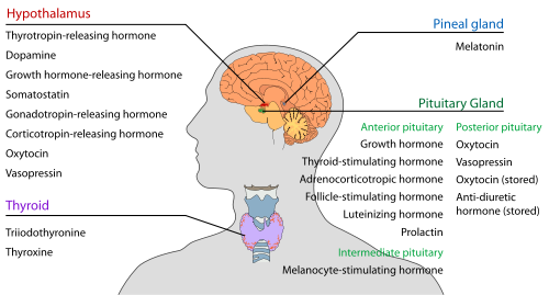
Endocrine glands in the human head and neck and their hormones

Alimentary system
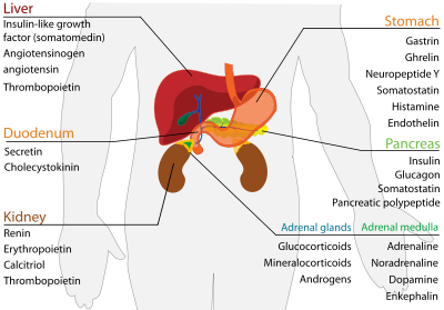
Reproductive
Woman
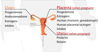
Man
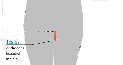

Calcium regulation
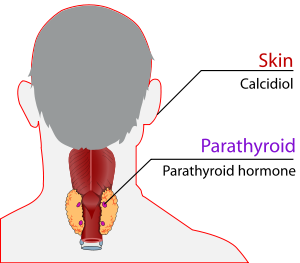

Targets
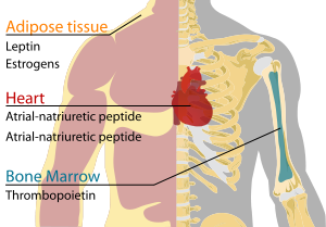
An Introduction to the Endocrine System
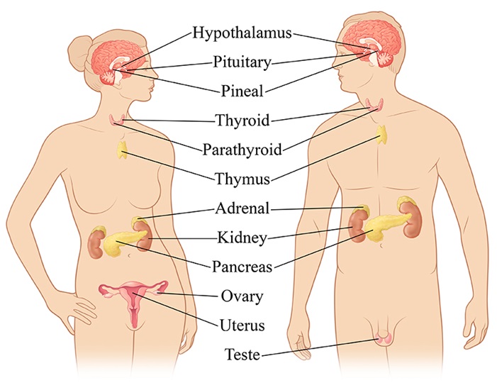
The endocrine system controls the way your body functions. It produces hormones that travel to all parts of your body to maintain your tissues and organs. Here are a few of the areas governed by the endocrine system:
Reproduction
Responses to stress and injury
Growth and sexual development
Body energy levels
Internal balance of body systems
Bone and muscle strength
Your endocrine system produces, stores and releases hormones. When everything goes smoothly, your body functions properly.
The Glands of the Endocrine System

Your endocrine system is made of many glands and organs. They have different functions, and work together to keep you healthy. These are the glands of your endocrine system:

Adrenal glands - influence the way your body uses energy, they also release a hormone called adrenaline when you are under stress
Hypothalamus - part of your brain that controls hormone production by releasing different chemicals to the pituitary gland
Ovaries - produce estrogen and progesterone in women, and also release egg cells
Pancreas - releases the insulin your body needs to metabolize sugar; problems with the pancreas can lead to diabetes
Parathyroid - located behind the thyroid gland, they are essential for proper bone development
Pineal gland - connects the endocrine system with the nervous system; produces several important hormones, including melatonin, important to sleep/wake cycles and sexual development
Pituitary gland – likely the most important gland in your body, it is crucial to growth, mental development and reproduction; influences or controls the rest of your endocrine system
Testes - produce the hormone testosterone; in men, testosterone maintains sperm production and bone mass
Thymus - crucial to normal immune function in childhood; once a child reaches puberty, its tissue is replaced by fat
Thyroid gland – located in the front of your neck, it releases hormones that control your metabolism and govern the way your body uses energy

Endocrine Organs

Several organs are vital to your endocrine system. They produce, store and excrete hormones to maintain the proper function of your body.

The placenta is the organ that provides the connection between a pregnant mother and the infant in her womb. It makes changes to a woman's body to better nourish her baby and prepare the mother for delivery.

Your skin, liver and kidneys work together to produce vitamin D. Vitamin D is produced when your skin is exposed to ultraviolet light. In the liver and kidneys, it becomes a chemical known as calcitriol, which maintains proper levels of calcium and phosphorus in your body.

Your stomach and small intestine secrete many hormones tied to eating and digestion. Disorders in the system can cause you to eat too little or too much food, and may lead to weight disorders.

Diabetes Symptoms

Urinating often
  • Feeling very thirsty

  • Feeling very hungry - even though you are eating

  • Extreme fatigue

  • Blurry vision

  • Cuts/bruises that are slow to heal

  • Weight loss - even though you are eating more (type 1)

  • Tingling, pain, or numbness in the hands/feet (type 2)

    Symptoms of Hypothyroidism

  • Abnormal menstrual cycles

  • Coarse, dry hair and dry skin

  • Constipation

  • Decreased libido

  • Depression

  • Fatigue

  • Hair loss

  • Increase in tongue size (severe cases)

  • Irritability

  • Jaundice (severe cases)

  • Memory loss

  • Muscle cramps and aches

  • Sensitivity to cold

  • Slowed speech (severe cases)

  • Weakness

  • Weight gain or difficulty losing weight (despite reduced food intake)
    Here are further guidelines.

  • Gastroenterology
    Digestive System
    Oral Health
    Mouth Disorders

    Mouth Disorders
    Oral human symptoms
    Oral health services or dental services and role of primary care physician.
    Oral Health
    Parts of the Mouth
    Questions a primary care physician should ask an individual claiming to be a dentist before any referral.
    Teeth Numbers Chart
    Toothpaste
    Teeth Conditions
    Oral health services or dental services and role of primary care physician.

    What type of physician takes care of oral health?
    Ideally, a primary care physician has to take care of oral health.
    In certain situations, a referral to a highly competent dentist may be required, depending on the diagnosis, recommended treatment and expertise of dentist.

    What is the human mouth made up of?
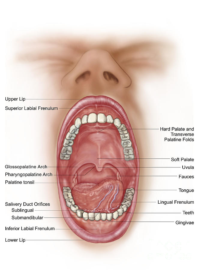

    Human mouth is made up of lips, tongue, gums, teeth, cheek lining, mouth floor, hard palate, soft palate, uvula, and tonsils.

    Parts of the Mouth

    Structure in/of the mouth

    Notes / Description

    1.

    Upper-lip

    The lips are fleshy folds of tissue around the opening of the mouth - covered with skin on the outside and a mucous membrane on the inside. The upper-lip is also known as the superior lip and is the lip closest to the nose (as opposed to closest to the chin).

    2.

    Lower-lip

    The lips are fleshy folds of tissue around the opening of the mouth - covered with skin on the outside and a mucous membrane on the inside. The lower-lip is also known as the inferior lip and is the lip closest to the chin (as opposed to closest to the nose).

    3.

    Hard Palate

    The hard palate extends over most of the roof of the mouth. The solid structure of the hard palate is formed by the maxillae and palatine bones.

    4.

    Soft Palate

    The soft palate is located posterior to the hard palate and consists mainly of muscle tissue.

    5.

    Uvula

    The uvula is a projection of soft tissue that hangs, some texts say "dangles", from the soft palate and moves upwards with the soft palate during swallowing. This prevents the food and/or liquid that is being swallowed from passing into the nasal cavity.

    6.

    Tonsils

    The tonsils consist of lymphoid tissue and are part of the immune system.
    There are three sets of tonsils in the oral cavity, the lingual tonsils towards the back of the tongue, the palatine tonsils shown in the diagram above and the pharyngeal tonsil in the pharynx.

    7.

    Tongue

    The tongue is a thick muscular organ that forms the floor of the mouth (= "oral cavity"). It consists of skeletal muscle covered with muscous membrane. The tongue also includes many taste buds - each of which consists of three types of epithelial cells: supporting cells, gustatory cells and basal cells. Some school biology courses include lessons about the zones of taste buds on tongue.

    8.

    Gums

    The sockets of the teeth (see below) are covered by dense fibrous connective tissue anchoring the teeth to the appropriate jaw bone (upper jaw bones = maxillae; lower jaw bone = mandible), keeping them in position and acting as a shock-absorber to cushion the impact of the teeth on the bones during active crunching actions.

    Teeth - Very brief notes about the teeth labelled in the diagram above. For more info see teeth and the digestive process.

    9.

    Incisors

    Incisors are teeth that are located at the front and mid-line of the mouth. They are relatively flat or "chisel-shaped" and suited for cutting into food.

    10.

    Canines

    Canine teeth are also known as cuspids (a useful term to avoid confusion with the use of the word "canine" to refer to dogs). They have a single pointed surface called a "cusp" which is adapted for tearing and shredding food.

    11.

    Premolars

    Premolars are also known as bicuspids. They are teeth that have two cusps with which to crush and grind food. They are located between the cuspids and the molars, as shown above. Premolar teeth generally have either one or two roots.

    12.

    Molars

    Molars are teeth that have three or four blunt cusps with which to crush and grind food. They are located at the back of the mouth (oral cavity). Molar teeth generally have two roots or three roots, two in the case of the molars in the lower-jaw and three in the case of the molars of the upper-jaw.


    How does your mouth help you?
    Your mouth helps you breathe, talk, swallow, taste, and chew. Symptoms involving the mouth are extremely common and affect people in all age groups.

    Oral human symptoms

    What can be oral symptoms of a human being?
    Bad breath
    Dry mouth or xerostomia
    Eroded enamel
    Eroded teeth
    Frictional keratosis
    Loose teeth
    Lichen planus
    Mouth sore
    Oral thrush (fungal infection in the mouth)
    Puffy gums
    Smoker's keratosis
    Sour taste in your mouth
    Swollen gum
    Toothache
    Toothache with swelling and/or fever (Abscess toothache)
    White patches in the mouth
    White or brown lines on childrens' teeth

    Oral human symptoms are focused around lips, teeth, gums, tongue, and buccal cavity. Throat symptoms are categorized separately.

    Bad breath

    What causes bad breath?
    Some types of bad breath, such as "morning mouth," are considered to be fairly normal, and they usually are not health concerns. The "morning mouth" type of bad breath occurs because the saliva that regularly washes away decaying food and odors during the daytime diminishes at night while you sleep. Your mouth becomes dry, and dead cells adhere to your tongue and to the inside of your cheeks. Bacteria use these cells for food and expel compounds that have a foul odor.

    In addition, bad breath can be caused by the following:

    Poor dental hygiene — Infrequent or improper brushing and flossing can leave food particles to decay inside the mouth.

    Infections in the mouth — Periodontal (gum) disease

    Respiratory tract infections — Throat infections, sinus infections, lung infections

    External agents — Garlic, onions, coffee, cigarette smoking, chewing tobacco

    Dry mouth (xerostomia) — This can be caused by salivary gland problems, medications or by "mouth breathing."

    Systemic illnesses — Diabetes, liver disease, kidney disease, lung disease, sinus disease, reflux disease and others

    Psychiatric illness — Some people may perceive that they have bad breath, but it is not noticed by oral-health-care professionals or others. This is referred to as "pseudohalitosis."

    Symptoms

    A person may not always know that he or she has bad breath. This phenomenon is because odor-detecting cells in the nose eventually become accustomed to the constant flow of bad smells from the mouth. Others may notice and react by recoiling as you speak.

    Other associated symptoms depend on the underlying cause of bad breath:

    Poor dental hygiene — Teeth are coated with film or plaque, food debris trapped between teeth, pale or swollen gums

    Infections in the mouth — Gums may be red, swollen and bleed easily, especially after brushing or flossing; pus may drain from between teeth; a pocket of pus (abscess) at the base of a tooth; loose teeth or a change in "fit" of a denture; painful, open sores on the tongue or gums

    Respiratory tract infections — Sore throat, swollen lymph nodes ("swollen glands") in the neck, fever, stuffy nose, a greenish or yellowish nasal discharge, a mucus-producing cough

    External agents — Cigarette stains on fingers and teeth, a uniform yellow "coffee stain" on teeth

    Dry mouth — Difficulty swallowing dry foods, difficulty speaking for a prolonged period because of mouth dryness, a burning sensation in the mouth, an unusually high number of dental caries, dry eyes (in Sjögren's syndrome)

    Systemic (bodywide) illnesses — Symptoms of diabetes, lung disease, kidney failure or liver disease

    Diagnosis

    A dentist or physician may notice the patient's bad breath while the patient is discussing his or her medical history and symptoms. In some cases, depending on the smell of the patient's breath, the dentist or physician may suspect a likely cause for the problem. For example, "fruity" breath may be a sign of uncontrolled diabetes. A urine-like smell, especially in a person who is at high risk of kidney disease, can sometimes indicate kidney failure.

    Your dentist will review your medical history for medical conditions that can cause bad breath and for medications that can cause dry mouth. Your dentist also will ask you about your diet, personal habits (smoking, chewing tobacco) and any symptoms, including when the bad breath was noticed and by whom.

    Your dentist will examine your teeth, gums, oral tissues and salivary glands. He or she also will feel your head and neck and will evaluate your breath when you exhale from your nose and from your mouth. Once the physical examination is finished, your dentist may refer you to your family physician if systemic problems are the most likely cause. In severe cases of gum disease, your dentist may recommend that you be seen by a periodontist (dentist who specializes in gum problems).

    You will need diagnostic tests if the doctor suspects a lung infection, diabetes, kidney disease, liver disease or Sjögren's syndrome. Depending on the suspected illness, these tests may include blood tests, urine tests, X-rays of the chest or sinuses, or other specialized testing.
    Here are further guidelines.
    http://www.qureshiuniversity.com//badbreath.html

    Oral thrush (fungal infection in the mouth)


    Oral thrush is an infection in the mouth caused by a yeast germ called Candida. It is not usually serious and can generally be cleared with treatment.

    What is oral thrush?
    Thrush is an infection caused by a yeast germ called Candida. The mouth is a common site where Candida causes infection. Candidal infection in the mouth is called oral thrush. Other common sites for thrush to develop are the vagina, nappy area, and nail folds. (See separate leaflets called Vaginal Thrush, Candidal Skin Infection and Nappy Rash for more details about these other types of thrush.)

    In adults, thrush can cause an uncomfortable burning sensation in the mouth and throat. Oral thrush is an infection of yeast fungus, Candida albicans, in the mucous membranes of the mouth.

    Strictly speaking, thrush is only a temporary candida infection in the oral cavity of babies.

    But we have, for this purpose, expanded the term to include candida infections occurring in the mouth and throat of adults, also known as candidiasis or moniliasis.

    Who develops oral thrush?
    Small numbers of Candida commonly live on healthy skin and in a healthy mouth. They are usually harmless. Healthy people do not normally develop oral thrush. However, certain situations or conditions may cause an overgrowth of Candida which can lead to a bout of oral thrush. These include:

    •Being a baby. Oral thrush is quite common in young babies.
    •Wearing dentures, especially if they are not taken out at night, not kept clean, or do not fit well and rub on the gums.
    •A course of antibiotic medicine. Antibiotics will kill harmless germs (bacteria) which live in the mouth. They do not kill Candida which may multiply more easily if there are fewer bacteria around. •Excessive use of antibacterial mouthwash (for similar reasons to above).
    •Taking steroid tablets or inhalers.
    •Having a dry mouth due to a lack of saliva. This may occur as a side-effect from certain medicines (such as antidepressants, antipsychotics, chemotherapy medicines), following radiotherapy to the head or neck, or as a symptom of Sjögren's syndrome.
    •Having diabetes.
    •Having severe anaemia.
    •Lacking iron, folate or vitamin B12.
    •Having a poor immune system.
    •Being frail or in generally poor health.
    •Smoking. Smokers are more likely to develop oral thrush.
    Oral thrush is not contagious. You cannot pass on oral thrush to other people.

    How do you get oral thrush?
    Candida is present in the oral cavity of almost half of the population. Everyone who wears dentures will have candida, without necessarily suffering any ill effects.

    Candida doesn't become a problem until there's a change in the chemistry of the oral cavity that favours candida over the other micro-organisms that are present.

    These changes can occur as a side-effect of taking antibiotics or drug treatment, such as chemotherapy.

    These changes can also be caused by certain conditions – such as diabetes, drug abuse, malnutrition – and as a consequence of immune deficiencies relating to old age or infection.

    Furthermore, people whose dentures don't fit well can sustain breaks in the mucous membranes in their mouth, which can act as a gateway for candida.

    People who suffer from this problem often have moist, pale pink spots on their lips, known as angular cheilitis, which is an indication of a candida infection.

    What are the symptoms of oral thrush?
    •The classical symptom is white spots that develop in your mouth. The spots may join together to form larger spots called plaques. They may become yellow or grey. If you wipe off a spot, the underlying tissue may be red but it is not usually sore or painful.
    •Often there are no white spots. Areas in your mouth may just become red and sore. This more typically occurs if you develop thrush after taking antibiotics or steroids.
    •Denture wearers may develop an area of persistent redness under a denture.
    •You may develop sore, cracked, red areas just outside your mouth. This mainly affects the angle where the upper and lower lips meet (angular stomatitis).
    •Some mild oral thrush infections are painless. However, sometimes oral thrush is quite sore and can make eating and drinking uncomfortable. Some babies with oral thrush may drool saliva, or not be able to feed properly because of soreness.
    •Taste can be affected in some people with oral thrush.
    White, cream-coloured, or yellow spots in the mouth. The spots are slightly raised.
    There is normally no pain in the area underneath the spots. If you scrape off these spots, they leave small wounds that bleed slightly.
    In adults, thrush can cause an uncomfortable burning sensation in the mouth and throat.

    Who is at special risk?
    Newborn babies.
    Denture users.
    Adults with diabetes or other metabolic disturbance.
    People with a dry mouth relating to side-effects of their medication (eg anti-psychotics) or medical conditions (eg Sjogren’s Syndrome).
    People undergoing antibiotic or chemotherapy treatment.
    People prescribed oral steroid medication or steroid metered dose inhalers.
    Drug users.
    People with poor nutrition.
    People with an immune deficiency.

    How does the doctor diagnose oral thrush?
    How is oral thrush diagnosed?

    In babies, thrush is usually diagnosed on the basis of the clinical picture. Occasionally, in order to make a diagnosis, the doctor will scrape the baby's tongue and send the sample for analysis.

    In adults, your doctor will usually diagnose oral thrush by your typical symptoms and the typical appearance in your mouth. No investigations are usually needed to diagnose oral thrush.

    However, your doctor may sometimes suggest a blood test to look for certain conditions that may make you more likely to develop oral thrush. For example, a blood test to see if you are lacking iron, vitamin B12 or folate.

    If oral thrush does not respond to treatment (see below), your doctor may suggest that they take a sample (swab) from inside your mouth. The swab is then sent to the laboratory to be examined under a microscope. They can also try to grow the Candida in the laboratory.

    Occasionally, a biopsy is needed to confirm a diagnosis of oral thrush. A small sample is taken from the white patches inside your mouth and this can be examined under a microscope.

    What is the treatment for oral thrush?
    In Babies, oral thrush may clear spontaneously without treatment and may be prevented by sterilising all feeding equipment and mouth toys.

    It's been suggested that by giving the child sterilised water immediately following a milk feed, residual milk in the mouth is rinsed away, reducing the population of candida within the oral cavity.

    In other circumstances, the condition that caused the thrush must be brought under control.

    Once the condition that caused the oral thrush has been treated, the thrush itself can be cured.

    Treatment is with antifungal medicines, in the form of pastilles that are sucked or oral suspensions that are held in the mouth before swallowing.

    These allow the antifungal agent to act locally in the mouth. Examples include nystatin (eg Nystan oral suspension), or miconazole (eg Daktarin oral gel).

    In certain complicated cases, or if the infection spreads, systemic treatment will be necessary in the form of antifungal tablets, or perhaps in the form of injections.

    Coping with the symptoms of oral thrush

    Thrush can make the mouth so sensitive that it's impossible to perform regular oral hygiene. Use a very soft toothbrush. It can often help to rinse the mouth with a diluted solution of 3 per cent hydrogen peroxide.

    Locally applied treatment
    For mild oral thrush, the usual treatment that is tried first is miconazole mouth gel for seven days. Sometimes a two-week course is needed. Nystatin drops are another option if miconazole gel cannot be used (for example, if you are known to be allergic to it).

    Follow the instructions in the packet:
    •The gel or drops should be used after you have eaten or drunk.
    •Smear a small amount of gel on to the affected areas, with a clean finger, four times a day.
    •With the drops, you use a dropper to place the liquid inside your mouth on to the affected areas four times a day.
    •Ideally, you should not eat or drink for about 30 minutes after using either the gel or the drops. This helps to prevent the medicine from being washed out of your mouth too soon.

    Anti-thrush tablets

    Tablets that contain a medicine called fluconazole can also help to clear fungal and thrush infections from the body. Tablets tend to be used in more severe or serious cases. For example, for people with a poor immune system who develop extensive oral thrush. Tablets are usually prescribed for seven days and this will usually clear oral thrush.

    Adjustment of other medication

    If you are taking other medication that may have caused oral thrush, such as steroids or antibiotics, your doctor may need to change this medication or reduce the dose to help clear up your thrush.

    Referral to a specialist
    Your doctor may suggest that he or she refer you to (or ask the advice of) a specialist if:
    •The above measures do not help to clear your oral thrush infection.
    •You have particularly severe infection or other health problems (for example, you are undergoing chemotherapy or are taking other medicines that weaken your immune system).
    The specialist may suggest other treatments for oral thrush. For example, medicines such as itraconazole, ketoconazole or amphotericin.

    Can oral thrush be prevented?
    It may be possible to alter one or more of the situations mentioned above to help prevent further bouts of oral thrush. For example:
    •If you have diabetes - good control of your blood sugar level reduces the risk of thrush and other infections.
    •If you use steroid inhalers - having a good inhaler technique and using a spacer device may reduce the risk of thrush. Also, rinse your mouth after using the inhaler, to help remove any medicine particles left in your mouth. Ask your doctor about reducing your dose of steroid in your inhaler to the lowest level needed to control your asthma.
    •If you wear dentures:
    •Leave your dentures out overnight, or for at least six hours daily. Constant wearing of dentures, and not taking them out at night, is thought to be one of the most common causes of oral thrush.
    •Clean and disinfect dentures daily. To clean, use soapy water and scrub the dentures with a soft nailbrush on the fitting surface - that is, the non-polished side. Then soak them in a disinfecting solution. The type of solution and the time they should be soaked for will be advised by your dentist. Rinse the dentures after disinfecting them, and then allow the dentures to air dry before wearing them again. Drying like this helps to kill any Candida that might be stuck to the dentures.
    •Clean the inside of your mouth (where the dentures sit) with a soft brush. •See a dentist if the dentures do not fit well.
    •If you take medication which causes a dry mouth - take frequent sips of water. (See separate leaflet called Dry Mouth for more details.)
    •Tips to prevent oral thrush in babies are included in the separate leaflet called Oral Thrush in Babies.
    •If you are found to have anaemia or low levels of vitamin B12, folate or iron, treating this may help to prevent oral thrush in the future.
    •If you are a smoker, quitting smoking may help to prevent further bouts of oral thrush. (See separate leaflet called Tips to Help You Stop Smoking for more details.)
    •Certain groups of people may be given anti-thrush tablets to help to prevent oral thrush. For example, people who are on medication to suppress their immune system or who are receiving chemotherapy.

    Toothache

    What causes a toothache?
    While tooth decay is often the primary cause of a toothache, it's important for you to have a complete oral examination to determine the cause. Other causes of a toothache can include the following:
    •Infection
    •Gum disease
    •Grinding teeth (bruxism)
    •Tooth trauma
    •An abnormal bite
    •Tooth eruption (in babies and school-age children)
    TMJ/TMD (Temporomandibular Joint Disorder), sinus or ear infections, and tension in the facial muscles can cause discomfort that resembles a toothache, but often these health problems are accompanied by a headache.

    Pain around the teeth and the jaws can be symptoms of heart disease such as angina. If your dentist suspects a medical illness could be the cause of your toothache, he or she may refer you to a physician.

    If you have a toothache, you may have a cavity or advanced gum disease. The first sign of decay may be the pain you feel when you eat something sweet, very cold or very hot. If the pulp – the inside of the tooth that has tissue and nerves – has become irritated, this can cause pain in your tooth.

    What symptoms accompany a toothache?
    Because the symptoms of a toothache may resemble other medical conditions or dental problems, it can be difficult to diagnose the cause without a complete evaluation by your dentist. If you notice pus near the source of the pain, your tooth may have become abscessed, causing the surrounding bone to become infected. Or the pus could indicate gum disease, which is usually characterized by inflammation of the soft tissue, bleeding gums and abnormal loss of bone surrounding the teeth.

    Contact your dentist immediately if you have any of the following symptoms:
    •Fever
    •Difficulty breathing or swallowing
    •Swelling around the tooth area
    •Pain when you bite
    •A foul-tasting discharge
    •Continuous lasting pain

    How do I alleviate the pain if I cannot see my dentist right away?
    Anyone with a toothache should see a dentist at once for diagnosis and treatment. If left untreated, a toothache and the condition that is causing it can worsen. However, if you are unable to schedule an emergency appointment, the following self-care treatment can temporarily alleviate pain and inflammation from a toothache:
    •Rinse with warm salt water
    •Gently floss teeth to dislodge any food particles trapped between teeth. •Take an over-the-counter pain reliever such as ibuprofen (Advil®) or acetaminophen (Tylenol®) to relieve pain.
    •Never put aspirin or any other painkiller against the gums near the aching tooth, as it may burn the gum tissue.
    •Apply an over-the-counter antiseptic containing benzocaine directly to the irritated tooth and gum to temporarily relieve pain. Direct application of oil of cloves (eugenol) may also help to numb the gums. The oil may be rubbed directly on the sore area, or soak a small piece of cotton and apply it to the sore tooth.
    •If there has been some trauma to the tooth, a cold compress may be applied on the outside cheek to relieve pain or swelling.

    How can my dentist help?
    Your dentist will conduct a complete oral examination to determine the location and cause of the toothache, looking for signs of swelling, redness and obvious tooth damage. He or she may also take x-rays to look for evidence of tooth decay between teeth, a cracked or impacted tooth or a disorder of the underlying bone.

    Your dentist also may prescribe pain medication or antibiotics to speed the healing of your toothache. If by the time you see your dentist your tooth has become infected, then treatment could require removal of the tooth or a root canal procedure, which involves removing the damaged nerve tissue from the middle of a tooth.

    Is there a way to prevent a toothache?
    The key to preventing toothaches is establishing a regular oral hygiene routine and sticking to it. For example, failure to brush and floss regularly after meals can significantly increase your risk of developing cavities, which can cause toothaches.

    Here are a few tips to help reduce your risk for developing a toothache:
    •Brush at least twice a day, preferably after meals and snacks.
    •Floss at least once a day to prevent gum disease.
    •Visit your dentist regularly for oral examinations and a professional cleaning.

    Why should a primary care physician be able to diagnose and treat all nonemergency medical conditions?
    There are thousands of medical conditions.
    It is impossible to have a separate physician for every medical condition.
    A primary care physician should be able to diagnose and treat all oral health medical conditions, in addition to other medical conditions.
    Dental services are actually oral health care services.
    A primary care physician can provide these services.

    Mouth disorders.

    What are other names of oral medical conditions?
    Mouth disorders.

    What are examples of mouth disorders?
    Take a look at this.

    Amalgam Tattoo
    Aspirin Burn
    Bad Breath
    Black Hairy Tongue
    Canker Sores
    Cavities, Abscesses, Discoloration
    Chipped Teeth
    Cold Sores
    Geographic Tongue
    Gum Disease
    Leukoplakia
    Lichen Planus
    Lie Bumps
    Oral Cancer
    Periodontitis
    Teeth Conditions
    Thrush
    TMJ

    Cold Sores

    Also called fever blisters, you don't get cold sores from fevers or colds but they can be triggered by them. The virus that causes cold sores is usually passed via a kiss, shared utensils, or other close contact. Over-the-counter creams and ointments may help discomfort and speed healing. Frequent sores may require a prescription. Cold sores are a top mouth problem. Other problems include canker sores, TMJ, bad breath, and mouth cancer.

    Thrush
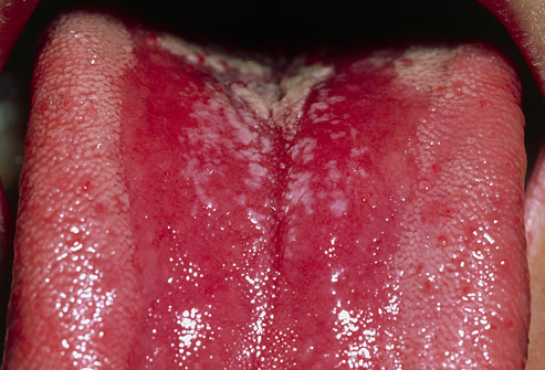
    Caused by candida yeast, thrush is most common in older adults or babies. But a weakened immune system, antibiotics, diabetes, or certain medications -- such as inhaled corticosteroids -- can give candida a chance to grow wild. Wiping away the patches will cause soreness. See a doctor for a firm diagnosis.

    Black Hairy Tongue
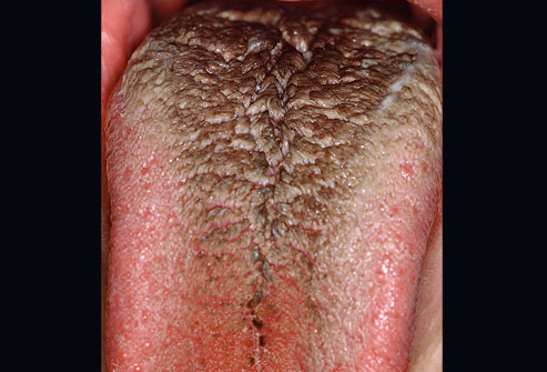
    This painless condition occurs when the little bumps on your tongue grow long and trap bacteria that live in your mouth -- making the tongue look black and hairy. Causes can include antibiotic use, poor oral hygiene, smoking, drinking a lot of tea or coffee, and not producing enough saliva. Brushing the tongue and using a tongue scraper is usually all you need to treat it, though sometimes medication is necessary.

    Canker Sores
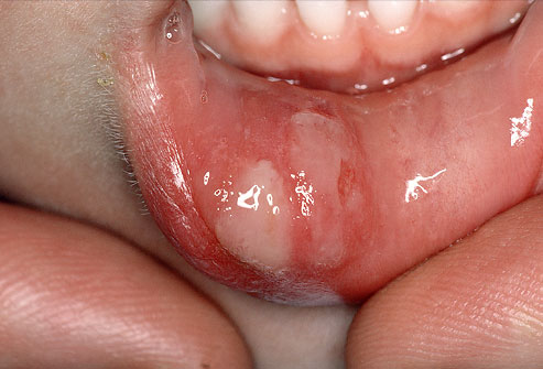
    No one knows what causes these small, painful blisters inside your mouth. Triggers include hypersensitivity, infection, hormones, stress, and not getting enough of some vitamins. Also called aphthous ulcers, canker sores can show up on the tongue, cheek, even your gums. They usually last a week or two. Persistent, severe canker sores can be treated with numbing creams, prescription drugs, or dental lasers.

    Leukoplakia
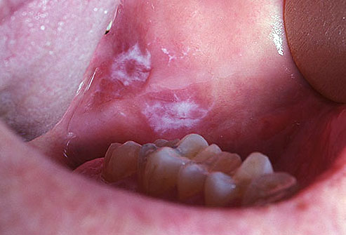
    Leukoplakia is a reaction to an irritant, like rough teeth, badly fitting dentures, smoking, and smokeless tobacco. It can show up as white patches or plaques in the mouth, is usually painless, and can't be scraped off. Leukoplakia can also be a precancerous condition. Persistent patches or other changes in your mouth need a dentist's evaluation.

    Lichen Planus
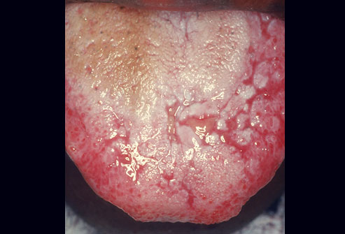
    A rare rash that shows up as lacy, white patches or red shiny bumps on the inside of the cheeks or tongue could be lichen planus. No one knows what causes it. Generally, mild lichen planus doesn't need any treatment. If it causes pain or ulcers, it can be treated with oral and topical medication. Oral lichen planus can be chronic and may increase the risk for oral cancer. Lichen planus can also affect skin, scalp, nails, and genitals.

    Geographic Tongue
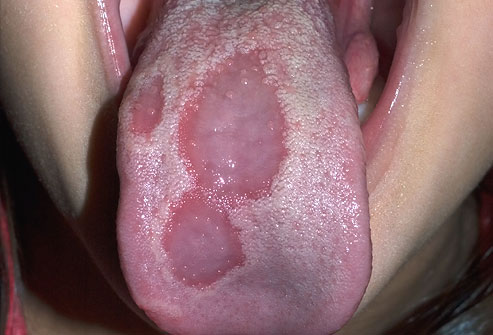
    When parts of your tongue are missing some of their small bumps, you end up with raised and lowered spots, giving your tongue a map-like appearance. The spots can change location, pattern, and size within minutes to hours. Geographic tongue is harmless and can come and go. It usually doesn't need any treatment. If there's pain, over-the-counter pain relievers and anti-inflammatory medications can help.

    Oral Cancer
    A mouth sore that doesn't go away. Unexplained numbness in the face, mouth, or neck. Problems chewing, speaking or swallowing. These are a few symptoms of oral cancer. Causes can include smoking cigarettes and using smokeless tobacco, drinking heavily, overexposure to the sun, and a family history of cancer. Oral cancer has also been linked to the human papillomavirus, or HPV. Don't let fear keep you from the doctor -- oral cancer that is caught early is treatable and curable.


    TMJ
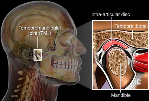
    A problem with the jaw called temporomandibular joint syndrome can cause severe pain in the jaw, face, ear, or neck. Clenching, tooth grinding, or injury can all cause TMJ syndrome, but the results are often the same: pain, headaches, dizziness, even trouble swallowing. Treatment may involve rest, moist heat, a mouth guard, medication, or surgery.

    Chipped Teeth
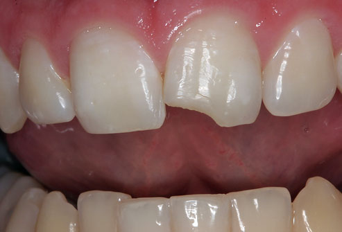
    Munching on ice or hard candies, grinding or clenching teeth, even exposing teeth to heat and cold can lead to chips, cracks, and breaks in your teeth. Tiny chips or cracks may not be a bother. But anything more could lead to pain or permanent tooth damage. Your dentist can offer dental bonding, tooth contouring, porcelain veneers, and crowns to fix badly damaged teeth.

    Amalgam Tattoo
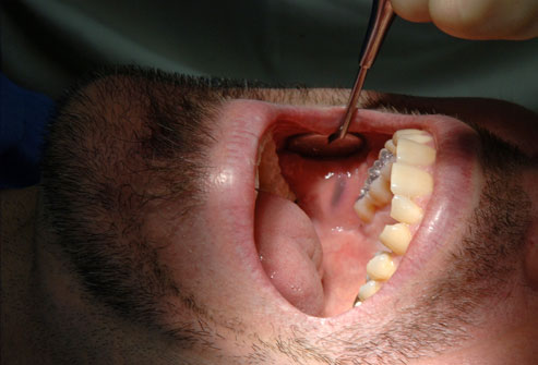
    Ever notice a small blue-gray "stain" in a soft part of your mouth after dental work? Called amalgam tattoos, they occur when a tiny piece of amalgam filling gets embedded in your cheek or gum. The silver in the amalgam leaches into your mouth's soft tissue, resulting in what looks a bit like a tiny tattoo. Amalgam tattoos pose no harm. But if the blue-gray spot grows or changes color, there is a good possibility it may not be an amalgam tattoo. Ask your dentist to check it out.

    Gum Disease
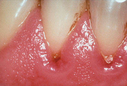
    When periodontal (gum) disease develops, bacteria in plaque accumulate along the gum line. Gingivitis is the first stage of gum disease. Symptoms include red, puffy, and bleeding gums. Proper oral hygiene can help prevent periodontal disease. Smoking, poor diet, and stress can make it worse.

    Periodontitis
    The next stage of gum disease is periodontitis, or gum infection. Increased inflammation causes the gums to recede, forming pockets between the teeth and gums. These pockets trap tartar, plaque, and food debris that eventually lead to infection and abscesses. Advanced gum disease damages the bone that supports teeth and is one of the leading causes of tooth loss in adults. See your dentist to treat receding gums.
    www.qureshiuniversity.com/periodontist.html

    Aspirin Burn
    Ever let an aspirin nestle in your cheek, near an aching tooth? While you may hope this relieves pain faster, instead the acid in the aspirin burns a white, rough lesion into your gums or cheek. Preventing aspirin burn is simple -- swallow those pain relievers! Treatment for aspirin burn is just as basic: Time. Simple burns should heal in about two weeks.

    Cavities, Abscesses, Discoloration
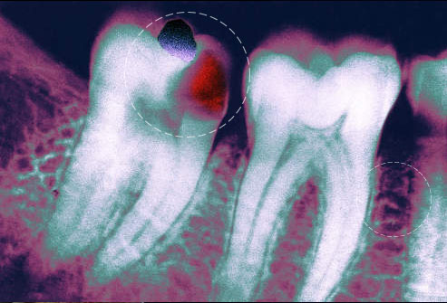
    Flossing and brushing daily and regular dental checkups help prevent problems like cavities, abscesses, and tooth discoloration. Don't mess around with a severe toothache. Dental infections can spread to the face, skull, and even to the bloodstream. See your dentist as soon as possible if your tooth aches or if you have a fever, earache, or pain when you open your mouth wide.

    Bad Breath
    Unbrushed teeth have food particles around them that promote bacteria and cause bad breath. Persistent bad breath or a bad taste in your mouth may be from continuous breathing through your mouth, dry mouth, tooth decay, a sign of gum disease, or even diabetes. Fight bad breath by brushing your teeth and tongue, drinking water, and avoiding food triggers. See your dentist if bad breath persists.

    "Lie" Bumps
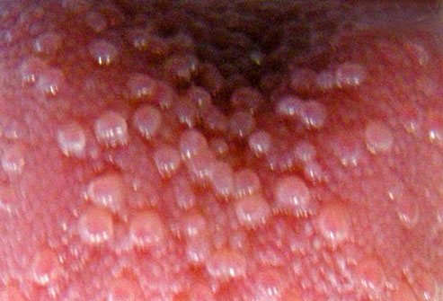
    According to an old wives' tale, telling a lie causes a bump on the tongue. So-called "lie bumps" or transient lingual papillitis are common even if you tell only the truth. These small, harmless bumps go away on their own after a few days, but they may be uncomfortable. Their cause is a mystery -- it could be a reaction to a food or a minor trauma like biting the tongue. You don't need to treat them, although oral anesthetics may relieve discomfort.
    Teeth Conditions

    Cavities (caries): Bacteria evade removal by brushing and saliva and damage the enamel and deeper structures of teeth. Most cavities occur on molars and premolars.
    Tooth decay: A general name for disease of the teeth, including cavities and caries.
    Periodontitis: Inflammation of the deeper structures of the teeth (periodontal ligament, jawbone, and cementum). Poor oral hygiene is usually to blame.
    Gingivitis: Inflammation of the surface portion of the gums, around and between the crowns of the teeth. Plaque and tartar buildup can lead to gingivitis.
    Plaque: A sticky, colorless film made of bacteria and the substances they secrete. Plaque develops quickly on teeth after eating sugary food, but can be easily brushed off.
    Tartar: If plaque is not removed, it mixes with minerals to become tartar, a harder substance. Tartar requires professional cleaning for removal.
    Overbite: The upper teeth protrude significantly over the lower teeth.
    Underbite: The lower teeth protrude significantly past the upper teeth.
    Teeth grinding (bruxism): Stress, anxiety, or sleep disorders can cause teeth grinding, usually during sleep. A dull headache or sore jaw are symptoms.
    Tooth sensitivity: When one or more teeth become sensitive to hot or cold, it may mean the dentin is exposed.

    Teeth Numbers Chart

    Teeth Numbers and Names: A First Step in Understanding Your Treatment Plan
    Teeth numbers diagram. The human teeth is composed of 16 upper teeth and 16 lower teeth. They are also divided into four quadrants.
    Upper Right Quadrant
    Teeth Numbers Teeth Names
    8
    7
    6
    5
    4
    3
    2
    1
    Incisor (Central)
    Incisor (Lateral)
    Canine (Eye tooth / Cuspid)
    Bicuspid (1st)
    Bicuspid (2nd)
    Molar (1st Molar)
    Molar (2nd Molar)
    Wisdom Tooth (3rd Molar)
    Upper Left Quadrant
    Teeth Numbers Teeth Names
    9
    10
    11
    12
    13
    14
    15
    16
    Incisor (Central)
    Incisor (Lateral)
    Canine (Eye tooth / Cuspid)
    Bicuspid (1st)
    Bicuspid (2nd)
    Molar (1st)
    Molar (2nd)
    Wisdom Tooth (3rd Molar)
    Lower Right Quadrant
    Teeth Numbers Teeth Names
    32
    31
    30
    29
    28
    27
    26
    25
    Wisdom Tooth (3rd Molar)
    Molar (2nd Molar)
    Molar (1st Molar)
    Bicuspid (2nd)
    Bicuspid (1st)
    Canine (Eye tooth / Cuspid)
    Incisor (Lateral)
    Incisor (Central)
    Lower Left Quadrant
    Teeth Numbers Teeth Names
    17
    18
    19
    20
    21
    22
    23
    24
    Wisdom Tooth (3rd Molar)
    Molar (2nd Molar)
    Molar (1st Molar)
    Bicuspid (2nd)
    Bicuspid (1st)
    Canine (Eye tooth / Cuspid)
    Incisor (Lateral)
    Incisor (Central)

    How many individuals each year have mouth disorders?
    On May 1, 2015, half of the American population had gum disease.
    Does that mean we need to have separate doctors for oral health?
    No.

    Who must primarily diagnose and treat oral medical conditions?
    Primary care physician.

    When is a referral to competent dentist required?
    Rarely is a referral needed to a dentist who must know everything about oral health.

    Why should oral health be taught under the pretext of the human digestive system?
    The mouth is part of the human digestive system.

    Which organs of the body are part of the human digestive system?
    The digestive system is composed of the digestive tube organs plus the digestive adnexal glands. The digestive tube is composed of the mouth, pharynx, esophagus, stomach, small intestine (duodenum, jejunum, ileum), large intestine (caecum, colon, rectum) and anus.

    If oral health or mouth disorders should be taught under pretext of digestive system, where do dentists fit in?
    Actually, oral or mouth issue diagnosis and treatment is the duty of a primary care physician.
    In recent years, all oral conditions have been tried to be categorized under separate specialties.

    For example, general dentist, endodontist, orthodontist, prosthodontist, pediatric dentist/pedodontist, and periodontist.

    If any individual declares he or she can diagnose and treat only one medical condition, he or she is not a physician. A physician should be able to diagnose and treat all medical conditions in various healthcare settings. If a physician knows any additional technique, that technique can be recognized after having expertise of a physician.

    Questions a primary care physician should ask an individual claiming to be a dentist before any referral.

    Can you reach a correct diagnosis and treatment of human beings in various healthcare settings?
    What technique or procedure do you know that can help the patient?

    How will this technique or procedure help the patient?

    If an individual claims he or she can do tooth extraction, primary care physicians can do tooth extraction.
    Within 5 minutes, tooth extraction can be done.
    Tooth extraction is not treatment for gingivitis, early periodontitis, moderate periodontitis, or advanced periodontitis. Tooth extraction in such situation only relieves the symptoms that can be relieved with medications.

    A loose tooth with gingivitis or periodontitis causes pain with gums.
    Tooth extraction prevents irritation of gums with loose teeth; thus, you do not feel pain. This can be treated with medications.

    What recommendations are there for manufacturers of oral healthcare products?
    Individuals are reporting they brush twice a day and do dental flossing, but still, there are reports of gum disease after proper oral hygiene.
    Antiseptic, anti-inflammatory content of toothpaste needs to be enhanced to prevent gum disease.
    A special toothpaste needs to be manufactured for gum disease with antiseptic, anti-inflammatory content that can be utilized after 30 years of age. Moreover, toothpaste should be effective against plaque and tartar.

    Bad breath
    Bridges and partial dentures
    Burning mouth syndrome
    Caring for my teeth
    Children's teeth
    Coldsores
    Cosmetic treatment
    Cracked teeth
    Crowns
    Dental care for older people
    Dental care for people with special needs
    Dental decay
    Dental erosion
    Denture cleaning
    Denture stomatitis (Thrush)
    Dentures
    Diet
    Different filling materials
    Dry mouth
    Fluoride
    Geographic tongue
    Gum disease
    Healthy gums and healthy body
    Implants
    Jaw problems and headaches
    Knocked out teeth
    Lichen planus
    Living with my brace
    Medical conditions and oral health
    Mouth ulcers
    Mouthguards
    Oral care products
    Orthodontic treatment (braces)
    Parts of the Mouth
    Pit and fissure sealants
    Preventive care and oral hygiene
    Relaxation and sedation
    Root canal treatment
    Sensitive teeth
    Smokeless tobacco
    Smoking and oral health
    Sugar-free chewing gum
    Teeth Conditions
    Teeth Treatments
    Teens teeth
    Tooth whitening
    Veneers
    What to do following an extraction
    White fillings
    Wisdom teeth
    X-rays
    Why is Oral Health Important?
    Oral health is essential to overall health. Good oral health improves a person’s ability to speak, smile, smell, taste, touch, chew, swallow, and make facial expressions to show feelings and emotions.

    Oral Health Assessment Tool for Dental Screening
    Patient: _________________________ Completed by: _______________________ Date: _____/_____/_____
    Scores: The final score is the sum of scores from the eight categories and can range from 0 (very healthy) to 16 (very unhealthy). While the cumulative score is important in assessing oral health, the score of each item should be considered individually. Symptoms that are underlined require immediate attention.
    *If any category has a score of 1 or 2, please arrange for the patient to be examined by a dentist.
    Category 0 = healthy 1 = changes * 2 = unhealthy * Category scores
    Lips Smooth, pink, moist Dry, chapped, or red at corners Swelling or lump, white/red/ulcerated patch; bleeding/ulcerated at corners
    Tongue Normal, moist, roughness, pink Patchy, fissured, red, coated Patch that is red and/or white, ulcerated, swollen
    Gums and tissues Pink, moist, smooth, no bleeding Dry, shiny, rough, red, swollen, one ulcer/sore spot under dentures Swollen, bleeding gums, ulcers, white/red patches, generalized redness or ulcers under dentures
    Saliva Moist tissues, watery and free-flowing saliva Dry, sticky tissues, little saliva present Tissues parched and red, very little/no saliva present, saliva very thick
    Natural teeth
    Yes/No
    No decayed or broken teeth/roots 1-3 decayed or broken teeth/ roots or teeth very worn down 4 or more decayed or broken teeth/roots, or fewer than 4 teeth, or very worn down teeth
    Dentures
    Yes/No
    No broken areas or teeth, dentures regularly worn 1 broken area/ tooth or dentures only worn for 1-2 hrs daily, or loose dentures More than 1 broken area/tooth, denture missing or not worn, needs denture adhesive
    Oral cleanliness Clean, no food particles or tartar in mouth or on dentures Food particles/ tartar/ plaque in 1-2 areas of the mouth or on small area of dentures or bad breath Food particles/tartar/plaque in most areas of the mouth or on most of dentures or severe halitosis (bad breath)
    Dental pain No behavioral, verbal, or physical signs of dental pain Verbal &/or behavioral signs of pain such as pulling at face, chewing lips, not eating, aggression Physical signs such as facial swelling, sinus on gum, broken teeth, large ulcers, and verbal and/or behavioral signs such as pulling at face, chewing lips, not eating, aggression

    Arrange for patient to be examined by a dentist.
    Patient or family/guardian refuses dental treatment.
    Review this patient’s oral health again on (date): ___/___/___
    TOTAL ____ SCORE: 16
    Here are further guidelines.
    http://www.qureshiuniversity.com/oralhealth.html
    Dental Cleanings
    Gum disease

    Medical conditions and oral health

    Dental caries (tooth decay)
    Gum conditions

    Symptoms of Gum conditions (Gum disorders)

    Gum pain
    Gum swelling
    Gum mass
    Gum ulceration
    Gum bleeding

    Gum conditions

    Gingivitis
    Periodontitis
    Plaque
    Tartar
    Calculus
    Dental scaling
    Root planing
    Oral hygiene
    Swollen gums
    Bleeding gums
    Red gums
    inflamed gums

    Cleft lip and palate
    Oral and facial pain

    Gum disease

    What is gum disease?
    Gum disease is described as swelling, soreness or infection of the tissues supporting the teeth. There are two main forms of gum disease: gingivitis and periodontal disease.

    What is gingivitis?
    Gingivitis means ‘inflammation of the gums'. This is when the gums around the teeth become very red and swollen. Often the swollen gums bleed when they are brushed during cleaning.

    What is periodontal disease?
    Long-standing gingivitis can turn into periodontal disease. There are a number of types of periodontal disease and they all affect the tissues supporting the teeth. As the disease gets worse the bone anchoring the teeth in the jaw is lost, making the teeth loose. If this is not treated, the teeth may eventually fall out.

    Am I likely to suffer from gum disease?
    Probably. Most people suffer from some form of gum disease, and it is a major cause of tooth loss in adults. However, the disease develops very slowly in most people, and it can be slowed down to a rate that should allow you to keep most of your teeth for life.

    What is the cause of gum disease?
    All gum disease is caused by plaque. Plaque is a film of bacteria which forms on the surface of the teeth every day. Many of the bacteria in plaque are completely harmless, but there are some that have been shown to be the main cause of gum disease. To prevent and treat gum disease, you need to make sure you remove all the plaque from your teeth every day. This is done by brushing and cleaning in between the teeth with ‘interdental' brushes or floss. See our leaflet ‘Tell me about Caring for my teeth and gums' for how to do this.

    How will smoking affect my gums and teeth?
    Smoking can also make gum disease worse. People who smoke are more likely to produce bacterial plaque, which leads to gum disease. The gums are affected because smoking causes a lack of oxygen in the bloodstream, so the infected gums don't heal. Smoking causes people to have more plaque and the gum disease to get worse more quickly than in non-smokers. Gum disease is still a major cause of tooth loss in adults.

    What happens if gum disease is not treated?
    Unfortunately, gum disease does not usually cause pain as it gets worse so you do not notice the damage it is doing. However, the bacteria are sometimes more active and this makes your gums sore. This can lead to gum abscesses, and pus may ooze from around the teeth. Over a number of years, the bone supporting the teeth can be lost. If the disease is left untreated for a long time, treatment can be more difficult.

    How do I know if I have gum disease?
    The first sign is blood on your toothbrush or in the rinsing water when you clean your teeth. Your gums may also bleed when you are eating, leaving a bad taste in your mouth. Your breath may also become unpleasant.

    What do I do if I think I have gum disease?
    The first thing to do is visit your dental team for a thorough check-up of your teeth and gums. They will measure the 'cuff' of gum around each tooth to see if there is any sign that periodontal disease has started. X-rays may also be needed to see the amount of bone that has been lost. This assessment is very important, so the correct treatment can be prescribed for you.

    What treatments are needed?
    Your dental team will remove all plaque and tartar from your teeth. You will also be shown how to remove plaque successfully yourself, cleaning all the surfaces of your teeth thoroughly and effectively. This may take a number of sessions with the dental team. A good oral care routine at home with brushing and interdental cleaning is the most important thing you can do to help prevent gum disease getting worse.

    What else may be needed?
    Once your teeth are clean, your dental team may need to treat the roots of the teeth to make sure that the last pockets of bacteria are removed. This is called ‘root planing'. You'll probably need the treatment area to be numbed before anything is done. Afterwards, you may feel some discomfort for up to 48 hours.

    Once I have had periodontal disease, can I get it again?
    There is no cure for periodontal disease, but it can be controlled as long as you keep up the home care you have been taught. Any further loss of bone will be very slow and it may stop altogether. However, you must make sure you remove plaque every day, and go for regular check-ups by the dental team.

    I have heard gum disease is linked with other health conditions – is this true?
    In recent years gum disease has been linked with general health conditions such as diabetes, strokes, cardiovascular disease, poor pregnancy outcomes and even dementia. More research is needed to understand how these links work but there is more and more evidence that having a healthy mouth and gums can help improve your general health and reduce the costs of medical treatment.

    Teeth Treatments

    Brushing teeth: Daily brushing of the teeth removes plaque and prevents cavities.

    Flossing teeth: Using floss or an approved dental gum cleaner cleans teeth below the gum line, where brushing cannot reach.

    Teeth cleaning: Professional teeth cleaning every six months may help prevent teeth and gum disease.

    Tooth filling: Drilling out the diseased part of a tooth and packing the space with a mineral filling can prevent a cavity from destroying the tooth.

    Root canal: The deep pulp of a tooth is drilled out, cleaned, and filled. A root canal is done when damage to the teeth has affected the deep pulp.

    Tooth extraction: If a tooth is too damaged to repair with a filling or root canal, it may be removed. Wisdom teeth are often extracted to prevent displacement of the other teeth.

    Braces: An artificial device or system that places teeth under tension for a long period of time. Eventually, braces can help crooked teeth become realigned.

    Mouth guard: A plastic mouthpiece can provide protection from teeth grinding and injury during sports.

    Dental sealants: A plastic sealant applied to the teeth can help block bacteria from hiding in crevices on teeth surfaces. Sealants can help prevent cavities.

    Teeth whitening: Over-the-counter and professional chemical treatments can bleach teeth to a brighter white. Tooth sensitivity is the most common side effect.
    Toothpaste

    What is Toothpaste? : Guide to Toothpaste

    Toothpaste is a gel or paste type of substance which is applied to a toothbrush to clean the teeth. This mainly white substance removes particles of food and plaque from your teeth and prevents bad breath (halitosis) as well.

    Toothpaste contains a range of ingredients, for example fluoride which helps to protect the teeth against tooth decay and gum disease.

    Toothpaste is an important part of a daily dental care routine and dentists recommend that people use it twice a day. Ideally, you would brush your teeth after every meal but failing that, twice a day will do.

    If you visiting this guide with the intention of finding out which is the best toothpaste to buy then you will find it helpful to start by looking at what toothpaste is made from.

    What is the role of toothpaste manufacturers in enhancing oral health?
    On May 1, 2015, Colgate toothpaste manufacturer did not know there are four stages of gum disease.

    What are the stages of gum disease?
    Gingivitis
    Early periodontitis
    Moderate periodontitis
    Advanced periodontitis

    You can manufacture quality oral health products if you know everything about human healthcare, including oral health.
    Here are further facts. What is in your toothpaste?
    You will find that a great many toothpastes contain the same ingredients but some will have an added emphasis on a particular ingredient. One example is a particular flavouring such as mint.

    Toothpaste is comprised of several ingredients which include:

    •A ‘binder’ (holds the ingredients together) such as sodium alginate.
    •Chalk or baking soda
    •A foam based compound which helps to remove food debris from the teeth, e.g. sodium lauryl sulphate.
    •An ingredient called a ‘humectant’ which ensures that the toothpaste is moist.
    •A flavouring such as mint
    •Fluoride
    •Water
    •Anti-bacterial agent (prevent gum disease)

    These ingredients are weighed, mixed together in a large vat and passed through a filling machine and into tubes.

    You may be interested to learn that toothpaste has a long history; one that goes back 4,000 years to be exact! People then used a wide variety of substances to clean their teeth which include sand, ground up fish bones and salt!

    It wasn’t until the late nineteenth century that toothpaste first appeared in tubes and the mid twentieth century when fluoride was added to toothpaste for the first time.

    Toothpaste has certainly moved on since then and is available as a gel, paste, with ‘stripes’ etc.

    Nowadays there is a brand of toothpaste to cater for everyone’s needs.

    Mouthrinses

    Mouthrinses are used for a variety of reasons: to freshen breath, to help prevent or control tooth decay, to reduce plaque (a thin film of bacteria that forms on teeth), to prevent or reduce gingivitis (an early stage of gum disease), to reduce the speed that tartar (hardened plaque) forms on the teeth, or to produce a combination of these effects. Most mouthrinses are available without a prescription.

    What ingredients are commonly found in mouthrinse?
    Basic ingredients include water, alcohol, cleansing agents, flavoring ingredients and coloring agents. Active ingredients vary depending on the type of mouthrinse, but they can be placed into four general groups:
    •Antimicrobial agents act directly on oral bacteria to help reduce plaque, decrease the severity of gingivitis and control bad breath.
    •Fluoride helps reduce tiny lesions (tooth decay) on tooth enamel and make teeth more resistant to decay.
    •Astringent salts can serve as temporary deodorizers that mask bad breath.
    •Odor neutralizers act by chemically inactivating odor causing compounds.

    What’s the difference between cosmetic and therapeutic mouthrinses?
    Cosmetic mouthrinses may temporarily control or reduce bad breath and leave the mouth with a pleasant taste. But they don’t deal with the causes of bad breath. They don’t kill the bacteria that cause bad breath or chemically inactivate odor causing compounds. Also, none of the cosmetic mouthrinses helps reduce plaque, gingivitis or cavities.

    Therapeutic mouthrinses, on the other hand, can help reduce plaque, gingivitis, cavities and bad breath. Some fight the bacteria present in plaque, a sticky film that forms on teeth and gums. Plaque bacteria create toxins that can damage the gums. Plaque that is not removed with daily brushing and flossing can cause gingivitis, an early stage of gum disease. If plaque is allowed to continue to accumulate, gingivitis can progress to advanced gum disease, called periodontitis, which only a dentist can treat. Plaque can also turn into tartar (or calculus), a hard substance that can only be removed during a professional cleaning. Some therapeutic mouthwashes contain agents that either fight bad breath bacteria or that chemically inactivate odor causing compounds. Therapeutic mouthrinses that contain fluoride help prevent or reduce tooth decay.

    Do I need a mouthrinse?
    Your dentist can advise you whether you need a mouthrinse depending on your oral health needs. Rinsing helps remove debris from the mouth. It can be done before or after brushing, but it is not a substitute for brushing or flossing. You may consider, or your dentist may recommend, using a mouthrinse with fluoride or antimicrobial agents as part of your daily oral hygiene routine.

    If you have difficulty brushing and flossing, a mouthrinse may provide additional protection against cavities and periodontal (gum) disease. Anti-cavity rinses with fluoride help protect tooth enamel. Dentists may prescribe special rinses for patients who have had periodontal surgery.

    What is an “anti-gingivitis or anti-plaque” mouth rinse?
    Anti-plaque, anti-gingivitis, antibacterial, antimicrobial or chemotherapeutic mouthrinses reduce bacterial count and inhibit bacterial activity that can cause gingivitis, a form of periodontal (gum) disease.

    Can a mouthrinse eliminate bad breath?
    Therapeutic mouthrinses can help reduce bad breath. However, if you must constantly use a mouthrinse or breath freshener to hide unpleasant mouth odor, it would be a good idea to visit your dentist to see if there is some underlying cause that can be corrected.

    How does a mouthrinse get the ADA Seal?
    A company earns the ADA Seal for its product by submitting scientific evidence that the product is safe and effective. The ADA Council on Scientific Affairs carefully evaluates the evidence according to objective guidelines. In the case of a mouthrinse, the Council may use any of the following guidelines, depending on the product’s intended use:
    •Chemotherapeutic Products for the Control of Gingivitis
    •Products Used in the Management of Oral Malodor
    A mouthrinse that claims to control gingivitis must substantiate that claim by demonstrating a statistically significant reduction in gingival inflammation. A mouthrinse that claims to control bad breath must substantiate that claim by showing that it works to reduce odors over a prolonged time frame. A mouthrinse that contains fluoride for reducing decay must either demonstrate effectiveness in clinical studies, or show that the formula is the same as a similar product that has been clinically proven. With any type of mouthrinse, a manufacturer must show that the product is safe and that it does not damage oral tissues or cause any internal problems.

    Should I brush, floss or rinse first?
    The sequence in which you brush, floss and rinse makes no difference as long as you do a thorough job and use quality products. Check the mouthrinse manufacturer’s label for recommendations on how and when to use the product, and look for products that have the ADA’s Seal of Acceptance.

    At what age can a child use a fluoride mouthrinse?
    The use of fluoride mouthrinses is not recommended for children six and younger because they may swallow the rinse. Always check the manufacturer’s label for precautions and age recommendations and talk to your dentist. Clinical studies indicate that regular use of a fluoride mouthrinse (daily or weekly, depending on the rinse) can provide additional protection against cavities over that provided by a fluoride toothpaste.

    White patches in the mouth

    What is a white patch in the mouth?
    A white patch is an area of thickening in the lining of the mouth (mucosa) and has a white appearance (also called leukoplakia).

    Why do people get white patches?
    Your mouth (and tongue) are lined with a special type of skin (mucosa), which looks (mostly) pink because it is thinner than your other skin (eg on your arms and legs). Some changes to your mouth lining can change its color to white:

    •It is common for people to have a line of white along the inside of their cheeks, this is where their teeth rub against the cheek (and is quite normal).

    •If you burn your mouth (eg after hot drinks or food) you can get white blisters, which usually peel off soon afterwards.

    •If you have been ill for a while or haven't moved your tongue around much, you can get a furry white tongue. This is because the skin on the taste buds has not been rubbed off normally. You can usually gently rub/scrape this off with a toothbrush.

    •Mouth ulcers can be white because of thicker layers of skin cells forming that are trying to heal the injury.

    •If people have poorly fitting dentures they can get ulcers or sometimes white patches forming.

    Dentists and doctors are most concerned about the white patches that can't be rubbed or scraped away. These show areas of thicker mouth lining, which can be signs of mouth conditions.

    Where are white patches found?
    A white patch can be situated anywhere in the mouth.

    How big are white patches?
    A white patch can be of any size.

    What causes mouth white patches?
    White patches in the mouth result from infections caused by bacteria, viruses and fungi. Infections of the skin around the mouth, mucous membranes inside the mouth, gums, and tongue are common causes of mouth white patches.
    White patches are almost always due to some form of irritation including infection:
    Alcohol use (long-term use)
    Bacterial infections
    Behcet’s syndrome (disease characterized by widespread inflammation of blood vessels)
    Candidiasis (fungal infection)
    Canker sores
    Celiac disease (severe sensitivity to gluten from wheat and other grains that causes intestinal damage)
    Chemical irritation: eg aspirin burns, smoking (smoker's keratosis);
    Chewing on the inside of cheeks
    Crohn’s disease (inflammatory bowel disease that can affect any part of the intestine)
    Dental appliances
    Herpes simplex virus
    Human Papilloma Virus infections
    Immune system diseases, such as systemic lupus erythematosus (disorder in which the body attacks its own healthy cells and tissues)
    Infection: eg fungal (candida)
    Keratosis pharyngis
    Koplik's spots
    Leukoplakia (precancerous condition of the mouth)
    Lichen planus
    Measles
    Oral candidiasis
    Salivary duct stone
    Trauma and friction: eg poorly fitting dentures, cheek biting, sharp teeth or fillings (frictional keratosis)
    Tonsillitis
    Tobacco use (chewing or smoking)
    Ulcerative colitis

    Questions for diagnosing the cause of mouth white patches

    To diagnose your condition, your doctor or licensed health care practitioner will ask you several questions related to your mouth white patches including:

    How long have you had the mouth white patches?
    Why: to determine if acute or chronic.

    Where exactly are the mouth white patches?
    Why: e.g. lips, gums, tongue, lining of the mouth, tonsils.

    Medications

    What medications has the patient been taking for the past week?
    What medication is the patient taking every day in recent months?
    Why: some medications increase the risk of oral Candida infection including steroids (including inhaled steroids), broad spectrum antibiotics and tumor chemotherapy; some medications may induce a lichen planus like eruption including ACE inhibitors, gold, streptomycin, tetracycline, quinine, penicillamine, hydrochlorothiazide and chlorpropamide.

    Does the patient have a fever?

    Pain

    Do You Have Pain?
    Where is the Pain?
    Is there more than one site?
    When did your pain start?
    How often does it occur?
    Has its intensity changed?
    How long does it last?
    What makes your pain worse?
    What makes your pain better?
    What previous treatment have you tried to relieve your pain?
    Were they effective?
    What does your pain feel like?
    What words would you use to describe your pain?
    Does the pain move anywhere?
    On a scale of 0 to 10 with O being no pain and 10 being the worst pain you can imagine, how much does it hurt right now?
    How much does it hurt at it’s worst?
    How much does it hurt at it’s best?
    If your medicine helps with the pain, how many hours of relief do you get?
    Is this acute or chronic pain?
    Pain Evaluation and Treatment
    Here are further guidelines.
    http://www.qureshiuniversity.com/pain.html

    Do you have dentures?
    Why: Poorly fitting dentures may cause white, raised folds of tissue in the mouth.

    Symptoms of lichen planus?
    Why: e.g. raised flat topped purple colored shiny itchy patches on the skin and milky white raised patches in the mouth. Mouth lesions may be painful if ulcers are present. Mouth lesions may occur on the lining of the mouth, lips, tongue or gums.

    Symptoms of measles?
    Why: e.g. fever, malaise, red watery eyes, runny nose, blotchy bright red rash, cough, Koplik's spots ( bluish white spots on a red base opposite the premolars inside the mouth).

    Symptoms of tonsillitis?
    Why: e.g. fever, sore throat, difficulty with swallowing due to pain, malaise, white pus spots on tonsils, enlarged neck lymph nodes.

    Symptoms of Chronic mucocutaneous candidiasis?
    Why: a distinct syndrome usually occurring in children comprising of recurrent or persistent oral thrush, finger or toe bed infection and skin infection.

    Symptoms of diabetes
    Why: e.g. frequent urination, excessive thirst, weight loss, fatigue - can cause recurrent mouth infections especially Candida (thrush).

    Symptoms of oral candidiasis?
    Why: e.g. creamy white curd-like patches in the mouth which are removed only with difficulty and leave a bleeding surface. If the infection spreads to the esophagus it may cause pain with swallowing.

    Sexual history

    Are you currently receiving care for other health problems?
    Do you smoke or use smokeless tobacco products?
    When did you first notice the white patches in your mouth?
    Have you recently been on antibiotic therapy?
    Do you have any other symptoms?
    What medications are you taking?

    How are white patches diagnosed?
    Often an experienced dentist or doctor can tell what kind of white patch you have by its appearance, position and by asking you questions about how long you have had it etc.

    Sometimes they will wish to take a biopsy of a white patch to make sure it is not something that requires more involved treatment.

    What will a biopsy involve?
    A biopsy is a sample of mouth tissue (eg lip, cheek, gum, tongue) that enables us to look at it under a microscope. We will use local anaesthetic to numb the area and sew it up afterwards using dissolvable stitches (sutures).

    The biopsy area might be a little sore for a couple of days afterwards and you should rinse your mouth with warm salted water for a week to keep the area clean.

    What can be done to help white patches heal?
    As described above, there are many causes of white patches. Your dentist (or doctor) might offer you the following advice:
    •Stop (or greatly reduce) smoking (smoker's keratosis);
    Or will offer you the following treatment to help it heal up (or prevent it forming):
    •Smoothing sharp edges on teeth (frictional keratosis);
    •Remaking or relining dentures;
    •Eradicating infection (such as fungal infections: candida);

    I have a sore white patch, what can I do to make it more comfortable?
    Occasionally, white patches can become sore. If this is the case, it is important to avoid anything that might make it worse, such as spicy or abrasive foods and alcohol.

    If it remains a problem, your dentist or doctor can prescribe special mouthwashes or ointments.

    Should I be worried about white patches?
    Most white patches are a reaction to an irritation and are not a cause for concern. Very rarely, they can be an early sign of more serious mouth conditions (including mouth cancer). This is why it is important to seek advice from an experienced professional (your dentist or doctor) so they can help you remove possible reasons for them forming and follow up on any unusual ones.

    I have a white patch that is being 'watched', how often will I need to return and what should I do between appointments?
    Some people have white patches that we have some concerns about and are not sure if they will repair on their own (or after we have removed some of the possible causes of irritation).

    We will explain that we are 'watching' the patch and would like you to return for a follow-up appointment (usually after 3-12 months).

    It is important that you attend these appointments so we can check that the white patch has not changed.

    Do heed any advice you are given about stopping smoking, and reducing a heavy alcohol intake, or cleaning the area.

    Do keep an eye on the patch yourself and if you think that it has changed, or if it becomes painful or forms an ulcer or lump, please ring the dentist or doctor who is caring for you. Explain to them what you have noticed and they might decide to bring your appointment forwards. It is better that they take a look at it to stop you worrying about it.

    What is leukoplakia?
    Leukoplakia just means white patch in Greek. It is a term used for white patches that do not fit into any of the above categories, most of which have causes that can be avoided or treated.

    If we are not sure of the cause and type of white patch we will often want to find out what is happening in the mouth or skin by taking a biopsy.

    Depending on the biopsy result, we might recommend that the white patch is completely removed or carefully watched by an experienced professional to make sure that it doesn't develop into anything more serious. These options will be discussed with you carefully. .

    What Is the Treatment for Leukoplakia?
    Treatment for leukoplakia, if needed, involves removing the source of irritation. For example, if leukoplakia is caused by a rough tooth or an irregular surface on a denture or a filling, the tooth will be smoothed and dental appliances repaired. If leukoplakia is caused by smoking, you will be asked to minimize or stop smoking or using other tobacco products.

    Leukoplakia is usually harmless, and lesions usually clear in a few weeks or months after the source of irritation is removed. If eliminating the source of irritation is ineffective in reducing leukoplakia, the lesion may need to be surgically removed. The lesion can be removed either by your general dentist or by an oral surgeon.

    Hairy leukoplakia requires treatment with an antiviral medication.
    Here are further guidelines.
    http://www.qureshiuniversity.com/Whitepatchesinthemouth.html

    GENERAL DENTISTRY

    General Dentistry
    Dental Hygiene
    Perio Protect
    Dental Floss
    Toothpaste
    Mouthwash

    COSMETIC DENTISTRY

    Smile Makeover
    Teeth Whitening
    Dental Veneers
    Dental Crowns
    Dental Bridges
    Dental Restorations
    Dental Implants
    Cerec dentistry
    Cosmetic Bonding
    Tooth Reshaping
    Full Mouth Reconstruction
    Cost of Cosmetic Dentistry
    COSMETIC ORTHODONTICS

    Orthodontics
    Teeth Straightening
    Invisalign
    Inman Aligner
    6 month smiles
    Damon Braces
    Lingual braces

    Here are further guidelines.

    Here are further guidelines.
    Digestive system diagram
    Human Anatomy Digestive System
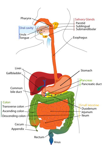
    Your digestive system is uniquely constructed to perform its specialized function of turning food into the energy you need to survive and packaging the residue for waste disposal. To help you understand how the many parts of the digestive system work together, here is an overview of the structure and function of this complex system.

    Mouth
    Esophagus
    Stomach
    Small intestine
    Pancreas
    Liver
    Gallbladder
    Colon (large intestine)
    Rectum
    Anus
    Part(s) Function
    Pancreas and salivary glands Produce digestive juices
    Stomach Digests food
    Liver Produces bile
    Small intestine Digest and absorb soluble food
    Large intestine Absorbs water from undigested food, producing faeces

    Digestive System

    1. Which organs of the body are part of the human digestive system?
    The digestive system, also known as “systema digestorium”, or gastrointestinal system, is composed of the digestive tube organs plus the digestive adnexal glands. The digestive tube is composed of mouth, pharynx, esophagus, stomach, small intestine (duodenum, jejunum, ileum), large intestine (caecum, colon, rectum) and anus.

    2. Which are the three parts of the small intestine?
    The small intestine is divided into three portions: duodenum, jejunum and ileum.

    3. How is extracellular digestion related to cellular and tissue specialization?
    A variety of specialized cells and tissues appeared with extracellular digestion to provide enzymes and special structures for the breaking down of dietary macromolecules.

    This phenomenon allowed other cells to be liberated for other tasks and differentiations while benefiting from nutrients distributed through the circulation.

    4. What is the difference between a complete digestive system and an incomplete digestive system? How are these types of digestive tubes associated or not to extracellular digestion?
    Animals with an incomplete digestive system are those in which the digestive tube has only one opening (cnidarians, platyhelminthes). Animals with a complete digestive system are those in which the digestive tube has two openings, mouth and anus (all other animal phyla, with the exception of poriferans, that do not have any digestive tube).

    In animals with incomplete digestive tubes the digestion is mixed, it begins in the extracellular space and finishes in the intracellular space. In animals with complete digestive systems extracellular digestion within the digestive tube predominates.

    5. What are some evolutionary advantages of animals with complete digestive tube?
    The complete digestive tube allows animals to continuously feed themselves without waiting for residuals to be eliminated before beginning the digestion of new foods. In this way the absorption of larger amount of nutrients is possible and therefore bigger and more complex species can develop. Digestive tubes with two openings also make digestion more efficient since they provide different sites with different physical and chemical conditions (mouth, stomach, bowels) for the action of different complementary digestive enzymatic systems.

    6. What is mechanical digestion? In molluscs, arthropods, earthworms, birds and vertebrates, in general, which organs respectively participate in this type of digestion?
    Mechanical digestion is the fragmentation of food aided by specialized physical structures, such as teeth, previous to extracellullar digestion. The mechanical fragmentation of food helps digestive enzymatic reactions because it provides a larger total area for the contact between enzymes and their substrates.

    In some molluscs, the mechanical fragmentation is done by the radula (a teeth-like structure). Some arthropods, like lobsters and dragonflies, have mouthparts that make mechanical digestion of food. In earthworms and birds, the mechanical digestion is made by an internal muscular organ. In mandibulate vertebrates there are mandibles and chewing muscles to triturate food previous to the chemical digestion.

    7. Concerning extracellular digestion what is meant by chemical digestion?
    Chemical digestion is the series of enzymatic reactions to break macromolecules into smaller ones.

    8. Which type of chemical reaction is the breaking of macromolecules into smaller ones that occurs in digestion? What are the enzymes that participate in this process called?
    The reactions of the extracellular digestion are hydrolysis reactions, i.e., breaking of molecules with the help of water. The enzymes that participate in digestion are hydrolytic enzymes.

    9. What is digestion?
    Digestion is the breaking down of larger organic molecules obtained from the diet, e.g. carbohydrates, fats, proteins, into smaller ones, like glucose, fatty acids, glycerol and amino acids.

    10. What are peristaltic movements? What is their role in human digestion?
    Peristalsis is the process of synchronized contractions of the muscular wall of the digestive tube. Peristaltic movements may occur from the esophagus until and including the bowels.

    The peristaltic movements are involuntary and they have the function of moving and mixing food along the digestive tube. Peristaltic movement deficiency, for example, in case of injuries of the innervation of the muscular wall of the digestive tube caused by Chagas’ disease, can lead to the interruption of the food traffic inside the bowels and to severe clinical consequences like megacolon (abnormal enlargement of the colon) and megaesophagus (enlargement of the esophagus).

    11. From the lumen to the external surface what are the tissues that form the digestive tube wall?
    From the internal surface to the external surface, the digestive tube wall is made of mucosa (epithelial tissue responsible for the intestinal absorption), submucosa (connective tissue beneath the mucous membrane and where blood and lymphatic vessels and neural fibers are located), muscle layers (smooth muscle tissue, two layers, one interior circular and other exterior longitudinal, structures responsible for the peristaltic movement), serous membrane (associated epithelial and connective tissue forming the external surface of the organ). In the bowels the serous membrane prolongs to form the mesentery, a serosa that encloses blood vessels and supports the bowels within the abdominal cavity.

    12. What is the location of the salivary glands in humans?
    There are 6 major salivary glands and they are located one in each parotid gland, two beneath the mandibles (submandibular) and two in the base of the tongue (sublingual). More than 700 other minor salivary glands exist dispersed on the lip mucosa, gingiva, palate and pharynx.

    13. What is the approximate pH of the salivary secretion? Is it an acid or basic fluid? What are the main functions of saliva?
    The saliva pH is approximately 6.8. It is thus a slightly acid pH.

    Saliva lubricates the food bolus and initiates the enzymatic extracellular digestion of food. It also works as a buffer for the mouth pH and it has an important role of having IgA antibodies (also present in tears, colostrum, mother’s milk and in the mucosae of the intestine and airways) that protect the organism against pathogens.

    14. What is the salivary digestive enzyme? Which type of food does it digest and into which smaller molecules does it transform the food?

    The salivary hydrolase is known as salivary amylase, or ptyalin. Ptyalin digests carbohydrates breaking starch and glycogen, glucose polymers, into maltose (a glucose disaccharide) and dextrin.

    15. Why doesn't the food enter the trachea instead of going to the esophagus?
    When food is swallowed the swallow reflex is activated and the larynx elevates and closes to avoid portions of the food bolus entering the trachea causing aspiration of strange material to the bronchi.

    16. Is the esophagus a muscular organ? Why even in a patient lying totally flat on a hospital bed can the swallowed food reach the stomach?
    The esophagus is a predominantly muscular organ so the assertion is correct. The esophagus is a muscular tube formed in its superior third of striated muscle tissue, in its middle third of mixed muscle tissue (striated and smooth) and in its lower third of smooth muscle tissue. The peristalsis of the esophagus provides the movement of the food towards the stomach even without gravitational help.

    17. What is the route of the ingested food from swallowing until the duodenum?
    Until reaching the duodenum the food enters the mouth, passes the pharynx, goes down the esophagus and passes the stomach.

    18. what is the valve that separates the stomach from the esophagus called? What is its function?
    The valve that separates the stomach from the esophagus is the cardia. It has the function of preventing acid gastric content from entering back into the esophagus. Insufficiency of this valve causes gastroesophageal reflux, a disease in which patients complain of bloating and heartburn (retrosternal burning).

    19. What is the valve that separates the duodenum from the stomach called? What is its function?
    The valve that separates the stomach from the duodenum is the pylorus. It has the function of keeping the food bolus within the gastric cavity for enough time to allow the gastric digestion to take place. It also has the function of preventing the intestinal content from going back into the stomach.

    20. What is the pH inside the stomach? Why is there a need to keep that pH level? How is it maintained? Which are the cells that produce that pH?
    The normal pH of the gastric juice is around 2. So it is an acid pH.

    It is necessary for the gastric pH to be kept acid for the activation of pepsinogen (a proenzyme secreted by the gastric chief cells) into pepsin, the digestive enzyme that acts only under low pH. This pH level is attained by the secretion of hydrochloric acid (HCl) by the parietal cells.

    21. Besides being fundamental for the activation of the main gastric digestive enzyme how does HCl also directly participate in digestion?
    With its corrosive effect, HCl also helps the rupture of the adhesion between food particles, facilitating the digestive process.

    22. How is the gastric mucosa protected from the acid pH of the stomach?
    The gastric epithelium is mucus secretory, i.e., it produces mucus. The mucus covers the stomach wall preventing corrosion by the gastric juice.

    23. What is the digestive enzyme that acts within the stomach? Which type of food does it digest? What are the cells that produce that enzyme?
    The digestive enzyme that acts in the stomach is pepsin. Pepsin has the function of breaking proteins into smaller peptides. The gastric cells that produce pepsinogen (the zymogen precursor of pepsin) are the chief cells.

    24. What name does the food bolus that passes from the stomach to the duodenum get?
    The partially digested and semifluid food bolus that leaves the stomach and enters the duodenum is called chyme.

    25. How different are intracellular and extracellular digestion? What is the evolutionary advantage of extracellular digestion?
    Intracellular digestion is that in which the breaking down of macromolecules takes place within the cell. Extracellular digestion is that in which macromolecules are broken down in places outside the cell (in the extracellular space, in the surrounds, in the lumen of digestive tubes, etc.)

    The advent of extracellular digestion in evolution allowed organisms to benefit from a greater variety of food. The breaking down of larger molecules into smaller ones outside the cell permitted the use of other foods than those that, due the size of their molecules, could not be interiorized by diffusion, phagocytosis or pinocytosis.

    26. By generally dividing food into carbohydrates, fats and proteins and considering the digestive process until the pylorus (exit of stomach), which of these mentioned types of food have already undergone chemical digestion?

    Until the exit of the stomach, carbohydrates, in the mouth, and proteins, in the stomach, have already undergone chemical breaking by digestive enzymes. Carbohydrates have suffered action of the salivary amylase (ptyalin) and proteins have suffered action of the enzyme pepsin of the gastric juice. Fats, until reaching the duodenum, do not undergo chemical digestion.

    27. What is the substance produced in the liver that acts in the small intestine during digestion? How does that substance act in the digestive process?
    Bile, an emulsifier liquid, is made by the liver and later stored within the gallbladder and released in the duodenum.

    Bile is composed of bile salts, cholesterol and bile pigments. Bile salts are detergents, amphiphilic molecules, i.e., molecules with a polar water-soluble portion and a non-polar fat-soluble portion. This feature allows bile salts to enclose fats inside water-soluble micelles in a process called emulsification for them to be in contact with intestinal lipases, enzymes that break fats into simpler fatty acids and glycerol.

    28. What is the adnexal organ of the digestive system in which bile is stored? How does this organ react to the ingestion of fat rich food?
    Bile is concentrated and stored in the gallbladder.

    When fat rich foods are ingested the gallbladder contracts to release bile inside the duodenum. (This is the reason why patients with gallstones must not ingest fatty food, the reactive contraction of the gallbladder may move some of the stones to the point of blocking the duct that drains bile into the duodenum, causing pain and possible severe complications.)

    29. What are the digestive functions of the liver?
    Besides making bile for release in the duodenum, the liver has other digestive functions.

    The venous network that absorbs nutrients from the guts, called mesenteric circulation, drains its blood content almost entirely to the hepatic portal vein. This vein irrigates the liver with absorbed material from the digestion. So the liver has the functions of storing, processing and inactivating nutrients.

    Glucose is polymerized into glycogen in the liver; this organ also stores many vitamins and the iron absorbed in the intestine. Some important metabolic molecules, like albumin and clotting factors, are made in the liver from amino acids of the diet. In the liver ingested toxic substances, like alcohol and drugs, are inactivated too.

    30. Besides the liver which is the other adnexal gland of the digestive system that releases substances in the duodenum participating in extracellular digestion?
    The other adnexal gland of the digestive system is the pancreas. This organ makes digestive enzymes that digest proteins (proteases), lipids (lipases) and carbohydrates (pancreatic amylases). Other digestive enzymes, like gelatinase, elastase, carboxipeptidase, ribonuclease and deoxyribonuclease are also secreted by the pancreas.

    31. How does the pancreatic juice participate in the digestion of proteins? What are the involved enzymes?
    The pancreas secretes trypsinogen that, undergoing action of the enzyme enterokinase secreted by the duodenum, is transformed into trypsin. Trypsin in its turn catalyzes the activation of pancreatic chymotrypsinogen into chymotrypsin. Trypsin and chymotrypsin are proteases that break proteins into smaller peptides. The smaller peptides are then broken into amino acids by the enzyme carboxipeptidase (also secreted by the pancreas in a zymogen form and activated by trypsin) helped by the enzyme aminopeptidase made in the intestinal mucous membrane.

    32. How does the pancreatic juice resume the digestion of carbohydrates? What is the involved enzyme?
    Carbohydrate digestion begins with the action of the salivary amylase (ptyalin) in the mouth and it continues in the duodenum by the action of the pancreatic juice. This juice contains the enzyme pancreatic amylase, or amylopsin, that breaks starch (amylum) into maltose (a disaccharide made of two glucose molecules).

    33. How does the pancreatic juice help the digestion of lipids? What is the involved enzyme?
    The enzyme pancreatic lipase is present in the pancreatic juice. This enzyme breaks triacylglycerol (triglyceride) into fatty acids and glycerol.

    34. Besides the pancreatic juice in the intestine there is the releasing of the enteric juice that contains digestive enzymes too. What are these enzymes and which type of molecule do each of these enzymes break?
    The enteric juice is secreted by the small intestine mucosa. The enzymes of the enteric juice and their respective functions are described as follows:

    Enterokinase: enzyme that activates trypsinogen into trypsin. Saccharase: enzyme that breaks sucrose (saccharose) into glucose and fructose. Maltase: enzyme that breaks maltose into two glucose molecules. Lactase: enzyme that breaks lactose into glucose and galactose. Peptidases: enzymes that break oligopeptides into amino acids. Nucleotidases: Enzymes that break nucleotides into its components (nitrogen-containing bases, phosphates and pentoses).

    35. Coming from the acid pH of the stomach which pH level does the chyme find when it enters the duodenum? Why is it necessary to maintain that pH level in the small intestine? What are the organs responsible for that pH level and how is it kept?
    Entering the duodenum the chyme meets the pancreatic juice under a pH of approximately 8.5. The neutralization of the chyme acidity is necessary to keep adequate pH level for the functioning of the digestive enzymes that act in the duodenum. Without the neutralization of the chyme acidity the mucous membrane of the intestine would be injured.

    When stimulated by the chyme acidity the duodenum makes a hormone called secretin. Secretin stimulates the pancreas to release the pancreatic juice and also the gallbladder to expel bile in the duodenum. The pancreatic secretion, rich in bicarbonate ions, is released in the duodenum and neutralizes the chyme acidity; this acidity is also neutralized by the secretion of bile in the duodenal lumen.

    36. What are the five human digestive secretions? Which of them is the only one that does not contain digestive enzymes?
    The human digestive secretions are: saliva, gastric juice, bile, pancreatic juice and enteric juice. Among these secretions only the bile does not contain digestive enzymes.

    37. Why do protease-supplying cells of the stomach and of the pancreas make only precursors of the active proteolytic enzymes?
    The stomach and the pancreas make zymogens of the proteases pepsin, chymotrypsin and trypsin and these zymogens are released into the gastric or duodenal lumen for activation. This happens to prevent the digestion of these organs' (stomach and pancreas) own cells and tissues by the active form of the enzymes. So the production of zymogens is a protective strategy against the natural effects of the proteolytic enzymes.

    38. After digestion the next step is absorption done by cells of the mucous membrane of the intestine. For this task a large absorption surface is an advantage. How is it possible in the small internal space of the body of a pluricellular organism to present a large intestinal surface?
    Evolution tried to solve this problem in two ways. The simplest is the long and tubular shape of the bowels (approximately eight meters in extension), making possible that numerous small intestine loops fold closely. More efficient solutions are the intestinal villi and the microvilli of the mucosal membrane cells.

    The intestinal wall is not smooth. The mucous membrane, together with its submucosa, projects inside the gut lumen like glove fingers forming invaginations and villi that multiply the available surface for absorption. In addition the epithelial cells that cover these villi have themselves numerous hairlike projections called microvilli on the external face (lumen face) of their plasma membrane. The absorptive area of the intestines is thus increased hundreds of times with these solutions.

    In the jejunum and ileum there are folds that have the function of increasing the absorption surface too.

    39. In which part of the digestive tube is water is chiefly absorbed? What about the mineral ions and vitamins?
    Most part of water, vitamins and mineral ions are absorbed by the small intestine. The large intestine, however, is responsible for the reabsorption of nearly 10% of the ingested water, an important amount that gives consistency to feces (colon diseases can cause diarrhea).

    40. From the intestinal lumen through to the tissues - what is the route of nutrients after digestion?
    Monosaccharides, amino acids, mineral salts and water are absorbed by the intestinal epithelium and collected by capillary vessels of the intestinal villi. From the capillaries, nutrients go to the mesenteric circulation, a system of vessels that drains the intestinal loops. The blood of the mesenteric circulation is drained to the portal hepatic vein and some nutrients are processed by the liver. From the liver, nutrients are gathered by the hepatic veins that discharge its blood content into the inferior vena cava. Blood from the inferior vena cava then gains the right chambers of the heart and is pumped to the lungs for oxygenation. From the lungs the blood then returns to the heart where it is pumped to the tissues distributing nutrients and oxygen.

    41. What is the special route that lipids follow during digestion? What are chylomicrons?
    Triglycerides emulsified by the bile within micelles suffer the action of lipases that break them into fatty acids and glycerol. Fatty acids, glycerol and cholesterol are absorbed by the intestinal mucosa. In the interior of the mucosal cells fatty acids and glycerol form again triglycerides that together with cholesterol and phospholipids are packed in small vesicles covered by proteins and called chylomicrons. The chylomicrons are released in minuscule lymphatic vessels not in blood vessels and they gain the lymphatic circulation. So the lymphatic system plays an important role in the absorption of lipids.

    The lymphatic circulation drains its content to the venous blood circulation. In that manner chylomicrons reach the liver where their lipid content is processed and released in the blood under the form of protein-containing complexes called lipoproteins, like HDL, VLDL and LDL.

    42. What are the so-called “good” and “bad” cholesterol?
    Lipoproteins are complexes made of lipids (triglycerides and cholesterol) and proteins. The lipoproteins present different densities according to the relationship between their protein and lipid quantities since lipids are less dense than proteins. Low-density lipoproteins (LDL) are those with a low protein/lipid relation; high-density lipoproteins (HDL) have a high protein/lipid relation; another group is the very low-density lipoproteins (VLDL) with very low protein/lipid relation.

    LDL is known as “bad cholesterol” because it transports cholesterol from the liver to the tissues and so induces the formation of atheroma plaques inside blood vessels, a condition called atherosclerosis (do not confuse with arteriosclerosis) that can lead to severe circulatory obstructions like acute myocardial infarction, cerebrovascular accidents and thrombosis. HDL is known as “good cholesterol” since it transports cholesterol from the tissues to the liver (to be eliminated with the bile) and elevation of the HDL blood level reduces the risk of atherosclerosis. (VLDL transforms into LDL after losing triglycerides in the blood).

    43. Why does the ingestion of vegetable fibers improve the bowel habit in people that suffer from hard stools?
    Some types of plant fibers are not absorbed by the intestine but play an important role in the functioning of the organ. They retain water inside the bowels and thus contribute to the softening of the fecal bolus. A softer fecal bolus is easier to be eliminated during defecation. People that eat less dietary fiber may suffer from hard stools and constipation.

    44. What are the main functions of the bacterial flora within the human gut?
    Bacteria that live inside the gut have great importance in digestion. Some polysaccharides like cellulose, hemicellulose and pectin are not digested by the digestive enzymes secreted by the body, instead, they are broken by enzymes released by bacteria of the gastrointestinal tract. The intestinal bacterial flora also make vital substances for the functioning of the bowels facilitating or blocking the absorption of nutrients and stimulating or reducing peristalsis. Some gut bacteria are the main source of vitamin K for the body and so they are essential for the blood clotting process.

    In the intestinal flora there are utile but also potentially harmful bacteria. It is estimated that more than 100 trillion bacteria live in a human gut. Some bacteria are useful too because they compete with other species preventing excessive proliferation of these bacteria.

    45. The releasing of digestive secretions is controlled by hormones. What are the hormones that participate in this regulation?
    The hormones that participate in the regulation of digestion are gastrin, secretin, cholecystokin and enterogastrone.

    46. How is it produced and what is the function of gastrin in the digestive process?
    The presence of food in the stomach stimulates the secretion of gastrin that in its turn triggers the releasing of the gastric juice.

    47. Where is it produced and what is the function of secretin in the digestive process?
    Secretin is made in the duodenum. The chyme acidity causes the duodenum to release this hormone that in its turn stimulates the secretion of the pancreatic juice.

    48. How is it produced and what is the function of cholecystokin in the digestive process?
    The fat level of the chyme detected in the duodenum stimulates the secretion of cholecystokin (CCK). CCK acts by stimulating the secretion of the pancreatic juice also, and the releasing of bile by the gallbladder.

    49. Where is it produced and what is the function of enterogastrone in the digestive process?
    When the chyme is too fatty there is a secretion of enterogastrone by the duodenum. This hormone reduces the peristalsis of the stomach thus slowing the entrance of food into the duodenum (as the digestion of fats takes more time).

    50. What are the special structures of the avian digestive tube and their respective functions?
    The digestive tube of birds has special structures, in this sequential order: the crop, the proventriculus and the gizzard.

    The crop has the function of temporary storage of ingested food and it is a more dilated area of the avian esophagus. The proventriculus is the chemical stomach of the birds where food is mixed with digestive enzymes. The gizzard is a muscular pouch that serves as a mechanical stomach where the food is ground to increase the exposure area of the food particles for the digestive enzymes to act.

    51. Compared to mammals do birds absorb more or less water in their digestive system? Why is this phenomenon an adaptation to flight?
    Bird feces are more liquid than mammal feces, i.e., less water is absorbed in the avian digestive system. The more frequent elimination of feces in birds due to their less solid feces is an adaptation to flight since their body weight is kept lower.

    52. What is meant by “mutualist exploration of cellulose digestion”, a phenomenon that occurs in some mammals and insects?
    Herbivorous animals eat great amounts of cellulose, a substance not digested by their digestive enzymes. In these animals regions of the digestive tube are colonized by microorganisms that digest cellulose. This mutualist ecological interaction between animals and microorganisms occurs, e.g., in horses, cows, rabbits and in some insects such as termites.

    53. Cows swallow their food once and then this food goes back to the mouth to be chewed again. How can this phenomenon be explained?
    The food ingested by cows and other ruminant animals passes first within two compartments of the digestive tube called the rumen and the reticulum. Within them the food suffers the action of digestive enzymes released by microorganisms that live there in mutualist ecological interaction. In the reticulum the food is divided in some food bolus too. After passing the reticulum the food (cud) is regurgitated to the mouth to be again chewed and swallowed in a process called rumination. The digesting food then enters the omasum where it is mechanically mixed. After that the food goes to the abomasum, the organ where the chemical digestion takes place. After leaving the abomasum (the true stomach) the food bolus gains the intestine.

    Mouth

    The mouth is the beginning of the digestive tract; and, in fact, digestion starts here when taking the first bite of food. Chewing breaks the food into pieces that are more easily digested, while saliva mixes with food to begin the process of breaking it down into a form your body can absorb and use.

    Esophagus

    Located in your throat near your trachea (windpipe), the esophagus receives food from your mouth when you swallow. By means of a series of muscular contractions called peristalsis, the esophagus delivers food to your stomach.

    Stomach

    The stomach is a hollow organ, or "container," that holds food while it is being mixed with enzymes that continue the process of breaking down food into a usable form. Cells in the lining of the stomach secrete a strong acid and powerful enzymes that are responsible for the breakdown process. When the contents of the stomach are sufficiently processed, they are released into the small intestine.

    Small intestine

    Made up of three segments — the duodenum, jejunum, and ileum — the small intestine is a 22-foot long muscular tube that breaks down food using enzymes released by the pancreas and bile from the liver. Peristalsis also is at work in this organ, moving food through and mixing it with digestive secretions from the pancreas and liver. The duodenum is largely responsible for the continuous breaking-down process, with the jejunum and ileum mainly responsible for absorption of nutrients into the bloodstream.

    Contents of the small intestine start out semi-solid, and end in a liquid form after passing through the organ. Water, bile, enzymes, and mucous contribute to the change in consistency. Once the nutrients have been absorbed and the leftover-food residue liquid has passed through the small intestine, it then moves on to the large intestine, or colon.

    Pancreas

    The pancreas secretes digestive enzymes into the duodenum, the first segment of the small intestine. These enzymes break down protein, fats, and carbohydrates. The pancreas also makes insulin, secreting it directly into the bloodstream. Insulin is the chief hormone for metabolizing sugar.

    Liver

    The liver has multiple functions, but its main function within the digestive system is to process the nutrients absorbed from the small intestine. Bile from the liver secreted into the small intestine also plays an important role in digesting fat. In addition, the liver is the body’s chemical "factory." It takes the raw materials absorbed by the intestine and makes all the various chemicals the body needs to function. The liver also detoxifies potentially harmful chemicals. It breaks down and secretes many drugs.

    Gallbladder

    The gallbladder stores and concentrates bile, and then releases it into the duodenum to help absorb and digest fats.

    Colon (large intestine)

    The colon is a 6-foot long muscular tube that connects the small intestine to the rectum. The large intestine is made up of the cecum, the ascending (right) colon, the transverse (across) colon, the descending (left) colon, and the sigmoid colon, which connects to the rectum. The appendix is a small tube attached to the cecum. The large intestine is a highly specialized organ that is responsible for processing waste so that emptying the bowels is easy and convenient.

    Stool, or waste left over from the digestive process, is passed through the colon by means of peristalsis, first in a liquid state and ultimately in a solid form. As stool passes through the colon, water is removed. Stool is stored in the sigmoid (S-shaped) colon until a "mass movement" empties it into the rectum once or twice a day. It normally takes about 36 hours for stool to get through the colon. The stool itself is mostly food debris and bacteria. These bacteria perform several useful functions, such as synthesizing various vitamins, processing waste products and food particles, and protecting against harmful bacteria. When the descending colon becomes full of stool, or feces, it empties its contents into the rectum to begin the process of elimination.

    Rectum

    The rectum (Latin for "straight") is an 8-inch chamber that connects the colon to the anus. It is the rectum's job to receive stool from the colon, to let the person know that there is stool to be evacuated, and to hold the stool until evacuation happens. When anything (gas or stool) comes into the rectum, sensors send a message to the brain. The brain then decides if the rectal contents can be released or not. If they can, the sphincters relax and the rectum contracts, disposing its contents. If the contents cannot be disposed, the sphincter contracts and the rectum accommodates so that the sensation temporarily goes away.

    Anus

    The anus is the last part of the digestive tract. It is a 2-inch long canal consisting of the pelvic floor muscles and the two anal sphincters (internal and external). The lining of the upper anus is specialized to detect rectal contents. It lets you know whether the contents are liquid, gas, or solid. The anus is surrounded by sphincter muscles that are important in allowing control of stool. The pelvic floor muscle creates an angle between the rectum and the anus that stops stool from coming out when it is not supposed to. The internal sphincter is always tight, except when stool enters the rectum. It keeps us continent when we are asleep or otherwise unaware of the presence of stool. When we get an urge to go to the bathroom, we rely on our external sphincter to hold the stool until reaching a toilet, where it then relaxes to release the contents.


    Gastroenterology

    Emergency medicine specialist training programs and relevance to primary care physician.
    Why should primary care physicians know about emergency medicine specialist training programs?
    Medical emergency room can report to you that because of the negligence of a primary care physician, a resident has to face a preventable medical emergency.
    You may occasionally have to work as emergency medical specialist depending on the needs of the healthcare system in the state.

    Here are further guidelines.
    http://www.qureshiuniversity.com/emergencymedicalservices.html

    What is primary health care?
    A health care resource advertises capabilities of screening hypertension and diabetes. That is not primary health care. That is quackery.

    Such activities are planned on April 20, 2012, at Near North Center, Chicago, Illinois.
    Heartland Alliance Chicago, Illinois, also is involved in similar quackery.

    Asian Human Services has declared an annual health fair on April 25, 2012, in Chicago, Illinois, to screen specific medical conditions.

    Primary Care

    In defining primary care, it is necessary to describe the nature of services provided to patients, as well as to identify who are the primary care providers. The domain of primary care includes the primary care physician, other physicians who include some primary care services in their practices, and some non-physician providers. However, central to the concept of primary care is the patient. Therefore, such definitions are incomplete without including a description of the primary care practice.

    The following five definitions relating to primary care should be taken together. They describe the care provided to the patient, the system of providing such care, the types of physicians whose role in the system is to provide primary care, and the role of other physicians, and non-physicians, in providing such care. Taken together they form a framework within which patients will have access to efficient and effective primary care services of the highest quality.

    Definition #1 - Primary Care

    Primary care is that care provided by physicians specifically trained for and skilled in comprehensive first contact and continuing care for persons with any undiagnosed sign, symptom, or health concern (the "undifferentiated" patient) not limited by problem origin (biological, behavioral, or social), organ system, or diagnosis.

    Primary care includes health promotion, disease prevention, health maintenance, counseling, patient education, diagnosis and treatment of acute and chronic illnesses in a variety of health care settings (e.g., office, inpatient, critical care, long-term care, home care, day care, etc.). Primary care is performed and managed by a personal physician often collaborating with other health professionals, and utilizing consultation or referral as appropriate.Primary care provides patient advocacy in the health care system to accomplish cost-effective care by coordination of health care services. Primary care promotes effective communication with patients and encourages the role of the patient as a partner in health care.

    Definition #2 - Primary Care Practice

    A primary care practice serves as the patient's first point of entry into the health care system and as the continuing focal point for all needed health care services. Primary care practices provide patients with ready access to their own personal physician, or to an established back-up physician when the primary physician is not available.

    Primary care practices provide health promotion, disease prevention, health maintenance, counseling, patient education, diagnosis and treatment of acute and chronic illnesses in a variety of health care settings (e.g., office, inpatient, critical care, long-term care, home care, day care, etc.).

    Primary care practices are organized to meet the needs of patients with undifferentiated problems, with the vast majority of patient concerns and needs being cared for in the primary care practice itself. Primary care practices are generally located in the community of the patients, thereby facilitating access to health care while maintaining a wide variety of specialty and institutional consultative and referral relationships for specific care needs. The structure of the primary care practice may include a team of physicians and non-physician health professionals.

    Definition #3 - Primary Care Physician

    A primary care physician is a generalist physician who provides definitive care to the undifferentiated patient at the point of first contact and takes continuing responsibility for providing the patient's care. Such a physician must be specifically trained to provide primary care services.

    Primary care physicians devote the majority of their practice to providing primary care services to a defined population of patients. The style of primary care practice is such that the personal primary care physician serves as the entry point for substantially all of the patient's medical and health care needs - not limited by problem origin, organ system, or diagnosis. Primary care physicians are advocates for the patient in coordinating the use of the entire health care system to benefit the patient.

    Definition #4 - Non-Primary Care Physicians Providing Primary Care Services

    Physicians who are not trained in the primary care specialties of family medicine, general internal medicine, or general pediatrics may sometimes provide patient care services that are usually delivered by primary care physicians. These physicians may focus on specific patient care needs related to prevention, health maintenance, acute care, chronic care or rehabilitation. These physicians, however, do not offer these services within the context of comprehensive, first contact and continuing care.

    The contributions of physicians who deliver some services usually found within the scope of primary care practice may be important to specific patient needs. However, the absence of a full scope of training in primary care requires that these individuals work in close consultation with fully-trained, primary care physicians. An effective system of primary care may utilize these physicians as members of the health care team with a primary care physician maintaining responsibility for the function of the health care team and the comprehensive, ongoing health care of the patient.

    Definition #5 - Non-Physician Primary Care Providers

    There are providers of health care other than physicians who render some primary care services. Such providers may include nurse practitioners, physician assistants and some other health care providers.

    These providers of primary care may meet the needs of specific patients. They should provide these services in collaborative teams in which the ultimate responsibility for the patient resides with the primary care physician.

    *In this document, the term physician refers only to doctors of medicine (M.D.) and osteopathy (D.O.).

    Types of Primary Care Physicians

    Your primary care physician serves as the entry point for substantially all of your medical and health care needs. He or she is your advocate in coordinating the use of the entire health care system to benefit you and takes continuing responsibility for providing your care. Primary care includes health promotion, disease prevention, health maintenance, counseling, patient education, diagnosis and treatment of acute and chronic illnesses. By regularly seeing a primary care practitioner, you can identify and control health risk factors before they become problems. Your primary care practitioner encourages you to take charge of your health and is a trusted partner in an environment where you can feel comfortable and known.

    All doctors are not alike. Many different health care professionals with different types of education can provide health care. Adults in your family could see an internist. Children could go to a pediatrician. Or the whole family could see a family medicine doctor. You could have a family medicine doctor and an obstetrician/gynecologist, or an internist and a geriatrician. If it's right for you, it's the right choice. Here's a guide to the five kinds of primary care doctors available at Lehigh Valley Health Network.

    If a physician on your care team is board-certified, he or she chose to obtain an additional credential after medical school and residency training. Board-certified physicians have completed additional education and passed an examination to earn this credential. If one of your caregivers is fellowship-trained, the physician has had additional training in a particular area of interest.

    Family medicine doctors: Also called family practice doctors, they see people of all ages, including infants, children, teens and seniors. They are educated and board-certified in their knowledge of the body at all ages.

    Internal medicine doctors: Also called internists, they generally care for people age 14 and up (the starting age can vary by practice). They are educated and board-certified in the understanding of the internal organs of the adult body. Some internal medicine and family medicine physicians also can provide care that you might not expect, like routine gynecologic care, dermatology and sports medicine.

    Gynecologist or obstetrician: For many women, this physician is a key health resource. Through the reproductive years and afterward, your obstetrician or gynecologist has the knowledge and experience to promote your well-being. They recommend tests and exams necessary at different stages of your life.

    Geriatricians: They have special education to address the specific needs of adults over age 60. They provide routine primary care, as well as medication management, physical and mental assessments related to falls, memory issues and other aspects of aging. Lehigh Valley Hospital-Cedar Crest has been nationally ranked by U.S. News & World Report as one of "America's Best Hospitals" in geriatrics, and LVH-Muhlenberg is ranked as high-performing.

    Pediatricians: Every child needs to see a doctor – in fact, more often than adults. Pediatricians are specially educated to care for children, ranging from newborns through age 21. If your child needs special care, our pediatricians work closely with our specialists so your child can receive the best care.

    MD or DO? So you've narrowed down the type or primary care doctor you'd like to see. Now you may have questions about whether you'd prefer an MD or a DO. Here is some information to help you choose.

    MD stands for medical doctor, a doctor who trained at a medical school. DO stands for doctor of osteopathic medicine, who trained at a college of osteopathic medicine. They represent two branches of medicine that started out completely separate but have grown more alike over 125 years. Both types of physicians spend the same number of years training, must pass a licensing exam, and are licensed to provide all types of medical care, including surgery.

    Most people are familiar with how medical doctors are trained in understanding the body and treating disease. Osteopathic medicine was founded in 1874 based on a "whole person" approach that emphasizes preventive care and wellness. Osteopaths also receive ongoing education in the musculoskeletal system, and some but not all osteopaths perform osteopathic manipulative treatments. Because of this "whole person" approach, about two-thirds of osteopaths choose to become primary care doctors.

    Today, the two branches of medicine seem more alike than different. MDs emphasize preventive care and wellness, and a growing number of osteopaths are choosing to become specialists.

    Can so many residents be screened for so many medical conditions in one day by a few medical doctors?
    No, they cannot.

    How many residents should be assigned to a medical doctor for primary health care?
    One handred patients per medical doctor.

    What should a health care resource be able to provide in primary health care?

    Why You Need a Primary Care Physician
    Your Primary Care Physician: The Go-To Person for Good Health and Excellent Medical Care

    Choosing a primary care physician and maintaining the relationship with regular visits is an important step you can take to protect your health, at any age. When you get ill or injured, your primary care doctor is the person in charge of seeing that you get the medical care you need and he/she is also focused on keeping you well and healthy so that you can live life the way you want to.

    What Do Primary Care Doctors Do?
    Your primary care doctor's responsibilities are divided into three areas:

    •Preventive care to help you stay healthy: Your primary care physician offers advice on how to live a healthy lifestyle and makes sure you get screenings and immunizations when you need them.

    •Treating illnesses and injuries: Primary care doctors diagnose and treat common medical conditions, assess the severity of medical problems, refer patients to specialists when necessary, treat illnesses and help you manage chronic diseases, such as diabetes or hypertension (high blood pressure).

    •Coordinating all your medical care: Medical care can get complicated, particularly as you age and if you have one or more chronic medical conditions. Your primary care doctor keeps track of your health, including treatments and medications, makes sure you see the right doctors at the right times and communicates with them about what's going on with your health, and serves as the point person to make sure that your medical treatments and medications won't interact dangerously with one another.

    What Are The Benefits of Having a Primary Care Doctor?
    Today's health care environment is complex. As your go-to health care provider, your primary care physician protects you and represents your interests in a variety of ways, including:

    •Overseeing your care. In his/her role as coordinator of your care, your primary care doctor keeps all doctors informed of your health status and treatments so you don't undergo unnecessary tests or procedures or have treatments you don't want or don't need.

    •Lower medical costs. Keeping an up-to-date medical record of all your care, tests and treatments, your primary care physician ensures that you aren't paying for care you don't need.

    •Keeping you healthy, particularly as you age. Your primary care doctor monitors health changes and, seeing you year after year, may detect changes or signs of developing disease at the earliest, most treatable stages. Advising you on necessary health screenings is another important service your primary care physician can provide.

    When Do You Need A Primary Care Doctor?
    Even when you are in good health it is important to have a good relationship with your primary care physician. He/she will keep you healthy with physical examinations, immunizations and health education information. Your primary care doctor will let you know whether or not you need to get a flu shot and whether you need certain tests, such as heart screenings and cancer screenings.

    When you get injured or are feeling unwell, your primary care doctor can diagnose and treat your problem. An important advantage of having a primary care doctor is that you will always know that your care is being directed by a doctor who knows and understands all of your medical needs. He/she should be your first stop for all your medical concerns, including undiagnosed, non-emergencies, such as:
    •Colds vs. Flu. If you have a sore throat or chest cold symptoms, your primary care doctor will help determine whether you are suffering a common cold, a virus or the flu.

    •Infections. Primary care doctors can diagnose and treat ear infections, eye infections and other common complaints.

    •Stomach upset. Your primary care doctor can help determine whether your digestive upset is caused by a stomach flu, a food-borne illness, or may be something more serious or complicated that requires a visit to a specialist.

    •Injuries. If you fall or are injured during your normal activities or even while playing sports, your primary care physician can assess your condition and determine whether you need an x-ray or to see an orthopedic specialist.

    •Chronic disease management. Your primary care doctor will also help you manage chronic conditions, such as diabetes, hypertension and high cholesterol.

    •Care Coordination. If you need to see a medical specialist, your primary care doctor coordinates and manages your care, making sure that treatments you receive are appropriate for you, that the drugs you are prescribed work well together, and that all the doctors who care for you know about other medical problems you may be having.

    Here are further guidelines.

    Primary care diseases and conditions with treatment.
    Lung Disease Alphabetical Listing
    Common Breast Conditions
    Common Eye Disorders
    Common Infectious Diseases
    Common Orthopedic Disorders
    Common Skin Disorders
    Developmental Disorders
    Common Endocrine Diseases
    Hypoxemia (low blood oxygen)…
    Minor Cuts, Scrapes, and Skin Wounds
    Sports Injuries
    Stress
    Common Oral Conditions in Older Persons
    Substance Abuse
    Common Breast Conditions

    1. Mastalgia (Breast Pain)

    2. Common Benign Lumps

    3. Fibrocystic Breast Changes

    4. Nipple Problems and Discharge

    5. Breast Infections and Inflammations

    6. Diagnosing Benign Breast Conditions

      Common Eye Disorders

      What are some common eye disorders?
      The following list provides a brief description of several common eye disorders. Consult your physician or an ophthalmologist or optometrist for more information.


    7. Age-Related Macular Degeneration is the breaking down, or degeneration, of the macula area of the retina of the eye.

    8. Amblyopia is reduction or dimming of vision in an eye that appears to be normal.

    9. Astigmatism is a condition in which an abnormal curvature of the cornea can cause two focal points to fall in two different locations - making objects up close and at a distance appear blurry.

    10. Blepharitis is an inflammation of the edges of the eyelids involving hair follicles and glands that open onto the surface.

    11. Cataract is a condition in which the lens of the eye becomes dense or opaque and does not properly transmit light.

    12. Chalazion is a small bump that develops on the upper or lower eyelid. It is caused by inflamed meibomian glands that produce the oil in tears.

    13. Conjunctivitis, sometimes called pink eye, is an inflammation of the blood vessels in the conjunctiva, the membrane that covers the sclera and inside of the eyelids. Conjunctivitis may be caused by bacteria or viruses, making it very contagious.

    14. Diabetic Retinopathy is a disorder of the retina resulting from changes in the eye blood vessels and found in some people who have diabetes.

    15. Dry Eye occurs when there is not enough moisture in the eye, causing it to feel dry, hot, sandy, and gritty. Dry eye may be caused by low humidity, smoke, aging, certain diseases, and certain medications (i.e., antihistamines, decongestants).

    16. Floaters appear as spots, dots, or lines and affect or interrupt vision. Floaters are usually caused by bits of debris in the vitreus humor.

    17. Glaucoma is a disease that impairs the optic nerve when fluid and pressure build up in the eye and damage the optic nerve.

    18. Hyperopia, or farsightedness, means a person has trouble seeing clearly up close.

    19. Iritis is an inflammation of the iris of the eye.

    20. Myopia, or nearsightedness, means a person has trouble seeing clearly at a distance.

    21. Presbyopia is another type of farsightedness and is caused when the center of the eye lens hardens making it unable to accommodate near vision.

    22. Retina Detachment is the separation of the retina from the back of the eye.

    23. Retinitis Pigmentosa is actually the name given to a group of hereditary eye disorders, all of which involve the eye's retina, the light-sensitive nerve layer that lines the back of the eye, and all of which cause a gradual, yet progressive, loss or reduction in visual ability.

    24. Strabismus is crossed eyes.

    25. Stye is a noncontagious, bacterial infection of one of the sebaceous glands of the eyelid. A stye looks like a small, red bump either on the eyelid or on the edge of the eyelid.

    26. Uveitis is a condition that occurs in the uvea, or the middle coat of the eye. Because the uvea contains the blood vessels that supply nutrients to the eye, any form of uveitis may be serious and may be a symptom for other serious conditions.

      The Primary Care Eye Exam: Evaluation of Ophthalmic Complaints
      Visual Acuity Chart or Card
      Fingers and hands
      Fluorescein and saline
      Pen Light or Transilluminator
      Direct Ophthalmoscope
      Possible Slit Lamp
      Most will have little to no magnification

    27. Abrasion

    28. Allergic

    29. Bacterial Conjunctivitis

    30. Chemical Conjunctivitis

    31. Chemical/Toxic Conjunctivitis

    32. Contact Lens Related

    33. Cranial Nerve III Palsy

    34. Double Vision

    35. Episcleritis

    36. Exposure

    37. Foreign Body

    38. Herpes Simplex Keratitis

    39. Migraine

    40. RED EYE

    41. Restrictions of Gaze

    42. Rust Ring

    43. Sub Conj Heme

    44. Uveitis or Iritis

    45. Viral Conjunctivitis
      Direct Ophthalmoscopy
      Normal Fundus
      Diabetic Retinopathy
      Hypertensive Retinopathy
      Macular Degeneration
      Glaucoma

      Common Infectious Diseases

      Many infectious diseases require clinical care by a physician or other health care professional. Listed in the directory below are some of these diseases, for which we have provided a brief overview.


    46. Chickenpox

    47. Chronic Fatigue Syndrome

    48. Common Cold

    49. Diphtheria

    50. E. coli

    51. Giardiasis

    52. Infectious Mononucleosis

    53. Influenza (Flu)

    54. Lyme Disease

    55. Malaria

    56. Measles

    57. Meningitis

    58. Mumps

    59. Poliomyelitis (Polio)

    60. Pneumonia

    61. Rocky Mountain Spotted Fever

    62. Rubella (German Measles)

    63. Salmonella Infections

    64. Severe Acute Respiratory Syndrome (SARS)

    65. Sexually Transmitted Diseases

    66. Shingles (Herpes Zoster)

    67. Sore throat & fever

    68. Tetanus

    69. Tuberculosis

    70. Viral Hepatitis Overview

    71. West Nile Virus

    72. Whooping Cough (Pertussis)
      Common Orthopedic Disorders

      There are many conditions that affect our body's musculoskeletal system, that require clinical care by a physician or other healthcare professional. Listed in the directory below are some, for which we have provided a brief overview.

    73. Arthritis

    74. Osteoarthritis

    75. Rheumatoid Arthritis

    76. Treatment for Arthritis

    77. Bursitis

    78. Elbow Pain and Problems

    79. Cubital Tunnel Syndrome

    80. Lateral Epicondylitis (Tennis Elbow)

    81. Medial Epicondylitis (Golfer's or Baseball Elbow)

    82. Fibromyalgia

    83. Foot Pain and Problems

    84. Fractures

    85. Low Back Pain

    86. Hand Pain and Problems

    87. Carpal Tunnel Syndrome

    88. Knee Pain and Problems

    89. Ligament Injuries to the Knee

    90. Torn Meniscus

    91. Kyphosis

    92. Neck Pain and Problems

    93. Osteoporosis

    94. Paget's Disease of the Bone

    95. Scoliosis

    96. Shoulder Pain and Problems

    97. Soft-Tissue Injuries

      Common Skin Disorders

      Many common skin disorders require the clinical care of a physician or other health care professional. Listed in the directory below are some, for which we have provided a brief overview.

    98. Acne

    99. Bed Sores

    100. Dry Skin

    101. Calluses and Corns

    102. Keratosis Pilaris

    103. Psoriasis

    104. Pityriasis Rosea

    105. Rosacea

    106. Sebaceous Cysts

      Developmental Disorders

      Developmental disorders are one category of mental health problems. The category identifies children who have difficulty accomplishing early developmental tasks such as language, communication, socialization, and motor skills. Treatment of developmental disorders usually includes medication and specialized training to assist the child in developing needed skills.

      Many different types of developmental disorders require clinical care by a doctor or other health care professional. Listed in the directory below are some, for which we have provided a brief overview.

    107. Pervasive Developmental Disorders

    108. Autistic Disorder

    109. Learning Disorders

    110. Communication Disorders

    111. Common Endocrine Diseases


    112. Diabetes (Type 1, 2, and Gestational)

      Minor Cuts, Scrapes, and Skin Wounds

      Listed in the directory below is some additional information about minor cuts, scrapes, and skin wounds, for which we have provided a brief overview.

    113. Minor Injuries Overview

    114. Abrasions

    115. Blisters

    116. Bruises

    117. Lacerations Without Stitches

    118. Lacerations With Stitches

    119. Puncture Wounds

    120. Small Cuts and Scrapes

    121. Splinters

      Sports Injuries

      Many sports injuries require clinical care by a physician or other health care professional. Listed below are some, for which we have provided a brief overview.

      Overview of Sports

      Injuries

    122. Exercise

    123. Exercise and the Aging Person

    124. Lumbar Strain (Weight Lifter's Back)

    125. Patellar Tendonitis (Jumper's Knee)

    126. Patellofemoral Stress Syndrome (Runner's Knee)

    127. Preventing Sports Injuries

    128. Sports and Fractures

    129. Shin Splints

      Substance Abuse

    130. Substance abuse is a pattern of repeated use of alcohol, drugs, or both, even though this use causes unpleasant or distressing events in the user's life.

      A person has a substance abuse problem if one or more of the following have occurred within the past 12 months.

      Substance use has interfered with the person's ability to meet his or her obligations at home, school, or work. The person may not show up for, have poor performance at, or be fired or expelled from work or school. The person may neglect family members or the home.

      Alcohol, drugs, or both have been used repeatedly in dangerous situations, such as while driving a car or operating machinery.

      The person has incurred legal problems related to substance abuse, such as arrests for driving under the influence (DUI) or disorderly conduct.

      The person continues to use alcohol, drugs, or both, despite social or personal problems caused by or made worse by use of the substance. This includes everything from arguments with a family member about drug use to physical fights with strangers.

      Someone who has a substance abuse problem may suffer serious withdrawal symptoms if he or she stops drinking alcohol or using a drug suddenly ("cold turkey"). After dependency develops, it may become very difficult to stop drinking or using a drug without outside help. Medical detoxification may be needed.

      Common Oral Conditions in Older Persons

      Condition Clinical presentation Treatment Comments
      Dental caries Coronal (above the gum) or root: painful brownish discoloration with cavitation Root caries may be treated with fluoride gels, varnishes, or toothpaste; effective for some shallow cariesInfection can be reduced with good oral hygiene and professional dental care; patients should avoid sugary foods and drinks; see Table 2 for risk factors
      Gingivitis Red, swollen, bleeding gums Good oral hygiene, including brushing and flossing daily
      Periodontitis Gingivitis, gingiva recession, loose or shifting teeth Good oral hygiene, including brushing and flossing daily; dental scaling performed by a dental health professional; adjunct antibiotic therapy Associated with cardiovascular disease, worsening diabetes, and aspiration pneumonia
      Xerostomia Swollen, dry, red tongue; burning sensation; difficulty with speech and swallowing; change in taste Saliva substitutes; sugar-free gum or pilocarpine (Salagen) and cevimeline (Evoxac) drops may stimulate saliva production See Table 3 for risk factors
      Candidiasis Acute pseudomembranous (thrush): adherent white plaques that can be wiped off

      Erythematous (denturestomatitis): red macular lesions, often with a burning sensation

      Angular cheilitis: erythematous, scaling fissures at the corners of the mouth
      Topical antifungals (e.g., nystatin oral suspension or troche [Mycostatin; brand no longer available in the United States]; clotrimazole troche [Mycelex])
      Or
      Systemic antifungals (e.g., fluconazole[Diflucan]; ketoconazole [Nizoral; brand no longer available in the United States]; itraconazole [Sporanox])
      Diagnosis can be confirmed with oral exfoliative cytology (stained with periodic acid-Schiff or potassium hydroxide), biopsy, or culture
      Denture stomatitis Varying erythema, occasionally accompanied by petechial hemorrhage; localized to the denture-bearing areas of the removable maxillary prosthesis; usually asymptomatic Removal of dentures at night; topical antifungals (see Candidiasis) placed inside the denture-fitting surface Dentures should be removed and cleaned at least once daily

      Diabetes (Type 1, 2, and Gestational)
      Endocrine diseases and disorders can be grouped into several different areas. Some endocrinologists focus on one or two areas, such as diabetes, pediatric disorders, thyroid, or reproductive and menstrual disorders. Others work in all areas of endocrinology. The major areas of endocrinology are described below.


      Diabetes

      Currently, there are approximately 23 million Americans with diabetes and an additional 57 million Americans have pre-diabetes. Patients with diabetes have too much sugar in their blood. Recent studies have found that controlling blood sugar helps prevent serious problems that can be caused by diabetes. These can include problems with the eyes, kidneys and nerves, which can lead to blindness, dialysis, or amputation. Endocrinologists treat diabetes with diet and medications, including insulin. They also work closely with patients to control blood sugar and monitor them so they can prevent health problems. (Click here for more info)


      Thyroid

      It's been estimated that up to 10% of Americans have some sort of thyroid disease. Patients with thyroid disorders often have problems with their energy levels. They may also have problems with muscle strength, emotions, weight control, and tolerating heat or cold. Endocrinologists treat patients with too much or too little thyroid hormone. They help patients reach a hormone balance by replacing or blocking thyroid hormone. Endocrinologists also receive special training to manage patients with thyroid growths or thyroid cancer, and enlarged thyroid glands.

      A thyroid nodule is a small swelling or lump in the thyroid gland. Thyroid nodules are common. These nodules represent either a growth of thyroid tissue or a fluid-filled cyst, which forms a lump in the thyroid gland. Almost half of the population will have tiny thyroid nodules at some point in their lives but, typically, these are not noticeable until they become large and affect normal thyroid size. Significant sized nodules, greater than a half inch across (about 1 centimeter), occur in about 5 percent of people. More than 90 percent of all thyroid nodules are not harmful or cancerous. In fact, thyroid cancer is found in only 8% of men and 4% of women.

      (Click here for more info)


      Bone

      Osteomalacia (rickets), which causes bones to soften, and osteoporosis are bone diseases that endocrinologists diagnose and treat. Osteoporosis is a disease that weakens your skeleton. Certain hormones act to protect bone tissue. When hormone levels are abnormal, bones can lose calcium and weaken. Menopause, loss of testicle function, and aging may put you at risk for bone fractures. Endocrinologists treat other disorders that can affect bones, such as too much parathyroid hormone and long term use of steroids like prednisone. (Click here for more info)


      Reproduction/Infertility

      About one in ten American couples are infertile. Endocrine research has helped thousands of couples to have children. Endocrinologists diagnose and treat hormone imbalances that can cause infertility, and also assess and treat patients with reproductive problems. They work with patients who need hormone replacement. Problems that they treat include menopause symptoms, irregular periods, endometriosis, polycystic ovary syndrome (PCOS), premenstrual syndrome, and impotence. (Click here for more info)


      Obesity and Overweight

      (Click here for more info)


      Pituitary Gland

      The pituitary is often called the master gland of the body because it controls other glands. The pituitary makes several important hormones. Over - or under - production of pituitary hormones can lead to infertility, menstrual disorders, growth disorders (acromegaly or short stature) and too much cortisol production (Cushing's syndrome). Endocrinologists control these conditions with medications and refer patients who need surgery. (Click here for more info)


      Growth

      Adults with growth hormone deficiency can experience emotional distress and fatigue. Safe and effective growth hormone replacement therapy is available for people whose growth hormone is abnormal. (Click here for more info)


      Hypertension

      Hypertension is high blood pressure, and it is a risk factor for heart disease. Up to 10% of people have hypertension because of too much aldosterone, a hormone produced in the adrenal glands. About half of these cases are caused by growths that can be removed with surgery. Conditions such as the metabolic syndrome or a rare adrenal growth called a pheochromocytoma also may cause hypertension. These conditions also can be treated successfully. (Click here for more info)


      Lipid Disorders

      Patients with lipid disorders have trouble maintaining normal levels of body fats. One of the most common lipid disorders is hyperlipidemia - high levels of total cholesterol, low-density lipoprotein cholesterol (known as "bad" cholesterol), and/or triglycerides in the blood. High levels of these fats are linked to heart (coronary) disease, strokes, and peripheral vascular disease (problems with circulation in the legs). Endocrinologists are trained to detect factors that may be related to lipid disorders, such as hypothyroidism, drug use (such as steroids), or genetic or metabolic conditions. Lipid disorders can be found in several conditions that require special management, including the metabolic syndrome, polycystic ovary syndrome (PCOS), and obesity. Special diets, exercise, and medications may be prescribed to manage hyperlipidemia and other lipid disorders. (Click here for more info)


      Diabetes

      What is type 1 diabetes?
      Type 1 diabetes may also be known by a variety of other names, including the following:
      Insulin-dependent diabetes mellitus (IDDM)

    131. Juvenile diabetes
      Brittle diabetes
      Sugar diabetes

      There are two forms of type 1 diabetes:
      Idiopathic type 1 diabetes. This refers to rare forms of the disease with no known cause.


      Immune-mediated diabetes. An autoimmune disorder in which the body's immune system destroys, or attempts to destroy, the cells in the pancreas that produce insulin.

      Immune-mediated diabetes is the most common form of type 1 diabetes, and the one generally referred to as type 1 diabetes. The information on this page refers to this form of type 1 diabetes.

      Type 1 diabetes accounts for 5 to 10 percent of all diagnosed cases of diabetes in the U.S. Type 1 diabetes usually develops in children or young adults, but can start at any age.

      What causes type 1 diabetes?
      The cause of type 1 diabetes is unknown, but it is believed that genetic and environmental factors (possibly viruses) may be involved. The body's immune system attacks and destroys the insulin producing cells in the pancreas. Insulin allows glucose to enter the cells of the body to provide energy.

      When glucose cannot enter the cells, it builds up in the blood, depriving the cells of nutrition. People with type 1 diabetes must take daily insulin injections and regularly monitor their blood sugar levels.

      What are the symptoms of type 1 diabetes?
      Type 1 diabetes often appears suddenly. The following are the most common symptoms of type 1 diabetes. However, each individual may experience symptoms differently. Symptoms may include:

      High levels of sugar in the blood when tested

      High levels of sugar in the urine when tested

      Unusual thirst

      Frequent urination

      Weight loss despite extreme hunger

      Blurred vision Nausea and vomiting

      Extreme weakness and fatigue

      Irritability and mood changes

      In children, symptoms may be similar to those of having the flu.

      The symptoms of type 1 diabetes may resemble other conditions or medical problems. Always consult your doctor for a diagnosis.

      What complications may be associated with type 1 diabetes? Type 1 diabetes can cause many different problems. However, the three key complications of diabetes include the following:

      1.Hypoglycemia. Low blood sugar; sometimes called an insulin reaction; occurs when blood sugar drops too low.

      2.Hyperglycemia. High blood sugar; occurs when blood sugar is too high, and can be a sign that diabetes is not well-controlled.

      3.Ketoacidosis. High blood sugar and a build up of acids in the blood due to untreated or undertreated diabetes.

      What is the treatment for type 1 diabetes?
      Specific treatment for type 1 diabetes will be determined by your doctor based on:

      Your age, overall health, and medical history
      Extent of the disease
      Your tolerance for specific medications, procedures, or therapies
      Expectations for the course of the disease
      Your opinion or preference
      People with type 1 diabetes must have daily injections of insulin to keep their blood sugar level within normal ranges. Other parts of the treatment protocol may include:

      Appropriate diet (to manage blood sugar levels)
      Exercise (to lower and help the body use blood sugar)
      Careful self-monitoring of blood sugar levels several times a day, as directed by your doctor
      Careful self-monitoring of ketone levels in the urine several times a day, as directed by your doctor

      Regular monitoring of the hemoglobin A1c levels

      The hemoglobin A1c test (also called HbA1c test) shows the average amount of sugar in the blood over the last three months. The result will indicate if the blood sugar level is under control. The frequency of HbA1c testing will be determined by your doctor. The American Diabetes Association (ADA) recommends that testing occur at least twice a year if the blood sugar level is in the target range and stable, and more frequently if the blood sugar level is unstable.

      Advances in diabetes research have led to improved methods of managing diabetes and treating its complications. However, scientists continue to explore the causes of diabetes and ways to prevent and treat the disorder. Other methods of administering insulin through inhalers and pills are currently being studied. Scientists are investigating gene involvement in type 1 and type 2 diabetes, and some genetic markers for type 1 diabetes have been identified. Pancreas and islet cell transplants are also being performed.

      What is type 2 diabetes?

    132. Type 2 diabetes is a metabolic disorder resulting from the body's inability to make enough, or to properly use, insulin. It used to be called non-insulin-dependent diabetes mellitus (NIDDM).

      Without adequate production or utilization of insulin, the body cannot move blood sugar into the cells. It is a chronic disease that has no known cure. It is the most common type of diabetes.

      What is prediabetes?
      Type 2 diabetes is commonly preceded by prediabetes. In prediabetes, blood glucose levels are higher than normal but not high enough to be defined as diabetes. However, many people with prediabetes develop type 2 diabetes within 10 years, states the National Institute of Diabetes and Digestive and Kidney Diseases. Prediabetes also increases the risk of heart disease and stroke. With modest weight loss and moderate physical activity, people with prediabetes can delay or prevent type 2 diabetes.

      What causes type 2 diabetes?
      The exact cause of type 2 diabetes is unknown. However, there does appear to be a genetic factor which causes it to run in families. Although a person can inherit a tendency to develop type 2 diabetes, it usually takes another factor, such as obesity or physical inactivity, to bring on the disease.

      Can type 2 diabetes be prevented or delayed?
      Type 2 diabetes may be prevented or delayed by following a program to eliminate or reduce risk factors--particularly losing weight and increasing exercise. Information gathered by the Diabetes Prevention Program, sponsored by the National Institutes of Health and the ADA, continues to study this possibility.

      What are the symptoms of type 2 diabetes?
      The following are the most common symptoms of type 2 diabetes. However, each individual may experience symptoms differently. Symptoms may include:

      Frequent infections that do not heal easily
      High levels of sugar in the blood when tested
      High levels of sugar in the urine when tested
      Unusual thirst
      Frequent urination
      Weight loss despite extreme hunger
      Blurred vision
      Nausea and vomiting
      Extreme weakness and fatigue
      Irritability and mood changes
      Dry, itchy skin
      Tingling or loss of feeling in the hands or feet

      Some people who have type 2 diabetes exhibit no symptoms. Symptoms may be mild and almost unnoticeable, or easy to confuse with signs of aging. Half of all Americans who have diabetes do not know it.

      The symptoms of type 2 diabetes may resemble other conditions or medical problems. Always consult your doctor for a diagnosis.

      What are the risk factors for type 2 diabetes?
      Risk factors for type 2 diabetes include:

      Age. People over the age of 45 are at higher risk for diabetes.

      Family history of diabetes

      Being overweight

      Not exercising regularly

      Race and ethnicity. Being a member of certain racial and ethnic groups, such as African-Americans, Hispanic Americans, and American Indians increases the risk for type 2 diabetes.

      Pregnancy

      History of gestational diabetes, or giving birth to a baby who weighed more than 9 pounds

      A low level HDL ("good") cholesterol
      A high triglyceride level

      What is the treatment for type 2 diabetes?
      Specific treatment for type 2 diabetes will be determined by your doctor based on:
      Your age, overall health, and medical history
      Extent of the disease
      Your tolerance for specific medications, procedures, or therapies
      Expectations for the course of the disease
      Your opinion or preference

      The goal of treatment is to keep blood sugar levels as close to normal as possible. Emphasis is on control of blood sugar (glucose) by monitoring the levels, regular physical activity, meal planning, and routine health care. Treatment of diabetes is an ongoing process of management and education that includes not only the person with diabetes, but also health care professionals and family members.

      Often, type 2 diabetes can be controlled through losing weight, improved nutrition, and exercise alone. However, in some cases, these measures are not enough and either oral or injected medications and/or insulin must be used. Treatment often includes:

      Proper diet

      Weight control

      An appropriate exercise program

      Regular foot inspections

      Oral medications, other medications, and/or insulin replacement therapy, as directed by your doctor. There are various types of medications that may be used to treat type 2 diabetes when lifestyle changes, such as diet, exercise, and weight loss are not effective. Oral medications of several different types are available, with each type working in a different manner to lower blood sugar. One medication may be combined with another one to improve blood sugar control. When oral medications are no longer effective, insulin may be required.

      New medications for treating diabetes are in development.

      Regular monitoring of the hemoglobin A1c levels. The hemoglobin A1c test (also called HbA1c test) shows the average amount of sugar in the blood over the last three months. The result will indicate if the blood sugar level is under control. The frequency of HbA1c testing will be determined by your doctor. It is recommended that testing occur at least twice a year if the blood sugar level is in the target range and stable, and more frequently if the blood sugar level is unstable.

      Untreated or inappropriately treated diabetes can cause problems with the kidneys, legs, feet, eyes, heart, nerves, and blood flow, which could lead to kidney failure, gangrene, amputation, blindness, or stroke. For these reasons, it is important to follow a strict treatment plan.

      What is gestational diabetes?
      Gestational diabetes is a condition in which the glucose level is elevated and other diabetic symptoms appear during pregnancy in a woman who has not previously been diagnosed with diabetes. All diabetic symptoms disappear following delivery.

      Unlike type 1 diabetes, gestational diabetes is not caused by a lack of insulin, but rather by the effects of hormones released during pregnancy on the insulin that is produced, a condition referred to as insulin resistance.

      What causes gestational diabetes mellitus (GDM)?
      Although the cause of GDM is not known, there are some theories as to why the condition occurs.


      The placenta supplies a growing fetus with nutrients and water, and also produces a variety of hormones to maintain the pregnancy. Some of these hormones (estrogen, cortisol, and human placental lactogen) can block the effects of insulin. This is called contra-insulin effect, which usually begins about 20 to 24 weeks into the pregnancy.

      As the placenta grows, more of these hormones are produced, and insulin resistance becomes greater. Normally, the pancreas is able to make additional insulin to overcome insulin resistance, but when the production of insulin is not enough to overcome the effect of the placental hormones, gestational diabetes results.

      What are the risks factors associated with GDM?
      Although any woman can develop GDM during pregnancy, some of the factors that may increase the risk include the following:

      Overweight or obesity

      Family history of diabetes

      Having given birth previously to a very large infant greater than nine pounds

      Age. Women who are age 25 or older are at a greater risk for developing gestational diabetes than younger women.

      Race. Women who are African-American, American Indian, Asian-American, Hispanic, or Pacific Islander have a higher risk.

      History of prediabetes

      Although increased glucose in the urine is often included in the list of risk factors, it is not believed to be a reliable indicator for GDM.

      How is GDM diagnosed?
      New Standards of Medical Care in Diabetes-2011 from the ADA recommend screening for undiagnosed type 2 diabetes at the first prenatal visit in women with diabetes risk factors. In pregnant women not known to have diabetes, GDM testing should be performed at 24 to 28 weeks of gestation.

      In addition, women with diagnosed GDM should be screened for persistent diabetes six to 12 weeks postpartum. Women with a history of GDM are now recommended to have lifelong screening for the development of diabetes or prediabetes at least every three years.

      What is the treatment for gestational diabetes?
      Specific treatment for gestational diabetes will be determined by your doctor based on:

      Your age, overall health, and medical history
      Extent of the disease
      Your tolerance for specific medications, procedures, or therapies
      Expectations for the course of the disease
      Your opinion or preference

      Treatment for gestational diabetes focuses on keeping blood glucose levels in the normal range. Treatment may include:

      Special diet
      Exercise
      Daily blood glucose monitoring
      Insulin injections

      What are possible complications for the baby?
      Unlike type 1 diabetes, gestational diabetes generally does not cause birth defects. Birth defects usually originate sometime during the first trimester (before the 13th week) of pregnancy. The insulin resistance from the contra-insulin hormones produced by the placenta does not usually occur until approximately the 24th week. Women with gestational diabetes generally have normal blood sugar levels during the critical first trimester.

      The complications of gestational diabetes are usually manageable and preventable. The key to prevention is careful control of blood sugar levels just as soon as the diagnosis of gestational diabetes is made.

      Infants of mothers with gestational diabetes are vulnerable to several chemical imbalances, such as low serum calcium and low serum magnesium levels, but, in general, there are two major problems of gestational diabetes: macrosomia and hypoglycemia.

      Macrosomia. Macrosomia refers to a baby that is considerably larger than normal. All of the nutrients the fetus receives come directly from the mother's blood. If the maternal blood has too much glucose, the pancreas of the fetus senses the high glucose levels and produces more insulin in an attempt to use this glucose. The fetus converts the extra glucose to fat. Even when the mother has gestational diabetes, the fetus is able to produce all the insulin it needs. The combination of high blood glucose levels from the mother and high insulin levels in the fetus results in large deposits of fat which causes the fetus to grow excessively large.

      Hypoglycemia. Hypoglycemia refers to low blood sugar in the baby immediately after delivery. This problem occurs if the mother's blood sugar levels have been consistently high, causing the fetus to have a high level of insulin in its circulation. After delivery, the baby continues to have a high insulin level, but it no longer has the high level of sugar from its mother, resulting in the newborn's blood sugar level becoming very low. The baby's blood sugar level is checked after birth, and if the level is too low, it may be necessary to give the baby glucose intravenously.
      Here are further guidelines.

    133. Muscle Symptoms
      Muscular system
      What are the signs of muscle problems?
      Muscle symptoms include muscle aches and pains, weakness, atrophy, twitching, stiffness, spasms, spasticity, cramps, decreased muscle tone, and paralysis.

      There are three types of muscles in your body:

      •Voluntary skeletal muscles (muscles attached to bone)
      •Involuntary smooth muscles (such as the muscles of the digestive tract)
      •Involuntary cardiac muscle (your heart muscle, which is also called the myocardium)

      Muscles respond to a command from the brain and nervous system or other stimulus, such as a tap from a reflex hammer during a physical examination. Muscles contract when stimulated, and relax after a contraction.

      Any type of muscle can produce symptoms when affected by various diseases, disorders and conditions. These include infection, trauma, autoimmune diseases, neurological and muscular disorders, malignancy (cancer), and even some medications. Muscle symptoms can also involve the ligaments, tendons and fascia, which is the soft tissue that connects the muscles, bones and organs.

      Muscle symptoms can affect a specific muscle of the body, such as your front thigh muscle (quadriceps), or you may feel muscle aches and pains all over, such as when you have the flu (influenza). Temporary muscle symptoms, such as temporary weakness or pain, can be caused by being sedentary for a period of time or a minor muscle strain, respectively. Muscle symptoms can also accompany serious conditions that can even be life threatening, such as severe dehydration, spinal cord injury, or stroke.

      Muscle symptoms can be a sign of a serious disease, disorder or condition, such as muscular dystrophy, an infection of the muscles, or a pinched nerve. Seek prompt medical attention if your muscle symptoms are persistent or causing you concern.

      What other symptoms might occur with muscle symptoms?
      Muscle symptoms may occur with other symptoms, which vary depending on the underlying disease, disorder or condition. Skeletal muscle symptoms that are due to an injury may be accompanied by bruising and swelling of the area. Muscle symptoms that are due to a disorder affecting your whole body, such as an infection, may be associated with fever and chills and other flu-like symptoms. Additional symptoms that may occur with muscle symptoms, such as pain, weakness, spasticity or stiffness include:

      •Depression
      •Diarrhea
      •Flu-like symptoms (fever, chills, sore throat, fatigue, headache, cough, aches and pains)
      •Inability to concentrate
      •Loss of appetite
      •Loss of muscle size (muscle wasting)
      •Numbness, tingling or burning sensations (called paresthesias)
      •Problems walking
      •Sleep disturbances
      •Swelling at site of injury
      •Unexpected weight loss
      •Vomiting

      Serious symptoms that might indicate a life-threatening condition

      In some cases, muscle symptoms may occur with other symptoms that might indicate a serious or life-threatening condition, such as a spinal cord injury, heart attack, meningitis, seizure or stroke. Seek immediate medical care if you, or someone you are with, have any of these symptoms, with or without obvious muscle symptoms:
      •Absent or diminished pulses
      •Change in consciousness, alertness or confusion
      •Changes in vision
      •Chest pain radiating to the arm, shoulder, neck or jaw
      •Difficulty breathing, wheezing, or shortness of breath
      •High fever (higher than 101 degrees Fahrenheit)
      •Inability to move any part of your body
      •Increased or decreased urine output
      •Irritability
      •Loss of bladder or bowel control
      •Progressive weakness and numbness
      •Redness, warmth or swelling
      •Seizure
      •Severe, unexplained pain
      •Slurred speech or difficulty understanding speech
      What causes muscle symptoms?
      Hundreds of different diseases, disorders and conditions can cause muscle symptoms, including inflammatory syndromes, neurological diseases, trauma, and infection.

      Skeletal muscle symptoms are most often due to injury or trauma, but can also be caused by a more serious disorder, such as fibromyalgia, infection, or drug reactions. Skeletal muscle cramps or spasms are often caused by muscle injury or overuse, particularly when you are dehydrated.

      Muscle twitching can be caused by drug side effects or nerve damage. Decreased muscle tone can be caused by myotonic disorders, such as muscular dystrophy. Other causes of general muscle symptoms include lupus, vitamin deficiencies, and hypothyroidism.

      In some cases, muscle symptoms may be a symptom of a serious or life-threatening condition, such as a heart attack, stroke, kidney failure, meningitis, or cancer.

      Traumatic causes of muscle symptoms

      Skeletal muscle symptoms can be due to any kind of injury or trauma including:
      •Blunt force trauma
      •Electrical injury
      •Muscle strain, pull or tear
      •Overuse or repetitive motion
      •Pinched nerve (nerve compression)
      Neuromuscular diseases, disorders and conditions

      A number of conditions that affect the muscles or nerves in the body can produce muscle symptoms. These include:

      •Amyotrophic lateral sclerosis (ALS, also known as Lou Gehrig’s disease; a severe neuromuscular disease that causes muscle weakness and disability)
      •Brain or spinal cord injury
      •Dermatomyositis (condition characterized by muscle inflammation and skin rash)
      •Guillain-Barre syndrome (autoimmune nerve disorder)
      •Lyme disease (inflammatory bacterial disease spread by ticks)
      •Multiple sclerosis (disease that affects the brain and spinal cord causing weakness, lack of coordination, balance difficulties, and other problems)
      •Muscle breakdown (rhabdomyolysis)
      •Muscle infection such as an abscess
      •Muscular dystrophy (inherited disorder that causes a progressive loss of muscle tissue and muscle weakness)
      •Myasthenia gravis (autoimmune neuromuscular disorder that causes muscle weakness)
      •Parkinson’s disease (brain disorder that impairs movement and coordination)
      •Peripheral neuropathy (possible pelvic mass)
      •Polymyalgia rheumatica (disease characterized by muscle pain and stiffness)
      •Stroke
      Other possible causes of muscle symptoms
      Muscle symptoms can be caused by a variety of other diseases, disorders and conditions including:
      •Alcoholism
      •Cancer
      •Depression
      •Fibromyalgia (chronic condition that causes pain, stiffness and tenderness)
      •Heart attack (myocardial infarction)
      •Hypothyroidism (underactive thyroid gland)
      •Influenza (flu) or other infection
      •Kidney failure
      •Pregnancy
      •Systemic lupus erythematosus (disorder in which the body attacks its own healthy cells and tissues)
      •Vitamin B12 deficiency
      •Vitamin D deficiency

      Medications and substances that cause muscle symptoms
      A variety of medications can cause muscle symptoms including:

      •ACE inhibitors for lowering blood pressure
      •Cocaine
      •Corticosteroids
      •Diuretics (water pills)
      •Estrogens
      •Herbal supplements
      •Statins for lowering cholesterol
      •Toxins such as nerve gas or insecticides
      •Zidovudine

      Questions for diagnosing the cause of muscle symptoms

      To help diagnose the underlying cause of your muscle symptoms, your _________ will ask you several questions related to your symptoms. Questions asked during your examination generally include:

      •Are you receiving hemodialysis?
      •What is your routine sleep posture?
      •Are you experiencing any other symptoms at the same time, such as a sore throat or fever?
      •How long have you had the condition?
      •Is the muscle symptom localized or all over?
      •What body parts are affected?
      •What makes your symptoms better or worse?
      •What medications, dietary supplements, herbal supplements, and over-the-counter medications, if any, are you currently taking?
      •What type of muscle symptom do you have?

      What are the potential complications of muscle symptoms?
      Complications associated with muscle symptoms depend on the underlying disease, disorder or condition. For example, muscle symptoms resulting from a degenerative condition such as multiple sclerosis can lead to inactivity and its associated complications. Fortunately, many types of skeletal muscle aches and pains can often be alleviated or minimized by physical therapy, basic self-help measures, and following the treatment plan outlined by your doctor. However, over time, muscle symptoms and the underlying cause can lead to complications including:

      •Chronic pain
      •Immobility and its associated complications (such as bed sores and blood clots)
      •Muscle atrophy
      •Muscle contracture
      •Nonrestorative sleep
      •Permanent muscle or nerve damage (due to a pinched nerve) including paralysis
      •Poor quality of life Muscular disorders

      1. Amyotrophy

      2. Anterior compartment syndrome of the lower leg

      3. Bimagrumab

      4. Cataplexy

      5. Central core disease of muscle

      6. Charley horse

      7. Chronic progressive external ophthalmoplegia

      8. Contracture

      9. Cramp

      10. Cricopharyngeal spasm

      11. Diastasis recti

      12. Distal spinal muscular atrophy type 2

      13. Exercise therapy for idiopathic inflammatory myopathies

      14. Exercise-associated muscle cramps

      15. Exertional rhabdomyolysis

      16. Fibrodysplasia ossificans progressiva

      17. Glycogen storage disease type XI

      18. Hereditary inclusion body myopathy

      19. Hypertonia

      20. IInclusion body myositis

      21. Laminopathy

      22. Macrophagic myofasciitis

      23. Metabolic myopathy

      24. Muscle atrophy

      25. Muscle fatigue

      26. Muscle weakness

      27. Myopathy

      28. Myopathy, X-linked, with excessive autophagy

      29. Myositis

      30. Myositis ossificans

      31. Myostatin-related muscle hypertrophy

      32. Orofacial myological disorders

      33. Paratonia

      34. Pelvic floor muscle disorder

      35. Psoas muscle abscess

      36. Pyomyositis

      37. Sarcoglycanopathy

      38. Sphincter paralysis

      39. Strain (injury)

      40. Thyrotoxic periodic paralysis

      41. Weakness

      42. Writer's cramp

      43. Zenker's degeneration

      Symptoms

      The main sign of muscular dystrophy is progressive muscle weakness. Specific signs and symptoms begin at different ages and in different muscle groups, depending on the type of muscular dystrophy.

      Duchenne muscular dystrophy

      About half of people with muscular dystrophy have this variety. Although girls can be carriers and mildly affected, the disease typically affects boys.

      About one-third of boys with Duchenne muscular dystrophy don't have a family history of the disease, possibly because the gene involved may be subject to sudden abnormal change (spontaneous mutation).

      Signs and symptoms typically appear between the ages of 2 and 3, and may include:

      •Frequent falls
      •Difficulty getting up from a lying or sitting position
      •Trouble running and jumping
      •Waddling gait
      •Walking on the toes
      •Large calf muscles
      •Muscle pain and stiffness
      •Learning disabilities

      Becker muscular dystrophy

      Signs and symptoms are similar to those of Duchenne muscular dystrophy, but typically are milder and progress more slowly. Symptoms generally begin in the teens but may not occur until the mid-20s or even later.

      Other types of muscular dystrophy

      Some types of muscular dystrophy are defined by a specific feature or by where in the body symptoms first begin. Examples include:

      •Myotonic. Also known as Steinert's disease, this form is characterized by an inability to relax muscles at will following contractions. Myotonic muscular dystrophy is the most common form of adult-onset muscular dystrophy. Facial and neck muscles are usually the first to be affected.
      •Facioscapulohumeral (FSHD). Muscle weakness typically begins in the face and shoulders. The shoulder blades might stick out like wings when a person with FSHD raises his or her arms. Onset usually occurs in the teenage years but may begin in childhood or as late as age 40.
      •Congenital. This type affects boys and girls and is apparent at birth or before age 2. Some forms progress slowly and cause only mild disability, while others progress rapidly and cause severe impairment.
      •Limb-girdle. Hip and shoulder muscles are usually the first affected. People with this type of muscular dystrophy may have difficulty lifting the front part of the foot and so may trip frequently. Onset usually begins in childhood or the teenage years.

      Lymphatic (immune) system
      Lymphatic (immune) system

      Respiratory System
      Human Respiratory System
      Lung Disease Alphabetical Listing
      What should a primary care physician, emergency medical specialist, or any other type of physician know about the human respiratory system?
      Annotation or definition of human respiratory system.
      Biology relevant to human respiratory system.
      Biology relevant to respiratory system.
      Breathing & Respiratory System
      Brief respiratory system medical history, physical examination, lab investigations.
      Diagnosis of medical emergencies relevant to human respiratory system.
      Diagnosis of medical nonemergency relevant to human respiratory system.
      Diagnosis relevant to human respiratory system.
      Follow-up of respiratory patient case.
      Human arterial blood gases.
      Human respiratory system quiz.
      Medications relevant to human respiratory conditions.
      Respiratory poisons (Human medicolegal cases)
      Pulse oximetry.
      Symptoms or complaints relevant to human respiratory system.

      Symptoms or complaints relevant to human respiratory system.
      Respiratory Symptoms
      Respiratory symptoms could mean you have a mild cold, or a life threatening condition.
      What are the main human respiratory symptoms?
      Painful Swallowing.
      Dysponea or breathlessness.
      Cough and sputum (acute/chronic cough in adults and acute/chronic cough in children).
      Haemoptysis.
      Chest pain.
      Wheeze ( bronchial asthma and wheezing in children); severe asthma may cause a silent chest with no wheeze.

      What causes pain on swallowing?
      Strep Throat
      Tonsillitis
      Gastroesophageal Reflux
      Esophagitis
      Oral Thrush
      Sore Throat
      Diphtheria
      Temporal Arteritis
      Other

      What can be other associated human respiratory symptoms?
      1. Abnormal respiration

      2. Asphyxia

      3. Breath-holding spell

      4. Bronchophony

      5. Cheyne–Stokes respiration

      6. Choking

      7. Cold air-provoked respiratory symptoms: the mechanisms and management

      8. Cough

      9. Crackles

      10. Dyspnea (Shortness of Breath)

      11. Egophony

      12. Hamman's sign

      13. Hemoptysis

      14. Hiccup

      15. Honeymoon rhinitis

      16. Hyperpnea

      17. Hyperventilation

      18. Hypocapnia

      19. Hypoventilation

      20. Hypoxemia (low blood oxygen)

      21. Merciful anosmia

      22. Mouth breathing

      23. Mucus

      24. Nasal congestion

      25. Nosebleed

      26. Orthopnea

      27. Pectoriloquy

      28. Phlegm

      29. Pleural friction rub

      30. Pleurisy

      31. Post-viral cough

      32. Respiratory arrest

      33. Respiratory distress

      34. Rhinorrhea

      35. Rhonchi

      36. Shortness of Breath (Dyspnea)

      37. Sneeze

      38. Sneeze

      39. Snoring

      40. Sore throat

      41. Sputum

      42. Stridor

      43. Tachypnea

      44. Trepopnea

      45. Velopharyngeal inadequacy

      46. Wheeze

      47. Whispered pectoriloquy

        What are the symptoms of upper respiratory infection?
        Cough (from laryngeal swelling and post nasal drip)
        Fever (more common in children).
        Malaise
        Nasal breathing
        Nasal congestion
        Nasal discharge (may change from clear to white to green)
        Painful swallowing (odynophagia)
        Runny nose (rhinorrhea)
        Sneezing
        Sore or scratchy throat

      Biology relevant to human respiratory system.
      Biology relevant to respiratory system.
      Biology of human respiration.

      What should you know about the biology of human respiration?
      In some regions, biology of human respiration is taught under the subjects of anatomy, physiology, and biochemistry of respiration.
      Human Anatomy, physiology, and biochemistry are branches of biology.
      There are many branches of biology.

      1. What are the organs that form the human respiratory system?
      The organs that are part of the human respiratory system can be divided into three groups: lungs, airway and respiratory muscles.

      The lungs are the right and the left lungs made of alveoli where gas exchange (entrance of oxygen and exit of carbon dioxide) takes place; the lungs are covered by the pleura (a serous membrane). The airway comprehends the nose, the pharynx, the larynx (including the vocal cords), the trachea, the bronchi and the bronchioles. The muscles upon which the breathing process depends are mainly the diaphragm and the intercostal muscles (muscles between the ribs).

      2. What is the chemical equation of the aerobic cellular respiration?
      The chemical equation of the aerobic cellular respiration is the following:
      C6H12O6 + 6 O2 + 36 ADP + 36 P --> 6 CO2 + 6 H2O + 36 ATP

      3. Considering the chemical equation of the aerobic cellular respiration which molecules does the cell need and which molecules does it liberate in the process?
      Considering the chemical equation of the aerobic cellular respiration it is observed that glucose and molecular oxygen are needed as reagents and carbon dioxide and water are released. The process also spends ADP and phosphate that turn into ATP.

      4. What are the different types of gas exchange that occur in animals?
      In beings from the kingdom Animalia the gas exchange may occur either by diffusion, tracheal respiration, cutaneous respiration, branchial respiration or pulmonary respiration.

      5. Oxygen comes from the environment and carbon dioxide in the end returns to the environment. How do small animals solve the problem of taking away and bringing these molecules from/to their cells? Why isn't that solution possible for larger animals?
      Small animals whose tissues make direct contact or are very close to the environment, like cnidarians and poriferans, make gas exchange by diffusion.

      Larger animals with cells without direct contact with the environment or far from it need special gas transportation systems. In these animals the respiratory and the circulatory systems play this role.

      6. Beings from four phyla of the animal kingdom “breath” (do gas exchange) by diffusion. Which are those phyla? How is this type of respiration associated to features present in those animals?
      The phyla of the animal kingdom whose beings do gas exchange by diffusion are the poriferans, the cnidarians, the platyelminthes (flatworms) and the nematodes (roundworms). This type of respiration in these beings is possible because their tissues and cells are relatively close to the exterior.

      7. Which animals make tracheal respiration? Is there a blood-like fluid that participates in this process?
      Insects and arachnids are the arthropod animals that make tracheal respiration. In the body surface of these animals there are many orifices called spiracles that communicate with small tubules, the tracheae, through which air penetrates and carbon dioxide is expelled. The tracheae ramify into tracheoles that reach all tissues of the animal.

      In the circulatory system of insects the blood only transports nutrients; gases are independently transported by the tracheal system.

      8. What is the difference between respiration by diffusion and cutaneous respiration? Does blood participate in cutaneous respiration?
      Cutaneous respiration is not as simple as diffusion. In diffusion the gases diffuse directly between the external environment and the cells. In cutaneous respiration molecular oxygen penetrates through the skin and it is collected by the blood circulation that then distributes the gas to the tissues. Carbon dioxide is also collected from the tissues by the blood and taken to the skin to be eliminated to the environment. So there is important participation of blood in cutaneous respiration.

      9. Which animals make cutaneous respiration?
      Terrestrial annelids and adult amphibians make cutaneous respiration (in amphibians there is also pulmonary respiration).

      The thin skin and the need for living in moist surrounds are typical features of these animals are.

      10. What are branchiae? What are examples of animals that “breath” through branchiae?
      Branchiae, also known as gills, are small portions of richly vascularized tissues internal or external to the body and in direct contact with the surrounding water. The gills are organs that make gas exchange in aquatic annelids, crustaceans, fishes and amphibian larvae (e.g., tadpoles).

      11. What is the difference between gills and lungs?
      Gills and lungs are richly vascularized organs that serve for gas exchange between the environment and the circulatory system.

      The lungs differentiate from gills in that they are saclike structures always internal to the organism and specialized in gas exchange in terrestrial environment. Branchiae, in their turn, are internal or external laminar structures in direct contact with water and specialized in gas exchange in aquatic environment.

      12. Besides vertebrates two invertebrate phyla contain species that make pulmonary respiration. Which are these phyla?
      Terrestrial molluscs and the arachnid arthropods are the invertebrates that present pulmonary-like respiration. Some terrestrial molluscs have a mantle cavity filled with air that makes contact with richly vascularized tissues that work as rudimentary lungs. Besides their tracheal respiration some arachnids have book lungs (thin folds resembling leaves of a book) that make gas exchange.

      13. What are the three types of respiration in which the circulatory system transports gases?
      The circulatory system has an important role in cutaneous respiration, branchial respiration and pulmonary respiration. The respiratory function of the blood is tailored for transportation of gases for exchange between tissues and respiratory surfaces in contact with the exterior (skin, gills, lungs).

      14. What are respiratory pigments? What are some respiratory pigments and in which animal groups can each of them be found?
      Respiratory pigments are molecules present in the blood that bind to oxygen transporting it to the tissues.

      In vertebrates the respiratory pigment is hemoglobin, reddish due to the iron of its composition. In crustacean and arachnid arthropods and in some molluscs the respiratory pigment is hemocyanin, blue due to the copper of its composition. Annelids have hemoglobin, hemerythrin and chlorocruorin as respiratory pigments.

      15. What is the difference between respiration meaning gas exchange and cellular respiration?
      Respiration meaning gas exchange is the process in which an organism absorbs from the environment gases necessary for its cellular metabolism and expels gases that are products of this metabolism. Cellular respiration (aerobic or anaerobic) is the chemical reaction in which organic molecules are degraded to make ATP molecules, the main energy source for the metabolism.

      Gas exchange is fundamental for cellular respiration since the supplying of some reagents (oxygen, in aerobic cellular respiration) and the expelling of some products (e.g., carbon dioxide) of this chemical reaction depends on gas exchange.

      16. What is the anatomical reason for the left bronchus to be more elevated than the right bronchus? Why in most cases of aspiration of foreign material by children is the object found in the right bronchus?
      The left bronchus is more elevated than the right bronchus because of the position of the heart in the left side of the chest, anterior and inferior to the left bronchus.

      Accidentally aspired objects are frequently found in the right bronchus because the inferior angle between the trachea and this bronchus is lower than the inferior angle between the trachea and the left bronchus since the left bronchus is more horizontalized. Therefore aspired objects tend to fall in the right side (bronchus) and not in the left.

      17. How does the body defend itself from microorganisms and other harmful substances that enter the airway during the breathing process?
      The epithelium of the airway is a ciliated epithelium and has mucus-secreting specialized cells. The secreted mucus covers the internal wall of the airway retaining organisms and foreign particles that then are swept by the cilia of the epithelium.

      In the mucous ciliated epithelium of the airway there is also intense activity of the immune system with antibodies and leukocytes inactivating and destroying foreign agents.

      Other defense mechanisms of the airway are the sneeze and the cough. They help the elimination of solid and semifluid particles like pathologic residuals (sputum) and accidentally aspired objects.

      18. Which are the respiratory muscles in mammals?
      In mammals the muscles that participate in the breathing process are the diaphragm and the intercostal muscles. In respiratory insufficiency other muscles can help the respiration, the muscles of the shoulders, neck, thorax and abdomen.

      19. How are inhalation and expiration carried out?
      The diaphragm (exclusive of mammals) and the intercostal muscles can contract or relax varying the volume of the thorax (the compartment where the lungs are located). The changing of the thorax volume forces inhalation or expiration.

      When the thorax volume is increased an internal pressure lower than the atmospheric pressure (external) is created and gases naturally enter the lungs. When the thorax volume is lowered the internal pressure rises above the external pressure and the air is expelled from the lungs.

      20. What is the difference between arterial and venous blood?
      Arterial blood is the oxygen-rich and carbon dioxide-poor blood that irrigates the tissues. Venous blood is the oxygen-poor and carbon dioxide-rich blood collected from the tissues.

      21. What is hematosis? In humans where does hematosis occur?
      Hematosis is the oxygenation of the blood. Venous blood (oxygen-poor) after hematosis is transformed into arterial blood (oxygen-rich).

      Respiratory History Taking
      Brief respiratory system medical history, physical examination, lab investigations.
      http://www.qureshiuniversity.com/pulmonarymedicineworld.html

      Presenting Complaint
      The guide below provides a framework to take a thorough respiratory history.

      Presenting Complaint
      What seems to be the complaint or problem?
      ________________________________________
      Begin Here

      Symptoms
      Diagnosis
      Self care

      1. Do you have a fever?

      No Go to Question 7.*
      Yes

      2. Do you have body aches, headache, cough or runny nose?
      Yes You probably have a COLD or FLU.
      Drink plenty of fluids and get plenty of rest. Children should be given nonaspirin medicine for the fever. If the cold lasts longer than two to three days, see your doctor.
      No

      3. Are you vomiting or do you have nausea or diarrhea?
      Yes You may have viral GASTROENTERITIS, also called STOMACH FLU.
      Drink plenty of fluids and get plenty of rest. Use an antinausea and/or antidiarrheal medicine . See your doctor if symptoms get worse, if they last longer than a week, or if you become dehydrated.
      No

      4. When you look at the back of your throat, do you see white patches on your tonsils?
      Yes You may have STREP THROAT or MONONUCLEOSIS.
      See your doctor.
      No

      5. Do you have a persistent cough or are you coughing mucus?
      Yes These symptoms may be from BRONCHITIS, PNEUMONIA or POST-NASAL DRIP.
      These illnesses need prescription treatments. See your doctor.
      No

      6. Is the person a child with a harsh barking cough?
      Yes A dry barking cough often means CROUP or, less commonly, EPIGLOTTITIS.
      Make sure the child is drinking plenty of fluids. Relieve fever and other discomfort with children's acetaminophen. See your doctor right away if there is shortness of breath. Croup and other respiratory infections may need treatment by your doctor.

      - 7. Do you have small, open sores on your tongue, inside your lips or on the sides or back of your mouth?
      Yes
      These sores are called CANKER SORES. They usually occur by themselves or with other viral illnesses.
      Most of these sores will heal in 7 to 14 days. Use an anesthetic spray or an analgesic medicine. If the sores are severe, last longer than expected, or are accompanied by other symptoms, see your doctor.
      No

      8. Is the skin in your mouth peeling, and are your tongue and gums swollen and red?
      Yes
      This may be from TRENCH MOUTH, an infection of the gums, teeth and other tissues. A rare drug reaction, STEVENS-JOHNSON REACTION, may also cause this. See your dentist or doctor. Poor dental hygiene may lead to this disease. Brush your teeth and floss as recommended by your dentist. Use over-the-counter pain medications to relieve discomfort.
      No

      9. Do you have white patches and redness on your tongue or on the sides or back of your mouth?
      Yes
      You may have ORAL THRUSH, a yeast infection in your mouth.
      This may be a simple infection, or it may come from another, more serious illness. You may be able to control the infection by eating unsweetened yogurt (with live cultures) or taking acidophilus. This may help restore normal bacteria in your body. See your doctor if it returns or doesn't go away.

      It’s important to use open questioning to elicit the patient’s presenting complaint

      “So what’s brought you in today?” or “Tell me about your symptoms”


      Allow the patient time to answer, trying not to interrupt or direct the conversation.


      Facilitate the patient to expand on their presenting complaint if required

      “Ok, so tell me more about that” “Can you explain what that pain was like?”

      The following questions may be useful in taking a respiratory history:
      Please describe the problem that caused you to come in today?
      How has this condition impacted your activities?
      How often does this occur
      How long has this been occuring?
      Do you have any chest pain with breathing? If so, what is the pain like, when does it occur, and what relieves it?
      Do you have a cough? If yes, what does the cough sound like, when does it occur, do you bring up any phlegm (sputum) when you cough, what does the phlegm look like?
      Normal sputum is thin, clear to white in color, and tasteless and odorless. Yellow-green colored sputum may indicate a bacterial infection and rust-colored sputum is characteristic of pneumonia.
      Are you ever short of breath? If so, does your shortness of breath occur at rest or with activity? Ask the patient specific questions about shortness of breath that impacts daily living, such as being able to carry groceries from a car, or being able to clean floors or do laundry. Do you have any problems breathing at night? If so, do you use pillows to help you get in a position to breathe easier?
      Do you have any allergies? If yes, how does your allergy affect your breathing? Do you smoke now or have you ever smoked? If yes, how many years did you smoke and how many packs of cigarettes did you smoke daily?
      What kind of work do you do/did you do? In your work are/were you exposed to substances such as asbestos, chemicals, or cigarette smoke?
      Do you have a personal or family history of asthma, tuberculosis, lung cancer, cystic fibrosis, bronchitis, emphysema, or any other lung disease?

      History of Presenting Complaint

      Onset - When did the symptom start?

      Acute or gradualDid it come on suddenly or has it gotten worse gradually?

      Duration? – minutes / hours / days / weeks / months / years

      Progression - Has it gotten worse/better or stayed same over the stated time frame?

      Intermittent or continuous? – Is the symptom always there or does it come and go?

      SOB – only on exertion or at rest? – how far can the patient walk before needing to rest?


      Painif pain is a symptom, clarify the details of the pain using SOCRATES

      • Site – where exactly is the pain / where is the pain worst
      • Onset – when did it start? / did it come on suddenly or gradually?
      • Character – what does it feel like? (sharp stabbing / dull ache / burning?)
      • Radiation – does the pain move anywhere else? (e.g. chest pain with left arm radiation)
      • Associations – any other symptoms associated with the pain (e.g. chest pain with SOB)
      • Time course – does the pain have a pattern (e.g. worse in the mornings)
      • Exacerbating / Relieving factors – anything make it particularly worse or better?
      • Severity – on a scale of 0-10, with 0 being no pain & 10 being the worst pain you’ve ever felt


      Associated symptoms:

      • Dyspnoea - only on exertion or at rest? – how far can the patient walk before needing to rest?
      • Coughproductive? – sputum (volume, colour, consistency, blood?)
      • Wheezeany particular time of day? – any triggers? (pets, cold air, work, pollen)
      • Haemoptysismalignancy, bronchiectasis, PE
      • Weight loss - chronic infection, malignancy
      • Chest pain - pleuritic – pneumothorax / pneumonia / PE
      • Orthopneaheart failure, COPD
      • PNDheart failure
      • Dizziness/ Tingling in finger & toes? – hypoxia


      Ideas, Concerns & Expectations

      Ideas – what are the patients thoughts regarding their symptoms?

      Concerns – explore any worries the patient may have regarding their symptoms

      Expectations – gain an understanding of what the patient is hoping to achieve from the consultation

      Summarising

      Summarise what the patient has told you about their presenting complaint.

      This allows you to check your understanding regarding everything the patient has told you.

      It also allows the patient to correct any inaccurate information & expand further on certain aspects.

      Once you have summarised, ask the patient if there’s anything else that you’ve overlooked.

      Continue to periodically summarise as you move through the rest of the history.

      Signposting

      Signposting involves explaining to the patient;

      • What you have covered – “Ok, so we’ve talked about your symptoms & your concerns regarding them”
      • What you plan to cover next - “Now I’d like to discuss your past medical history and your medications”
      Past Medical History

      Any similar symptoms in the past?

      Medical conditions - asthma, pneumonia, COPD, PE, DVT, malignancy, heart failure, tuberculosis?

      Any operations?lobectomy etc

      Any acute hospital admissions? – when and why?


      Regular medication:

      • Beta-Blockers & NSAIDS (bronchoconstriction)
      • ACE inhibitors (dry cough)
      • Cytotoxic agents – (diffuse lung disease)
      • Oestrogen – e.g. contraceptive pill /HRT – increased risk of PE
      • Amiodarone & Methotrexate (pleural effusions)

      Over the Counter drugs / Herbal remedies?

      Home oxygen?patient may have end stage COPD with Cor-pulmonale


      ALLERGIES?

      Family History
      Ask – Are there any diseases that run in your family?
      Drug history (DH)
      Are you taking any doctor prescribed medication at the moment? What is it?
      What medications are you currently on?
      What dose do you take?
      How many times a day do you take it/them and at what times of day?
      How do you take it/them? (oral or injection etc.)
      Have you any allergies?
      Find out what medications the patient is taking, including dosage and how often they are taking them e.g. once-a-day, twice-a-day, etc.
      At this point it is a good idea to find out if the patient has any allergies.
      Review of systems (ROS)
      Gather a short amount of information regarding the other systems in the body that are not covered in your HPC.

      These are the main systems you should cover:
      •CVS
      •Respiratory
      •GI
      •Neurology
      •Genitourinary/renal
      •Musculoskeletal
      •Psychiatry
      Summary of history
      Complete your history by reviewing what the patient has told you. Repeat back the important points so that the patient can correct you if there are any misunderstandings or errors.

      You should also address what the patient thinks is wrong with them and what they are expecting/hoping for from the consultation. A good acronym for this is ICE – Ideas, Concerns and Epectations.

      Social History
      Smoking - How many cigarettes do they smoke a day? How many years have they smoked for?
      Alcohol - How many units a week? – Be specific about type / volume / strength of alcohol.
      Drug use – Cannabis – increased risk of lung cancer
      Diet - Overweight? Fatty foods? Salt intake? – significant cardiovascular risk factors
      Exercise levels – gives an idea regarding baseline level of patients normal activity

      Living Situation:
      House/bungalow? – the presence of stairs is important – will the patient manage?
      Who lives with the patient? – are they a source of support? Any carer input? - what level of care do they receive?
      Activities of Daily Living – Does illness impact patients ADL’s? e.g. stairs, walking etc
      Occupation:
      Shipyard/Construction/Plumbers - Asbestos
      Miners – Pneumoconiosis
      Farmer – Allergic extrinsic alveolitis
      Hobbies - Bird Fancier - Allergic extrinsic alveolitis
      Living Situation
      How are you coping at home?
      Are you able to cook/clean/wash/go shopping on your own or do you need help?
      Do you need help to move around?
      Do you need a walking stick/wheelchair?
      Do you have stairs to climb?
      Do you have any carers? How often do they come?
      Who lives with you? – do they provide support?
      Any carers? - important factor when discharging a patient
      Travel History
      Where have you traveled since birth?
      Within the state details.
      Outside the state details.

      How long did you stay at each location?

      What was the purpose of travel?

      Did you face any incident or situation, during travel that was harmful or had adverse consequences or should not have happened?
      Systemic Enquiry
      Systemic enquiry (S/E)
      At this stage, in order to conclude the history, it is important to ask about symptoms from systems not yet enquired about in the history of the presenting complaint (HPC):
      General: fever, weight loss, loss of appetite, lethargy Cardiovascular system: chest pain, palpitations, shortness of breath, paroxysmal nocturnal dyspnoea (sudden breathlessness during the night), orthopnoea (breathlessness on lying flat), leg swelling, nausea, sweating, dizziness, loss of consciousness Respiratory system: shortness of breath, cough, haemoptysis, wheeze, chest pain Gastrointestinal system: nausea and vomiting, haematemesis, dysphagia, heartburn, jaundice, abdominal pain, change in bowel habit, rectal bleeding, tenesmus (sensation of incomplete bowel emptying) Genito-urinary system: dysuria (pain on passing urine), frequency, terminal dribbling, urethral discharge Gynaecological system: pelvic pain, vaginal bleeding, vaginal discharge, LMP Neurological system: headaches, dizziness, loss of consciousness, fits, faints, funny turns, numbness, tingling, weakness, problems speaking, change in vision, seizures, incontinence. Dermatology: Rashes / Skin breaks / Ulcers Musculoskeletal: Bone & Joint pain / Muscle pain / Joint swelling / Difficulty mobilising
      Patient questions/feedback
      During or after taking their history, the patient may have questions that they want to ask you. It is very important that you don’t give them any false information. As such, unless you are absolutely sure of the answer it is best to say that you will ask your seniors about this or that you will go away and get them more information (e.g. leaflets) about what they are asking.

      The respiratory exam has conventionally been split into different stages:
      Position of the patient and the environment.
      Inspection of the patient and respiratory effort.
      Palpation of the patient's anterior and posterior chest.
      Percussion of the patient's anterior and posterior chest walls.
      Auscultation of the patient's anterior and posterior chest walls.

      Inspection

      The examiner then observes the patient's respiratory rate, which is typically conducted under the pretext of some other exam, so that patient does not subconsciously increase their baseline respiratory rate. Signs of respiratory distress may include:
      Cyanosis, a bluish tinge of the extremities (peripheral cyanosis), or of tongue (central cyanosis). Pursed-lip breathing
      Accessory muscle use,including the scalene and intercostal muscles.
      Diaphragmatic breathing, paradoxical movement of the diaphragm outwards during inspiration. Intercostal indrawing.
      Decreased chest-chest movement on the affected side.
      An increased Jugular venous pressure, indicating possible right heart failure

      Chest wall abnormalities are also examined, and may include:

      Kyphosis, abnormal anterior-posterior curvature of the spine
      Scoliosis, abnormal lateral curvature of the spine
      Barrel chest, - chest wall increased anterior-posterior; normal in children; typical of hyperinflation seen in COPD
      Pectus excavatum - sternum sunken into the chest
      Pectus carinatum - sternum protruding from the chest

      As well as the patient's respiratory rate, the pattern of breathing is also noted:

      An acidotic patient will have more rapid breathing to compensate, known as Kussmaul breathing. Cheyne–Stokes respiration, which is alternating breathing in high frequency and low frequency from brain stem injury. It can be seen in newborn babies which is sometimes physiological (normal). Also observe for retractions seen in asthmatics. Retractions can be supra-sternal, where the accessory muscles of respirations of the neck are contracting to aid inspiration. Retractions can also be intercostal, in which there is visible contraction of the intercostal muscles (between the ribs) to aid in respiration. These are signs of respiratory distress. The physician then typically inspects the fingers for cyanosis and clubbing.

      Tracheal deviation is also examined.

      Palpation

      The physician then places both palms on the posterior lung fields, asking the patient to count 1 to 10. The physician aims to feel for vibrations and compare the right/left lung fields. If the patient has a consolidation, (for example caused by pneumonia), the vibration will be louder at that part of the lung. This is because sound travels faster through denser material than air.

      If a patient has pneumonia, palpation may reveal increased vibration and dullness on percussion. If there is pleural effusion, palpation should reveal decreased vibration and there will be 'stony dullness' on percussion.

      The examiner then tests for

      Tracheal deviation, whether trachea is in centred or not, indicating enlargement or collapse of a lung field.

      Tactile fremitus, with the patient asked to say boy-O-boy or ninety-nine, and the physician using the ulnar aspect of their hand to feel changes in sound conduction. Respiratory expansion, indicating whether lung expansion is equal. the position of the apex beat to check if there has been deviation of the heart. Percussion[edit]Main article: Percussion (medicine) The physician attempts to examine changes in density of the lung fields by examining its resonance.

      Specifically, percussion is performed with the middle finger striking the middle phalanx of the other middle finger of the other hand. The sides of the chest are compared. This is performed symmetrically on all lung fields, on the anterior and posterior chest walls.

      Examples of alterations in density may include pleural effusion and pneumothorax. The sound is described as tympanic if there is a pneumothorax because air will stretch the pleural membranes. Conversely, if there is fluid between the pleural membranes, the percussion will be dampened and sound muffled.
      dullness indicates consolidation hyper-resonance (as can be simulated by percussing the inflated cheek) suggests a pneumothorax (can be related to COPD or a pleural effusion) diaphragmatic excursion - normal is 3 to 6 cm.

      Auscultation

      The physician then auscultates the respiratory sounds over the lung fields, listening to the fields through a stethoscope. This is conducted while the patient is breathing, noting normal breath sounds and any abnormalities including:
      Wheezes, describing a continuous musical sound on expiration or inspiration. A wheeze is the result of narrowed airways. Common causes include asthma and emphysema
      Rhonchi (an increasingly obsolete term) characterised by low pitched, musical bubbly sounds heard on inspiration and expiration. Rhonchi are the result of viscous fluid in the airways.

      Crackles or rales. Intermittent, non-musical and brief sounds heard during inspiration only. They may be described as fine (soft, high-pitched) or coarse (louder, low-pitched). These are the result of alveoli opening due to increased air pressure during inspiration. Common causes include congestive heart failure.


      Stridor a high-pitched musical breath sound resulting from turbulent air flow in the larynx or lower in the bronchial tree. It is not to be confused with stertor. Causes are typically obstructive, including foreign bodies, croup, epiglottitis, tumours, infection and anaphylaxis.
      Appropriate ratio of inspiration to expiration time (expiration time increased in COPD) Bronchial or vesicular breath sounds.
      Lastly an assessment of transmitted voice sounds is performed.

      Egophony
      Bronchophony
      Whispered pectoriloquy

      Follow-up.
      What do you have to do?
      Answer these questions.

      What is the name, date of birth, location of patient?

      -------------------------------------------------

      What was the day, date, time, and location these readings were taken?

      -------------------------------------------------

      Who took these readings and verified these readings?

      -------------------------------------------------

      What are the readings of these vitals?
      Respiratory rate: ________
      Pulse rate: ________
      Consciousness: ________
      Blood pressure: ________
      Temperature: ________
      Emotional response: ________
      Behavior: ________
      Oxygen saturation by pulse oximetry: ________
      Mobility: How many minutes does the individual walk every day?
      Not at all.
      Walk up to washroom and back.
      5 minutes walk every day.
      15 minutes walk every day.
      More than 30 minutes walk every day.
      Do you have any complaints or problems now?
      -----------------------------------------

      What complaints or problems do you have now?
      -----------------------------------------


      Follow-up of respiratory patient case.
      What do you have to do?
      Answer these questions.

      What is the name, date of birth, location of patient?

      -------------------------------------------------

      What was the day, date, time, and location these readings were taken?

      -------------------------------------------------

      Who took these readings and verified these readings?

      -------------------------------------------------

      What are the readings of these vitals?
      Respiratory rate: ________
      Pulse rate: ________
      Consciousness: ________
      Blood pressure: ________
      Temperature: ________
      Emotional response: ________
      Behavior: ________
      Oxygen saturation by pulse oximetry: ________
      Mobility: How many minutes does the individual walk every day?
      Not at all.
      Walk up to washroom and back.
      5 minutes walk every day.
      15 minutes walk every day.
      More than 30 minutes walk every day.

      Diagnosis of medical emergencies relevant to human respiratory system.
      Diagnosis of medical nonemergency relevant to human respiratory system.
      Diagnosis relevant to human respiratory system.
      Conditions affecting the Respiratory System
      1. Upper Respiratory Disorders

        Upper Respiratory Infection (URI, or Common Cold)
        Sinusitis
        Allergic Rhinitis
        Stridor
        Congenital Laryngeal Stridor / Laryngomalacia
        Pharyngitis/Tonsillitis
        Influenza (Flu)
        Epiglottitis
        Whooping Cough (Pertussis)
        Croup

      2. Lung Diseases Affecting the Airways

      3. Lung Diseases Affecting the Air Sacs (Alveoli)

      4. Lung Diseases Affecting the Interstitium

      5. Lung Diseases Affecting Blood Vessels

      6. Lung Diseases Affecting the Pleura

      7. Lung Diseases Affecting the Chest Wall

      Lung Diseases Affecting the Airways

      The trachea (windpipe) branches into tubes called bronchi, which in turn branch to become progressively smaller tubes throughout the lungs. Diseases that affect the airways include:

      •Asthma: The airways are persistently inflamed, and may occasionally spasm, causing wheezing and shortness of breath. Allergies, infections, or pollution can trigger asthma's symptoms.

      •Chronic obstructive pulmonary disease (COPD):
      Lung conditions defined by an inability to exhale normally, which causes difficulty breathing.

      •Chronic bronchitis: A form of COPD characterized by a chronic productive cough.

      •Emphysema: Lung damage allows air to be trapped in the lungs in this form of COPD. Difficulty blowing air out is its hallmark.

      •Acute bronchitis: A sudden infection of the airways, usually by a virus.

      •Cystic fibrosis: A genetic condition causing poor clearance of mucus from the bronchi. The accumulated mucus results in repeated lung infections.

      Lung Diseases Affecting the Air Sacs (Alveoli)

      The airways eventually branch into tiny tubes (bronchioles) that dead-end into clusters of air sacs called alveoli. These air sacs make up most of the lung tissue. Lung diseases affecting the alveoli include:

      •Pneumonia: An infection of the alveoli, usually by bacteria.

      •Tuberculosis: A slowly progressive pneumonia caused by the bacteria Mycobacterium tuberculosis. •Emphysema results from damage to the fragile connections between alveoli. Smoking is the usual cause. (Emphysema also limits airflow, affecting the airways as well.)
      COPD vs. Emphysema: What’s the Difference?
      Chronic obstructive pulmonary disease (COPD) is an umbrella term that includes emphysema, chronic bronchitis, and sometimes asthma.

      •Pulmonary edema: Fluid leaks out of the small blood vessels of the lung into the air sacs and the surrounding area. One form is caused by heart failure and back pressure in the lungs' blood vessels; in another form, direct injury to the lung causes the leak of fluid.

      •Lung cancer has many forms, and may develop in any part of the lungs. Most often this is in the main part of the lung, in or near the air sacs. The type, location, and spread of lung cancer determines the treatment options.

      •Acute respiratory distress syndrome (ARDS): Severe, sudden injury to the lungs caused by a serious illness. Life support with mechanical ventilation is usually needed to survive until the lungs recover.

      •Pneumoconiosis: A category of conditions caused by the inhalation of a substance that injures the lungs. Examples include black lung disease from inhaled coal dust and asbestosis from inhaled asbestos dust.

      The interstitium is the microscopically thin, delicate lining between the lungs' air sacs (alveoli). Tiny blood vessels run through the interstitium and allow gas exchange between the alveoli and the blood. Various lung diseases affect the interstitium:

      •Interstitial lung disease (ILD): A broad collection of lung conditions affecting the interstitium. Sarcoidosis, idiopathic pulmonary fibrosis, and autoimmune disease are among the many types of ILD. •Pneumonias and pulmonary edemas can also affect the interstitium.

      Lung Diseases Affecting Blood Vessels

      The right side of the heart receives low-oxygen blood from the veins. It pumps blood into the lungs through the pulmonary arteries. These blood vessels can suffer from disease, as well.

      •Pulmonary embolism (PE): A blood clot (usually in a deep leg vein, deep vein thrombosis) breaks off, travels to the heart, and is pumped into the lungs. The clot lodges in a pulmonary artery, often causing shortness of breath and low blood oxygen levels.

      •Pulmonary hypertension: Various conditions can lead to high blood pressure in the pulmonary arteries. This can cause shortness of breath and chest pain. When no cause is identified, the condition is called idiopathic pulmonary arterial hypertension.

      Lung Diseases Affecting the Pleura

      The pleura is a thin lining that surrounds the lung and lines the inside of the chest wall. A tiny layer of fluid allows the pleura on the lung's surface to slide along the chest wall with each breath. Lung diseases of the pleura include:

      •Pleural effusion: Fluid collects in the normally tiny pleura space between the lung and the chest wall. Pneumonia or heart failure is usually responsible. If large, pleural effusions can impair breathing, and should be drained.

      •Pneumothorax: Air may enter the space between the chest wall and the lung, collapsing the lung. To remove the air, a tube is typically inserted through the chest wall.

      •Mesothelioma: A rare form of cancer that forms on the pleura. Mesothelioma tends to emerge several decades after asbestos exposure.

      Lung Diseases Affecting the Chest Wall

      The chest wall also plays an important role in breathing. Muscles connect the ribs to each other, helping the chest to expand. The diaphragm descends with each breath in, also causing chest expansion.

      •Obesity hypoventilation syndrome: Extra weight on the chest and abdomen makes it difficult for the chest to expand. Serious breathing problems can result.

      •Neuromuscular disorders: Poor function in the nerves controlling the respiratory muscles causes difficulty breathing. Amyotrophic lateral sclerosis and myasthenia gravis are examples of neuromuscular lung disease.

      COPD (Chronic Obstructive Pulmonary Disease)
      What is chronic obstructive pulmonary disease (COPD)?
      COPD is often a mix of two diseases:
      •Chronic bronchitis (say "bron-KY-tus"). In chronic bronchitis, the airways that carry air to the lungs (bronchial tubes ) get inflamed and make a lot of mucus. This can narrow or block the airways, making it hard for you to breathe.
      •Emphysema (say "em-fuh-ZEE-muh"). In a healthy person, the tiny air sacs in the lungs are like balloons. As you breathe in and out, they get bigger and smaller to move air through your lungs. But with emphysema, these air sacs are damaged and lose their stretch. Less air gets in and out of the lungs, which makes you feel short of breath.

      Chronic Obstructive Pulmonary Disease (COPD) is a serious lung disease that usually causes breathing to get worse over time. COPD includes chronic bronchitis, emphysema, or both. COPD also includes episodes called flare-ups (also known as exacerbations).

      As COPD gets worse, you may be short of breath even when you do simple things like get dressed or fix a meal. It gets harder to eat or exercise, and breathing takes much more energy. People often lose weight and get weaker.

      At times, your symptoms may suddenly flare up and get much worse. This is called a COPD exacerbation (say "egg-ZASS-er-BAY-shun"). An exacerbation can range from mild to life-threatening. The longer you have COPD, the more severe these flare-ups will be.

      What causes COPD?
      COPD is almost always caused by smoking.
      Is that correct?
      No.

      These issues need further research.

      Respiratory poisons (Human medicolegal cases)

      A woman presents with respiratory insufficiency.
      She is neither a smoker nor has consumed any passive smoke.

      What are the causes of her chronic obstructive pulmonary disease/respiratory insufficiency?
      Respiratory poisoning.
      She was given pesticide intentionally to cause poisoning.

      Who initiated and/or got involved in her respiratory poisoning?

      These issues need further Investigations/research.

      What harms can respiratory poisoning cause?
      Immediate harms.
      Chronic harms leading to respiratory insufficiency.

      What are the symptoms?
      The main symptoms are:
      •A long-lasting (chronic) cough.
      •Mucus that comes up when you cough.
      •Shortness of breath that gets worse when you exercise.

      How is COPD diagnosed?
      To find out if you have COPD, a doctor will:
      •Do a physical exam and listen to your lungs.

      •Ask you questions about your past health and whether you smoke or have been exposed to other things that can irritate your lungs.
      •Have you do breathing tests, including spirometry, to find out how well your lungs work. •Do chest X-rays and other tests to help rule out other problems that could be causing your symptoms.
      If there is a chance you could have COPD, it is very important to find out as soon as you can. This gives you time to take steps to slow the damage to your lungs.

      How is it treated?
      How is COPD Treated?

      COPD has no cure yet. However, lifestyle changes and treatments can help you feel better, stay more active, and slow the progress of the disease.

      The goals of COPD treatment include:

      •Relieving your symptoms
      •Slowing the progress of the disease
      •Improving your exercise tolerance (your ability to stay active)
      •Preventing and treating complications
      •Improving your overall health

      To assist with your treatment, your family doctor may advise you to see a pulmonologist. This is a doctor who specializes in treating lung disorders.

      Lifestyle Changes

      Quit Smoking and Avoid Lung Irritants

      Quitting smoking is the most important step you can take to treat COPD. Talk with your doctor about programs and products that can help you quit.

      If you have trouble quitting smoking on your own, consider joining a support group. Many hospitals, workplaces, and community groups offer classes to help people quit smoking. Ask your family members and friends to support you in your efforts to quit.

      Also, try to avoid secondhand smoke and places with dust, fumes, or other toxic substances that you may inhale.

      Other Lifestyle Changes

      If you have COPD, you may have trouble eating enough because of your symptoms, such as shortness of breath and fatigue. (This issue is more common with severe disease.)

      As a result, you may not get all of the calories and nutrients you need, which can worsen your symptoms and raise your risk for infections.

      Talk with your doctor about following an eating plan that will meet your nutritional needs. Your doctor may suggest eating smaller, more frequent meals; resting before eating; and taking vitamins or nutritional supplements.

      Also, talk with your doctor about what types of activity are safe for you. You may find it hard to be active with your symptoms. However, physical activity can strengthen the muscles that help you breathe and improve your overall wellness.

      Medicines

      Bronchodilators

      Bronchodilators relax the muscles around your airways. This helps open your airways and makes breathing easier.

      Depending on the severity of your COPD, your doctor may prescribe short-acting or long-acting bronchodilators. Short-acting bronchodilators last about 4–6 hours and should be used only when needed. Long-acting bronchodilators last about 12 hours or more and are used every day.

      Most bronchodilators are taken using a device called an inhaler. This device allows the medicine to go straight to your lungs. Not all inhalers are used the same way. Ask your health care team to show you the correct way to use your inhaler.

      If your COPD is mild, your doctor may only prescribe a short-acting inhaled bronchodilator. In this case, you may use the medicine only when symptoms occur.

      If your COPD is moderate or severe, your doctor may prescribe regular treatment with short- and long-acting bronchodilators.

      Combination Bronchodilators Plus Inhaled Glucocorticosteroids (Steroids)

      If your COPD is more severe, or if your symptoms flare up often, your doctor may prescribe a combination of medicines that includes a bronchodilator and an inhaled steroid. Steroids help reduce airway inflammation.

      In general, using inhaled steroids alone is not a preferred treatment.

      Your doctor may ask you to try inhaled steroids with the bronchodilator for a trial period of 6 weeks to 3 months to see whether the addition of the steroid helps relieve your breathing problems.

      Bronchodilators

      Bronchodilators are used for COPD treatment because they open up the airway tubes and allow air to more freely pass in and out of the lung tissue. There are both short-term (several hours) and long-term (12 or more hours) types of bronchodilators.

      Some short-term medications include:

      albuterol (Ventolin, Proventil),
      metaproterenol (Alupent),
      levalbuterol (Xopenex), and
      pirbuterol (Maxair).

      Some long-term bronchodilators include:

      salmeterol (Serevent),
      formoterol (Foradil),
      arformoterol (Brovana), and
      indacaterol (Arcapta).

      Anticholinergic bronchodilators include:

      ipratropium (Atrovent),
      tiotropium (Spiriva), and
      aclidinium (Tudorza).
      Other bronchodilators such as theophylline are occasionally used but are not favored because of unwanted side effects including anxiety, tremors, seizures, and arrhythmias.

      Albuterol Oral Inhalation
      Why is this medication prescribed?
      How should this medicine be used?
      Other uses for this medicine
      What special precautions should I follow?
      What should I do if I forget a dose?
      What side effects can this medication cause?
      What should I know about storage and disposal of this medication?
      In case of emergency/overdose

      What other information should I know?
      Brand names
      Other names .Why is this medication prescribed?.Albuterol is used to prevent and treat wheezing, shortness of breath, coughing, and chest tightness caused by lung diseases such as asthma and chronic obstructive pulmonary disease (COPD; a group of diseases that affect the lungs and airways). Albuterol inhalation aerosol is also used to prevent breathing difficulties during exercise. Albuterol is in a class of medications called bronchodilators. It works by relaxing and opening air passages to the lungs to make breathing easier.

      How should this medicine be used?.Albuterol comes as a solution (liquid) to inhale by mouth using a nebulizer (machine that turns medication into a mist that can be inhaled) and as an aerosol to inhale by mouth using an inhaler. When the inhalation aerosol is used to treat or prevent symptoms of lung disease, it is usually used every 4 to 6 hours as needed. When the inhalation aerosol is used to prevent breathing difficulty during exercise, it is usually used 15 to 30 minutes before exercise. The nebulizer solution is usually used three or four times a day. Follow the directions on your prescription label carefully, and ask your doctor or pharmacist to explain any part you do not understand. Use albuterol exactly as directed. Do not use more or less of it or use it more often than prescribed by your doctor.

      Call your doctor if your symptoms worsen or if you feel that albuterol inhalation no longer controls your symptoms. If you were told to use albuterol as needed to treat your symptoms and you find that you need to use the medication more often than usual, call your doctor.

      Albuterol controls symptoms of asthma and other lung diseases but does not cure them. Do not stop using albuterol without talking to your doctor.

      If you are using the inhaler, your medication will come in canisters. Each canister of albuterol aerosol is designed to provide 200 inhalations. After the labeled number of inhalations has been used, later inhalations may not contain the correct amount of medication. Throw away the canister after you have used the labeled number of inhalations even if it still contains some liquid and continues to release a spray when it is pressed.

      Your inhaler may come with an attached counter that keeps track of the number of sprays you have used. If you have this type of inhaler, you should not try to change the numbers or remove the counter from the canister. When the number that shows on the counter is 020, you should call your doctor or pharmacist to refill your prescription. When the number that shows on the counter is 000, you should not use that canister anymore. Throw away the empty canister.

      If your inhaler does not come with an attached counter, you will need to keep track of the number of inhalations you have used. You can divide the number of inhalations in your inhaler by the number of inhalations you use each day to find out how many days your inhaler will last. Do not float the canister in water to see if it still contains medication.

      The inhaler that comes with albuterol aerosol is designed for use only with a canister of albuterol. Never use it to inhale any other medication, and do not use any other inhaler to inhale albuterol.

      Be careful not to get albuterol inhalation into your eyes.

      Do not use your albuterol inhaler when you are near a flame or source of heat. The inhaler may explode if it is exposed to very high temperatures.

      Before you use albuterol for the first time, read the written instructions that come with the inhaler or nebulizer. Ask your doctor, pharmacist, or respiratory therapist to show you how to use it. Practice using the inhaler or nebulizer while he or she watches.

      If your child will be using the inhaler, be sure that he or she knows how to use it. Watch your child each time he or she uses the inhaler to be sure that he or she is using it correctly. To use the aerosol inhaler, follow these steps: 1.Remove the protective dust cap from the end of the mouthpiece. If the dust cap was not placed on the mouthpiece, check the mouthpiece for dirt or other objects. Be sure that the canister is fully and firmly inserted in the mouthpiece.

      2.If you are using the inhaler for the first time or if you have not used the inhaler in more than 14 days, you will need to prime it. You may also need to prime the inhaler if it has been dropped. Ask your pharmacist or check the manufacturer's information if this happens. To prime the inhaler, shake it well and then press down on the canister 4 times to release 4 sprays into the air, away from your face. Be careful not to get albuterol in your eyes.

      3.Shake the inhaler well.

      4.Breathe out as completely as possible through your mouth.

      5.Hold the canister with the mouthpiece on the bottom, facing you and the canister pointing upward. Place the open end of the mouthpiece into your mouth. Close your lips tightly around the mouthpiece.

      6.Breathe in slowly and deeply through the mouthpiece.At the same time, press down once on the container to spray the medication into your mouth.

      7.Try to hold your breath for 10 seconds. remove the inhaler, and breathe out slowly.

      8.If you were told to use 2 puffs, wait 1 minute and then repeat steps 3-7.

      9.Replace the protective cap on the inhaler.

      To inhale the solution using a nebulizer, follow these steps;
      1.Remove one vial of albuterol solution from the foil pouch. Leave the rest of the vials in the pouch until you are ready to use them.

      2.Look at the liquid in the vial. It should be clear and colorless. Do not use the vial if the liquid is cloudy or discolored.

      3.Twist off the top of the vial and squeeze all of the liquid into the nebulizer reservoir. If you are using your nebulizer to inhale other medications, ask your doctor or pharmacist if you can place the other medications in the reservoir along with albuterol.

      4.Connect the nebulizer reservoir to the mouthpiece or face mask.

      5.Connect the nebulizer to the compressor.

      6.Place the mouthpiece in your mouth or put on the face mask. Sit in an upright, comfortable position and turn on the compressor.

      7.Breathe in calmly, deeply, and evenly for about 5-15 minutes until mist stops forming in the nebulizer chamber.

      Clean your inhaler or nebulizer regularly. Follow the manufacturer's directions carefully and ask your doctor or pharmacist if you have any questions about cleaning your inhaler or nebulizer.If you do not clean your inhaler properly, the inhaler may become blocked and may not spray medication. If this happens, follow the manufacturer's directions for cleaning the inhaler and removing the blockage.

      Other uses for this medicine

      Inhaled albuterol is also sometimes used to treat or improve muscle paralysis (inability to move parts of the body) in patients with a condition that causes attacks of paralysis. Talk to your doctor about the possible risks of using this medication for your condition.

      This medication may be prescribed for other uses; ask your doctor or pharmacist for more information.

      What special precautions should I follow?
      Before using albuterol inhalation,
      •tell your doctor and pharmacist if you are allergic to albuterol (Vospire ER, in Combivent, in Duoneb), levalbuterol (Xoponex), or any other medications.
      •tell your doctor and pharmacist what prescription medications, vitamins, nutritional supplements, and herbal products you are taking or plan to take. Be sure to mention any of the following: beta blockers such as atenolol (Tenormin), labetalol (Normodyne), metoprolol (Lopressor, Toprol XL), nadolol (Corgard), and propranolol (Inderal); digoxin (Lanoxin); diuretics ('water pills'); epinephrine (Epipen, Primatene Mist); other inhaled medications used to relax the air passages such as metaproterenol (Alupent) and levalbuterol (Xoponex); and medications for colds. Also tell your doctor or pharmacist if you are taking the following medications or have stopped taking them within the past 2 weeks: antidepressants such as amitriptyline (Elavil), amoxapine (Asendin), clomipramine (Anafranil), desipramine (Norpramin), doxepin (Adapin, Sinequan), imipramine (Tofranil), nortriptyline (Aventyl, Pamelor), protriptyline (Vivactil), and trimipramine (Surmontil); and monoamine oxidase (MAO) inhibitors, including isocarboxazid (Marplan), phenelzine (Nardil), selegiline (Eldepryl, Emsam), and tranylcypromine (Parnate). Your doctor may need to change the doses of your medications or monitor you carefully for side effects.
      •tell your doctor if you have or have ever had an irregular heartbeat, heart disease, high blood pressure, hyperthyroidism (condition in which there is too much thyroid hormone in the body), diabetes, or seizures.
      •tell your doctor if you are pregnant, plan to become pregnant, or are breast-feeding. If you become pregnant while using albuterol, call your doctor.
      •you should know that albuterol inhalation sometimes causes wheezing and difficulty breathing immediately after it is inhaled. If this happens, call your doctor right away. Do not use albuterol inhalation again unless your doctor tells you that you should.

      What should I do if I forget a dose?
      If you have been told to use albuterol inhalation on a regular schedule, use the missed dose as soon as you remember it. However, if it is almost time for the next dose, skip the missed dose and continue your regular dosing schedule. Do not use a double dose to make up for a missed one.

      What side effects can this medication cause?
      Albuterol may cause side effects. Tell your doctor if any of these symptoms are severe or do not go away:
      •uncontrollable shaking of a part of the body
      •nervousness
      •headache
      •nausea
      •vomiting
      •cough
      •throat irritation
      •muscle, bone, or back pain
      Some side effects can be serious. If you experience any of the following symptoms, call your doctor immediately:

      •fast, pounding, or irregular heartbeat
      •chest pain
      •rash
      •hives
      •itching
      •swelling of the face, throat, tongue, lips, eyes, hands, feet, ankles, or lower legs
      •increased difficulty breathing
      •difficulty swallowing
      •hoarseness

      Albuterol may cause other side effects. Call your doctor if you have any unusual problems while using this medication.

      If you experience a serious side effect, you or your doctor may send a report to the Food and Drug Administration's (FDA) MedWatch Adverse Event Reporting program online [at __________] or by phone [__________].

      What should I know about storage and disposal of this medication?
      Keep this medication in the container it came in, tightly closed, and out of reach of children. Keep unused vials of nebulizer solution in the foil pouch until you are ready to use them. Store the medication at room temperature and away from excess heat and moisture (not in the bathroom). Throw away any medication that is outdated or no longer needed. If you are using the nebulizer solution, throw away vials one week after you remove them from the foil pouch. If you are using the inhaler with an attached counter, throw away the inhaler 2 months after you remove it from the foil pouch. Talk to your pharmacist about the proper disposal of your medication. Do not puncture the aerosol canister, and do not discard it in an incinerator or fire.

      In case of emergency/overdose.
      In case of overdose, call your local poison control center at 1-800-222-1222. If the victim has collapsed or is not breathing, call local emergency services at ________. Symptoms of overdose may include:
      •seizures
      •chest pain
      •fast, irregular or pounding heartbeat
      •nervousness
      •headache
      •uncontrollable shaking of a part of the body
      •dry mouth
      •nausea
      •dizziness
      •excessive tiredness
      •lack of energy
      •difficulty falling asleep or staying asleep

      What other information should I know?
      Keep all appointments with your doctor.
      Do not let anyone else use your medication. Ask your pharmacist any questions you have about refilling your prescription.

      It is important for you to keep a written list of all of the prescription and nonprescription (over-the-counter) medicines you are taking, as well as any products such as vitamins, minerals, or other dietary supplements. You should bring this list with you each time you visit a doctor or if you are admitted to a hospital. It is also important information to carry with you in case of emergencies.

      Brand names

      •Accuneb®
      •Proair® HFA
      •Proventil® HFA
      •Ventolin® HFA
      .Other names

      •Salbutamol

      Pneumococcal Vaccine

      This vaccine lowers your risk for pneumococcal pneumonia (NU-mo-KOK-al nu-MO-ne-ah) and its complications. People who have COPD are at higher risk for pneumonia than people who don't have COPD. Talk with your doctor about whether you should get this vaccine. Pulmonary Rehabilitation

      Pulmonary rehabilitation (rehab) is a broad program that helps improve the well-being of people who have chronic (ongoing) breathing problems.

      Rehab may include an exercise program, disease management training, and nutritional and psychological counseling. The program's goal is to help you stay active and carry out your daily activities.

      Your rehab team may include doctors, nurses, physical therapists, respiratory therapists, exercise specialists, and dietitians. These health professionals will create a program that meets your needs.

      Oxygen Therapy

      If you have severe COPD and low levels of oxygen in your blood, oxygen therapy can help you breathe better. For this treatment, you're given oxygen through nasal prongs or a mask.

      You may need extra oxygen all the time or only at certain times. For some people who have severe COPD, using extra oxygen for most of the day can help them:
      •Do tasks or activities, while having fewer symptoms
      •Protect their hearts and other organs from damage
      •Sleep more during the night and improve alertness during the day
      •Live longer

      How can you live well with COPD?
      There are many things you can do at home to stay as healthy as you can.

      •Avoid things that can irritate your lungs, such as smoke and air pollution.
      •Use an air filter in your home.
      •Get regular exercise to stay as strong as you can.
      •Eat well so you can keep up your strength. If you are losing weight, ask your doctor or dietitian about ways to make it easier to get the calories you need.

      Staying Healthy and Avoiding Exacerbations

      An acute exacerbation of COPD is a flare-up or episode when your breathing gets worse than usual and you become sick. It is most often linked to an infection. Exacerbations can be serious, causing you to call your health care provider, go to the emergency room, or stay overnight in the hospital. Having exacerbations often can actually cause your COPD to progress faster, so it’s important to help prevent them as much as possible.

      You can learn to avoid acute exacerbations by recognizing early warning signs and then taking action to stop them in their tracks! The best way to do this is to work with your health care provider on an action plan so you know what to do to treat an exacerbation before it becomes serious. Show your health care provider the following list of early warning signs and ask, “When do you want me to call you?”

      Early warning signs of an acute exacerbation:

      •Wheezing, or more wheezing than what’s normal for you
      •Coughing more than usual
      •Shortness of breath that is worse than usual
      •An increase in the amount of mucus
      •Change in the color of your mucus to yellow, green, tan, or bloody
      •Shallow or rapid breathing, more than what’s normal for you
      •Fever
      •Confusion or excessive sleepiness
      •Swelling in your feet or ankles
      Here are some tips to help you avoid acute exacerbations:

      •See your health care professional at your regularly scheduled appointment even if you feel fine
      •Get your flu shot every year. Local pharmacies and grocery stores often offer these shots for free at the start of flu season
      •Check if you are due for a pneumonia and pertussis shot
      •Wash your hands often for 20 seconds with warm water and mild soap
      •Carry a small bottle of hand sanitizer for when you cannot wash your hands
      •Avoid touching your mouth, eyes, and nose in public to help prevent germs from entering your body
      •Stay away from crowds, especially during cold and flu season
      •Use your own pen, especially when signing in at your HCP’s office or other health appointments
      •Get plenty of sleep. When your body is tired, you’re more likely to get sick
      •Drink plenty of water. Thick sticky mucus is more likely to get stuck in your lungs and cause problems

      COPD (Chronic Obstructive Pulmonary Disease) Quiz

      COPD (chronic obstructive pulmonary disease) is the same as adult-onset asthma.
      • True
      • False
      The correct answer is: • False
      Explanation:
      Chronic obstructive pulmonary disease (COPD) is comprised primarily of three related conditions: 1) chronic bronchitis, 2) chronic asthma, and 3) emphysema. With each of these three conditions there is a chronic obstruction of air flow through the airways and out of the lungs. The obstruction generally is permanent and may progress over time.

      Patients with COPD are often classified by the symptoms they are experiencing at the time of an increase of the symptoms of the disease. For example, if a patient is experiencing primarily shortness of breath, they may be referred to as a patient with emphysema. If the patient is primarily experiencing a cough and mucus production, he or she is referred to as having chronic bronchitis. Actually, it is preferable to refer to these patients as having COPD, since they can experience a variety of lung symptoms. Q:COPD is almost always caused by ____________.APollution BDust CSmoking DExposure to asbestos Q:COPD is almost always caused by ____________.The correct answer is: C Smoking Explanation: In the United States, tobacco use, especially smoking, is a key factor in the development and progression of COPD, but asthma, exposure to air pollutants in the home and workplace, genetic factors, and respiratory infections also play a role in the development of COPD. In less developed parts of the world, indoor air quality is thought to play a larger role in the development and progression of COPD than it does in the United States.

      In rare cases, COPD may be caused by alpha-1 antitrypsin deficiency. Alpha-1 antitrypsin deficiency is an inherited disorder that can cause lung disease in adults and liver disease in adults and children.

      Q:Out of 100 smokers, how many will likely develop COPD?
      A15 B20 C25 D30
      Q:Out of 100 smokers, how many will likely develop COPD?The correct answer is: A 15
      Explanation:
      Out of 100 smokers, about 15 of them will develop COPD. Cigarette smoking and tobacco use account for as much as 90% of the risk for the development of COPD.

      Q:People usually experience COPD symptoms after...ASmoking 10 cigarettes BSmoking cigarettes for 2 years CSmoking their first cigarette DSmoking for more than 20 years
      Q:People usually experience COPD symptoms after...The correct answer is: D Smoking for more than 20 years
      Explanation:
      Most people with COPD have smoked at least 10 to 20 cigarettes per day for 20 or more years before experiencing any symptoms. Thus, COPD is typically not diagnosed until the fifth decade of life (in people aged 40 to 49 years).

      Medically speaking, breathlessness or being short of breath is called _____________.
      A Dyspnea
      B Asthma C Bronchitis
      D Winded
      The correct answer is: A Dyspnea
      Explanation:
      Medically speaking, breathlessness or shortness of breath is called dyspnea. Dyspnea also refers to difficult or labored breathing. Dyspnea is a sign of serious disease of the airway, lungs, or heart, and is the most significant symptom of COPD. It does not usually occur until the sixth decade of life (in people aged 50 to 59 years).

      The onset of shortness of breath should not be ignored. If you or someone you know experience these symptoms, see a health care professional.
      Q:Cyanosis is also a symptom of COPD.
      • True
      • False

      Q:Cyanosis is also a symptom of COPD.
      The correct answer is: • True
      Explanation:
      In addition to shortness of breath, cyanosis is also a symptom of COPD. Cyanosis refers to a condition where the skin begins to look bluish in color due to insufficient oxygen in the blood.

      In addition to dyspnea (breathlessness) and cyanosis, other symptoms of COPD are wheezing, chronic cough, and frequent respiratory infections.

      Q:How many American adults have COPD?
      A12 million B15 million C27 million D32 million

      Q:How many American adults have COPD?
      The correct answer is: A 12 million
      Explanation:
      About 12 million American adults are estimated to have COPD, and about 120,000 die from it every year. Many more people are believed to have COPD that is undiagnosed. Estimates range as high as 12 million undiagnosed individuals.

      Q:Is it possible to have both asthma and COPD?
      • Yes • No Q:Is it possible to have both asthma and COPD?The correct answer is: • Yes
      Explanation:
      It is possible to have both COPD and asthma, and the diseases share several characteristics. Many people with COPD also suffer from asthma, but most people with asthma do not have COPD.

      One difference between the two is that asthma sufferers can be nearly free of symptoms between increases of severity of asthma symptoms with proper treatment. Moreover, the symptoms of asthma (such as wheezing, shortness of breath, cough) are usually caused by obvious triggers, such as allergens, cold air, or exercise. The onset of asthma generally occurs in younger non-smokers, while COPD is more often linked to older age and smoking. Airway obstruction with asthma is typically reversible, while people with COPD develop permanent lung damage that progresses over time.

      Q:What is true about people with COPD?APeople with COPD experience more headaches. BPeople with COPD are infertile. CPeople with COPD burn more calories. DPeople with COPD tend to be short in height.
      Q:What is true about people with COPD?The correct answer is: C People with COPD burn more calories. Explanation:
      People with COPD may burn up to 10 times more calories than people without COPD. Moreover, in people with COPD who are overweight, the heart and lungs have to work even harder to breathe properly. Underweight people with COPD are more susceptible to complications from other illnesses. Maintaining proper weight and achieving proper nutrition are important factors in coping with COPD.

      Q:Medical treatments for COPD can include:APulmonary rehabilitation BMedications COxygen therapy DAll of the above
      Q:Medical treatments for COPD can include:The correct answer is: D All of the above Explanation: There is no cure for COPD, but there are effective treatments and lifestyle changes that can improve the symptoms and slow the progression of the disease.

      Pulmonary rehabilitation involves exercise, education about nutritional needs, and sometimes psychological counseling. A health care professional also may prescribe inhaler medicines that relax the airways and helps make breathing easier. If a person has severe COPD, he or she may require the use of supplemental oxygen.

      Sore throat
      Throat Problems

      Diagnosis

      Cold and Flu
      Gastroenteritis, also called Stomach Flu
      Strep Throat or Mononucleosis/Acute pharyngitis/Pharyngitis - sore throat
      Bronchitis, Pneumonia or Post Nasal Drip
      Croup
      Epiglottitis
      Canker Sores
      Trench mouth
      Drug Reactions
      Oral thrush

      Begin Here

      Symptoms
      Diagnosis
      Self care

      1. Do you have a fever?
      No Go to Question 7.*
      Yes

      2. Do you have body aches, headache, cough or runny nose?
      Yes You probably have a COLD or FLU.
      Drink plenty of fluids and get plenty of rest. Children should be given nonaspirin medicine for the fever. If the cold lasts longer than two to three days, see your doctor.
      No

      3. Are you vomiting or do you have nausea or diarrhea?
      Yes You may have viral GASTROENTERITIS, also called STOMACH FLU.
      Drink plenty of fluids and get plenty of rest. Use an antinausea and/or antidiarrheal medicine . See your doctor if symptoms get worse, if they last longer than a week, or if you become dehydrated.
      No

      4. When you look at the back of your throat, do you see white patches on your tonsils?
      Yes You may have STREP THROAT or MONONUCLEOSIS.
      See your doctor.
      No

      5. Do you have a persistent cough or are you coughing mucus?
      Yes These symptoms may be from BRONCHITIS, PNEUMONIA or POST-NASAL DRIP.
      These illnesses need prescription treatments. See your doctor.
      No

      6. Is the person a child with a harsh barking cough?
      Yes A dry barking cough often means CROUP or, less commonly, EPIGLOTTITIS.
      Make sure the child is drinking plenty of fluids. Relieve fever and other discomfort with children's acetaminophen. See your doctor right away if there is shortness of breath. Croup and other respiratory infections may need treatment by your doctor.

      - 7. Do you have small, open sores on your tongue, inside your lips or on the sides or back of your mouth?
      Yes
      These sores are called CANKER SORES. They usually occur by themselves or with other viral illnesses.
      Most of these sores will heal in 7 to 14 days. Use an anesthetic spray or an analgesic medicine. If the sores are severe, last longer than expected, or are accompanied by other symptoms, see your doctor.
      No

      8. Is the skin in your mouth peeling, and are your tongue and gums swollen and red?
      Yes
      This may be from TRENCH MOUTH, an infection of the gums, teeth and other tissues. A rare drug reaction, STEVENS-JOHNSON REACTION, may also cause this. See your dentist or doctor. Poor dental hygiene may lead to this disease. Brush your teeth and floss as recommended by your dentist. Use over-the-counter pain medications to relieve discomfort.
      No

      9. Do you have white patches and redness on your tongue or on the sides or back of your mouth?
      Yes
      You may have ORAL THRUSH, a yeast infection in your mouth.
      This may be a simple infection, or it may come from another, more serious illness. You may be able to control the infection by eating unsweetened yogurt (with live cultures) or taking acidophilus. This may help restore normal bacteria in your body. See your doctor if it returns or doesn't go away.

      Sore throat

      Why will a patient with a sore throat go to the medical emergency room?
      What should you know about prescriptions relevant to this treatment?
      Does this treatment recommendation need a physician’s prescription?
      What will a label of treatment recommendations prescription look like?
      What is a Sore Throat?
      What are the symptoms of a sore throat?
      What causes sore throats?
      What is tonsillitis?
      What is mononucleosis?
      What tests may be used to find the cause of my sore throat?
      What is the treatment for a sore throat caused by bacteria?
      What is the treatment for a sore throat caused by a virus?
      What about a sore throat that's caused by allergies?
      If I have tonsillitis, will I need a tonsillectomy?
      How can I avoid catching or passing a sore throat?
      Sore Throat | Questions to Ask Your Doctor
      What is strep throat?

      Sore throat
      Why will a patient with a sore throat go to the medical emergency room?
      Pain of sore throat will trouble the patient.
      Pain due to sore throat brings patient to medical emergency room.
      Keep phenol spray (generic name: phenol, brand name: Chloraseptic) ready.

      This will give immediate pain relief to the patient.

      Here are further guidelines.

      What should you know about prescriptions relevant to this treatment?
      Not all such treatment recommendations need a prescription.
      Certain pharmacies and pharmacists may ask for a prescription.
      Forward this question to pharmacist.

      Does this treatment recommendation need a physician’s prescription?
      If a prescription is required, it will look like this.

      Dosage for adults

      Amoxicillin
      Take one capsule by mouth 3 times a day.
      Continue this medication for 10 days from the day of prescription.

      Advil PM
      Take one tablet by mouth 2 times a day.
      Continue this medication for 10 days from the day of prescription.
      If you are working 9AM-5PM, take one tablet in the evening and one tablet at night by mouth for 10 days.

      Alka-Seltzer Plus Night Severe Cold + Flu

      CONTAINS
      Acetaminophen (pain reliever-fever reducer)
      Phenylephrine Hydrochloride (nasal decongestant)
      Dextromethorphan Hydrobromide (cough suppressant)
      Guaifenesin (expectorant)

      Take throat lozenges by mouth 3 times a day.

      Gargle with warm salt water (1 teaspoon of salt per 1 cup [8 ounces] of water) 3 times a day.

      Take phenol spray (generic name: phenol, brand name: Chloraseptic) 3 times a day.
      Direction for use: Test one spray on hand for safety. If it does not hurt or irritate, go ahead like this. Shake the phenol spray. Open your mouth. Focus on area of throat that is painful; spray on both sides of the throat.

      or Strepsils Plus Throat Spray 20ml

      What will a label of treatment recommendations prescription look like?
      Prescription number:
      Name, address of the patient:
      Name, address of the Physician: Dr. Asif Qureshi
      5042 N. Winthrop Ave. #237, Chicago, Illinois 60640.
      Fill date: March 30, 2015
      Name of Medication: Amoxicillin 500 mg capsule.
      Take one capsule by mouth 3 times a day.
      Quantity: 20
      Refills: 0
      Use by April 10, 2015.
      This label will be on one of your medication.

      If prescription is not required, pharmacist will give the above medication to you without a prescription.
      Name and address of the patient and physician will not be required.
      You can take these medications from over the counter.

      What is a Sore Throat?
      A sore throat refers to pain, itchiness, or irritation of the throat. You may have difficulty swallowing food and liquids, and the pain may get worse when you try to swallow. Throat pain is the primary symptom of a sore throat. However, other symptoms may include a dry throat, swollen glands in the neck, white patches on the tonsils, and hoarseness.

      A sore throat can affect people of all ages—however, the risk of a sore throat is higher in some people. This includes children, smokers, allergy sufferers, and people with a compromised immune system. Sharing a close space with others also increases the risk of upper respiratory infections that can present initially as a sore throat.

      What are the symptoms of a sore throat?
      A sore throat means that your throat hurts and is irritated, swollen, or scratchy. It usually hurts worse when you swallow. Depending on the cause of your sore throat, you may have other symptoms in addition to sore throat, such as the following:
      •Fever
      •Headache
      •White patches in your throat or on your tonsils
      •Red and swollen tonsils
      •Abdominal pain (usually in children)
      •Vomiting (usually in children)

      What causes sore throats?
      Causes of a Sore Throat
      Many things can cause sore throat. Viruses (such as those that cause colds, the flu, and mononucleosis) can lead to a sore throat. Bacteria (such as those that cause strep throat) can also cause a sore throat, as can smoking, mouth breathing at night while you sleep, breathing polluted air and allergies to pet dander, pollens and molds.

      There are several causes of a sore throat.
      Viral Infection
      The majority of sore throats are triggered by a viral infection. These are infections caused by a virus, such as the cold and flu.

      Other types of viral infections include:
      •mononucleosis: infectious disease typically transmitted through saliva
      •measles: contagious illnesses characterized by a distinct rash and fever
      •chickenpox: infection that causes skin sores
      •croup: infection of the larynx
      Bacterial Infection
      A bacterial infection can also cause a sore throat. These types of infections include:
      •strep throat: inflammation of the throat caused by the Streptococcal bacteria
      •diphtheria: infectious disease that causes throat inflammation
      •whooping cough: disease of the respiratory mucous membrane

      Environmental Factors

      Not all sore throats are viral or bacterial. There are several other causes of throat pain. If you’re allergic to mold, pet dander, pollen, or other irritants, exposure to these allergens can trigger post-nasal drip. This is when excess mucus accumulates in the back of your throat. This accumulation can irritate your throat and cause pain or inflammation. Dry air can also make your throat feel raw and scratchy. Smoking cigarettes or exposure to cigarette smoke can trigger persistent sore throats, as well as throat strain from yelling or too much talking.

      GERD

      Gastroesophageal reflux disease may also cause your sore throat. This is a digestive condition characterized by the back flow of stomach acid into the esophagus. This condition causes an array of symptoms, such as a sore throat, hoarseness, heartburn, and nausea. Other Causes

      In very rare cases, a sore throat may be a sign of HIV or throat cancer.

      Diagnosing a Sore Throat

      Most sore throats do not require medical attention. However, see a doctor if your sore throat lasts for longer than one week and if you experience:
      •difficulty breathing
      •joint pain
      •difficulty swallowing
      •an earache
      •a rash
      •fever over 101 degrees F
      •bloody mucus
      •a lump in the throat
      •hoarseness for longer than two weeks

      Determining the cause of your sore throat can help your doctor treat your symptoms. Your doctor will do a physical examination and examine your throat with a lighted instrument. He or she will look for signs of inflammation or white patches, which might indicate strep throat. Your doctor will also feel your neck for swollen glands and check your breathing.

      Because strep throat is a common cause of sore throats, your doctor may swab the back of your throat and examine the sample for the Streptococcal bacteria. He or she may also run a blood test to determine whether you have a viral or bacterial infection.

      If your doctor is unable to diagnose your sore throat, he or she will refer you to an allergist or an ear, nose, and throat specialist. These specialists will determine whether allergens or a throat disorder is the cause of your sore throat.

      Note that it can be difficult to diagnose a sore throat in infants and toddlers. In this age group, refusal to eat is a common sign of throat irritation.

      What is tonsillitis?
      Tonsillitis is when the tonsils (at the back of your mouth on each side of your throat) become infected by bacteria or a virus. It causes the tonsils to swell and can cause a sore throat and other symptoms. Signs of strep throat and tonsillitis are often alike.

      What is mononucleosis?
      Mononucleosis (mono) is a viral infection caused by the Epstein-Barr virus. One of the main signs of mono is a sore throat that may last for 1 to 4 weeks. Other symptoms include large swollen glands in your neck and armpits, fever, headache and feeling tired.

      What tests may be used to find the cause of my sore throat?
      Your doctor may do a rapid strep test, a throat culture or both. A rapid strep test will give results fast (usually within about 15 minutes). But the test won't tell if your sore throat is caused by a bacterium other than Streptococcus or if it's caused by a virus. A throat culture takes longer (between 24 and 48 hours) but it's more accurate. If your doctor thinks you may have mono, he or she will probably do a blood test.

      What is the treatment for a sore throat caused by bacteria?
      How to Treat a Sore Throat

      If your sore throat is caused by bacteria, your ________ doctor will probably prescribe an antibiotic. You will most likely begin to feel better in a few days, but it is very important to take all the antibiotics your doctor prescribes. This reduces the risk that your sore throat will return and also helps prevent antibiotic resistance.

      The treatment for a sore throat depends on the cause. However, you can treat many sore throats at home. Home treatment options include:
      •gargling with warm salt water
      •drinking plenty of warm fluids, such as teas, soup, and water
      •avoiding allergens and irritants, such as smoke and chemicals
      •taking throat lozenges
      •reducing inflammation with ibuprofen or acetaminophen

      Phenol spray

      Generic Name: phenol (FEE-nole)
      Brand Name: Chloraseptic
      Treating sore throat pain, sore mouth, pain associated with canker sores, and minor mouth irritation. It may also be used for other conditions as determined by your doctor.

      Phenol spray is an oral anesthetic and analgesic combination. It works by numbing the painful or irritated areas.

      If a bacterial infection causes your sore throat, your doctor will prescribe a course of antibiotics to kill the infectious organisms. You should take your medication for 10 days or as prescribed by your doctor to treat the bacterial infection. A sore throat may recur if you stop treatment early. If you have a viral infection, your doctor may want to let the virus run its course. During that time, he or she may prescribe medications, such as decongestants and pain relievers, to ease your symptoms. In some cases, your doctor may want to try an antiviral drug to fight the virus. Complications of a Sore Throat

      In the case of persistent bacterial throat infections, your doctor may recommend a tonsillectomy to surgically remove the tonsils. This is a last resort treatment that should only be considered when sore throats do not respond to antibiotics.

      What is the treatment for a sore throat caused by a virus?
      Antibiotics don't work against viruses. Infections caused by viruses usually just have to run their course. Most symptoms caused by a cold- or flu-type virus go away in a week to 10 days.

      Symptoms caused by mono can last for 4 weeks or more. If you have mono, your doctor will probably suggest that you get plenty of rest and that you not exercise too hard.

      What about a sore throat that's caused by allergies?
      If a sore throat is a symptom of hay fever or another allergy, your doctor can help you figure out how to avoid the things that trigger your allergies. You may also need to take medicine for your allergies.

      Easing the pain of a sore throat

      •Take acetaminophen (one brand name: Tylenol), ibuprofen (one brand name: Motrin), or naproxen (one brand name: Aleve) to relieve pain. Children should not take aspirin. Aspirin can cause a serious illness called Reye's syndrome when it is given to children younger than 18 years of age.
      •Gargle with warm salt water (1 teaspoon of salt per 1 cup [8 ounces] of water).
      •Suck on throat lozenges or hard candy.
      •Suck on flavored frozen desserts (such as Popsicles).
      •Use a humidifier in your bedroom or other rooms you spend lots of time in.
      •Drink lots of liquids. They help keep your throat lubricated and prevent dehydration.

      If I have tonsillitis, will I need a tonsillectomy?
      Tonsillectomy is a surgery that removes the tonsils. Most people who have tonsillitis don't need a tonsillectomy. You might need a tonsillectomy if you get severe tonsillitis often or if your tonsils are too large and cause problems with your breathing. Your doctor can tell you if a tonsillectomy is needed.

      How can I avoid catching or passing a sore throat?
      How to Prevent a Sore Throat

      The best ways to avoid catching or passing the viruses and bacteria that can lead to a sore throat are to wash your hands regularly, avoid touching your eyes or mouth and cover your mouth when coughing or sneezing.

      Many underlying causes of sore throats are infectious, and there are certain steps you can help you prevent future infection. Repeatedly washing your hands throughout the day kills germs and bacteria that can cause viral and bacterial infections. Additional steps that you can take to prevent a sore throat include:
      •Do not share drinking glasses or utensils with others.
      •Use hand sanitizers whenever soap and water are not available.
      •Limit contact with commonly touched surfaces.
      •Reduce exposure to allergens, such as pollen, dust, and mold.
      •Avoid cigarette smoke.
      •Keep a humidifier in your house to eliminate dryness.

      Sore Throat | Questions to Ask Your Doctor

      When should I go to the doctor with a sore throat?
      What is causing my sore throat?
      Is there anything I can do to make myself more comfortable?
      I have a fever and a sore throat. Could I have strep throat?
      Are cold mist humidifiers better for me than warm mist humidifiers?
      How long will it take before I know what is causing my sore throat?
      Should I go to work if I have a sore throat?


      What is strep throat?
      Strep throat is caused by a type of bacteria called streptococcus. The pain of strep throat often feels much like sore throats caused by other bacteria or by viruses. What's important and different about strep throat is that if it isn't treated it can sometimes result in kidney inflammation or rheumatic fever. Rheumatic fever can lead to a rash, inflamed joints and, in severe cases, damage to the valves of the heart.

      What causes it?
      Strep throat is caused by streptococcal (strep) bacteria, most often by group A beta-hemolytic streptococcus (GABS). Other types of strep that can sometimes infect the throat are groups C and G strep bacteria.

      A strep infection causes the throat (pharynx) and the tonsils or adenoids to become irritated, inflamed, and painful.

      How is strep throat treated?
      Antibiotics such as amoxicillin, cephalexin, or penicillin are used to treat strep throat. Antibiotics work only against bacterial infections such as strep throat. They will not help sore throats caused by allergies or viral infections such as colds.

      Antibiotics are commonly used to:

      •Kill the bacteria and shorten the time you are contagious. You are typically no longer contagious 24 hours after you start antibiotics.

      •Prevent rare complications. Although uncommon, strep bacteria can spread to other parts of your body, causing ear or sinus infections or an abscess behind or around the tonsils (peritonsillar abscess). Antibiotics may also prevent the infection from triggering your immune system to attack itself and cause serious conditions such as rheumatic fever.

      •Relieve discomfort and speed healing to some degree.

      Antibiotic treatment can begin immediately if a strep infection is confirmed by a rapid strep test. But there is no harm in waiting for the results of a throat culture to confirm strep throat before starting antibiotic treatment. In fact, it is better to wait until strep throat has been confirmed so that antibiotics are not used unnecessarily. Overuse of antibiotics can make them ineffective.

      Although waiting to treat strep throat may prolong the time you have the illness, delaying treatment for a few days doesn't increase the risk of rheumatic fever or other complications.1

      Your doctor also may recommend nonprescription medicines such as acetaminophen or anesthetic throat sprays to help relieve the pain and discomfort caused by strep throat. Acetaminophen will also reduce fever.

      Can I prevent strep throat?
      What are the symptoms?
      What happens when I get strep throat?
      What increases my risk of getting strep throat?
      Who is affected by strep throat?
      Who can diagnose strep throat?
      How is strep throat diagnosed?
      Should I take antibiotics for a sore throat?
      What medicines will I need to take?
      What complications can develop?
      What can I do at home to relieve symptoms of strep throat?
      When should I call my doctor?
      Here are further guidelines.

      Lung Disease Alphabetical Listing
      Acute Bronchitis
      Acute Respiratory Distress Syndrome (ARDS)
      Asbestosis
      Asthma
      Bronchiectasis
      Bronchiolitis
      Bronchiolitis Obliterans Organizing Pneumonia (BOOP)
      Bronchopulmonary Dysplasia
      Byssinosis
      Chronic Bronchitis
      Coccidioidomycosis (Cocci)
      COPD
      Cryptogenic Organizing Pneumonia (COP)
      Cystic Fibrosis
      Emphysema
      Hantavirus Pulmonary Syndrome
      Histoplasmosis
      Human Metapneumovirus
      Hypersensitivity Pneumonitis
      Influenza
      Lung Cancer
      Lymphangiomatosis
      Mesothelioma
      Middle Eastern Respiratory Syndrome
      Nontuberculosis Mycobacterium
      Pertussis
      Pneumoconiosis (Black Lung Disease)
      Pneumonia
      Primary Ciliary Dyskinesia
      Primary Pulmonary Hypertension
      Pulmonary Arterial Hypertension
      Pulmonary Fibrosis
      Pulmonary Vascular Disease
      Respiratory Syncytial Virus
      Sarcoidosis
      Severe Acute Respiratory Syndrome
      Silicosis
      Sleep Apnea
      Sudden Infant Death Syndrome
      Tuberculosis
      Fingertip Pulse Oximeter

      What does SpO2 mean?
      What is the normal blood oxygen level?

      SpO2 stands for Peripheral capillary oxygen saturation. It is an estimation of the oxygen saturation level.

      Oxygen saturation is a term referring to the concentration of oxygen in the blood. It measures the percentage of hemoglobin binding sites in the bloodstream occupied by oxygen.

      Normal blood oxygen levels are considered 95-100 percent. Between 90 and 95 percent, your blood oxygen level is considered low but it is not necessarily indicative of a health issue.

      If the level is below 90 percent, it is considered low resulting in hypoxemia. The causes of hypoxemia can be sleep apnea, asthma crisis, pulmonary infection, etc. In order to confirm that the value is reliable, we recommend you to take another measure while making sure you respect the good conditions described here. If the low value is confirmed, we recommend you contact and see your doctor to perform further analysis.

      Blood oxygen levels below 80 percent may compromise organ function, such as the brain and heart, and should be promptly addressed. Continued low oxygen levels may lead to respiratory or cardiac arrest.

      Oxygen therapy may be used to assist in raising blood oxygen levels. Oxygenation occurs when oxygen molecules (O2) enter the tissues of the body. For example, blood is oxygenated in the lungs, where oxygen molecules travel from the air and into the blood. Oxygenation is commonly used to refer to medical oxygen saturation.

      Everyone’s oxygen saturation fluctuates, especially when changing activities throughout the day. To determine your normal oxygen range, simply check your oxygen saturation 4 times a day for 5 days using your personal Nonin GO2 brand fingertip oximeter. Record each measurement in the activity log and be sure to also record what you were doing prior to checking.

      Oxygen saturation measures how much oxygen the blood is carrying compared with its full capacity.

      An SpO2 of greater than 95% is generally considered to be normal.
      An SpO2 of 92% or less (at sea level) suggests hypoxemia.
      In a patient with acute respiratory illness (e.g., influenza) or breathing difficulty (e.g, an asthma attack), an SpO2 of 92% or less may indicate a need for oxygen supplementation.

      In a patient with stable chronic disease (e.g., COPD), an SpO2 of 92% or less should prompt referral for further investigation of the need for long-term oxygen therapy.

      Pulse oximetry can be a useful aid in decision-making, but is not a substitute for a clinical assessment nor sufficient for diagnosis by itself. Arterial blood gas measurements, obtained by arterial puncture, remain the gold standard for measurement of oxygen saturation.

      When Do I Take Action?
      If the saturation drops below 90%, a variety of symptoms may begin to occur such as increased shortness of breath with activity, increased heart work (reflected by an increase in pulse rate), the presence of bluish lips or nail beds, headaches, clumsiness, and confusion.

      Warning Signs

      A sudden drop in your oxygen level—for example during a severe cold or the flu—can be a sign of trouble.

      If you are on Long Term Oxygen Therapy, call your doctor if your normal oxygen setting is no longer maintaining your saturation and you feel sick. Also, call your supplier if you feel your oxygen system is not working.

      A high resting pulse rate of greater than 100 or a low pulse of less than 40 (check with your doctor to determine your individual pulse ranges) are also reasons to call your doctor.*

      During a severe breathing attack, it is possible to have a normal oxygen level. Seek medical help if you have severe shortness of breath, wheezing, or increased pulse rate, even if your oxygen saturation is normal.

      *Please be sure to check with your doctor to determine your own pulse rate ranges.

      Practice Pursed Lip Breathing

      The act of pursed lip breathing, i.e., exhaling slowly against pursed lips as in the act of whistling, is useful to your breathing efficiency in many ways. Pursed lip breathing teaches you to breathe in a deeper, slower fashion. In addition, it helps you to empty your lungs more completely. This is particularly important in emphysema/COPD, where over-inflation of the lungs is a problem.

      Some patients like to find out how long their oxygen saturation remains above 90% when their oxygen is turned off. It gives them a feeling of confidence when their oxygen flow is stopped for a short period.*

      *Check with your physician before trying this on your own.

      After you master pursed lip breathing, try doing this with exercise. Use your oximeter as you walk around the house and later outside and around the block. Try to walk at least 100 yards using pursed lip breathing. Use of a pedometer is helpful here. You will probably find out that pursed lip breathing will relieve your shortness of breath as well as improve your oxygen saturation, both at rest and during exercise.

      [TIP: When practicing pursed lip breathing, take a full breath, shape your mouth as though you are whistling, and breathe out slowly to resist the speed of the air leaving your lungs.]

      Reduce Shortness of Breath

      Overinflation of the lungs puts the breathing muscles at a mechanical disadvantage, adding increased load to the breathing. This is often interpreted as an increased effort to breathe or "dyspnea," which is an unpleasant sensation of breathing. Often patients can reduce their shortness of breath by slow, deep breathing and exhaling. Practice using your oximeter with pursed lip breathing, using two or three seconds to breathe in, and four, five or six seconds to exhale. Find a comfortable breathing rate and pattern and watch your oxygen saturation increase at a given oxygen setting. With your doctor's approval, you might also be interested in trying this while breathing room air. Your oxygen saturation while breathing room air will be achieved by stopping your oxygen for 10 to 20 minutes. It takes this long to use up the residual oxygen in your lungs (the amount of air left after you exhale).*

      Peripheral capillary oxygen saturation.

      What should you always watch and measure in a patient with lung disease?
      Peripheral capillary oxygen saturation.

      What equipment is available to measure peripheral capillary oxygen saturation?
      Pulse oximeter.

      What are other names for Peripheral capillary oxygen saturation?
      SpO2.
      SpO2 stands for Peripheral capillary oxygen saturation.
      SpO2 must be 95% or more.

    Respiratory poisons
    Human medicolegal cases
    Cell Respiration
    Respiratory poisons

    A wide variety of different compounds act as respiratory poisons, and inhibit the oxidation of metabolic fuels linked to the phosphorylation of ADP to ATP.

    Cyanide and carbon monoxide, well known poisons.
    Sodium azide - another well-known poison
    Antimycin - one of a family of antibiotics produced by Streptomyces spp., some of which are used as fungicides against fungi that are parasitic on rice.
    Amytal (amobarbital) - a barbituric acid derivative widely used as a sedative or hypnotic drug. Rotenone - the main insecticidal compound in derris powder, extracted from the root of the leguminous plant Lonchocarpus nicou.
    Oligomycin - one of a family of antibiotics produced by Streptomyces spp., which is of little or no therapeutic use.
    Atractyloside - a toxic alkaloid from the rhizomes of the Mediterranean thistle Atractylis gummiferra. It competes with ADP for binding to the adenine nucleotide transporter.
    Bongkrekic acid - a toxic antibiotic formed by Pseudomonas cocovenans growing on coconut. It anchors the adenine nucleotide transporter at the inner face of the membrane, so that ATP cannot be transported out, nor ADP in.

    Cell Respiration Explained

    1. How does the poison cyanide act upon the aerobic respiration?
    Cyanide is a poison that inhibits the last cytochrome of the respiratory chain, interrupting the ATP formation and thus leading the cell to death.

    2. What is the compound that is phosphorylated for ATP formation? What is the resulting compound when ATP liberates energy?
    ATP, or adenosine triphosphate, is formed after the binding of one phosphate (phosphorylation) to one ADP (adenosine diphosphate) molecule. This is a process that stores energy into the produced ATP molecule.

    When ATP gives energy to the cellular metabolism it loses one of its phosphates and ADP reappears.

    ADP can also lose more phosphates and generate AMP (adenosine monophosphate) or even non-phosphorylated adenosine. Adenosine production from ATP is a solution used in tissues that need urgent oxygen supply, for example, in the heart during myocardial infarction (heart attack), since adenosine has a local vasodilator effect thus providing faster vasodilation than other physiological methods.

    3. What are the types of cell respiration?
    There are two types of cell respiration: aerobic cell respiration, a reaction with participation of molecular oxygen (O2), and anaerobic cell respiration, without participation of molecular oxygen but with other inorganic molecules as oxidant. There are several varieties of anaerobic cell respiration, the main one is fermentation.

    4. Under which conditions do aerobic cells carry out fermentation?
    Some cells that usually obtain energy from aerobic cellular respiration can carry out fermentation when oxygen is not available.

    There are bacteria and fungi that under absence of oxygen use their anaerobic metabolic capability for energetic supply. Muscle cells carry out fermentation too when oxygen is scarce.

    5. What is the difference between aerobic and anaerobic beings?
    Aerobic organisms are those whose cells do not survive without oxygen since they depend on aerobic cell respiration to obtain energy for ATP production. Anaerobic organisms are those that live or can live under oxygen-lacking environments.

    6. What is the difference between facultative anaerobic beings and obligate anaerobic beings?
    Facultative anaerobic beings, like the fungi Saccharomyces cerevisiae, a brewing yeast, can survive under oxygen-poor environments carrying out fermentation. However when oxygen is available these beings carry out aerobic respiration.

    Obligate anaerobic beings are those that cannot survive when oxygen is present. Some fungi, some bacteria (like the agent of botulism Clostridium botulinum, and the agent of tetanus, Clostridium tetani) and some protozoans are examples of obligate anaerobes.

    7. What are the two types of fermentation? What are their chemical equations?
    The two main types of fermentation are alcoholic fermentation and lactic fermentation.

    In alcoholic fermentation pyruvic acid, an intermediate molecule, is converted into ethanol with liberation of carbon dioxide. The alcoholic fermentation equation is as follows:

    C6H12O6 + 2 ADP + P --> 2 C2H5OH + 2 CO2 + 2 ATP

    In lactic fermentation pyruvic acid is transformed into lactic acid and there is no production of carbon dioxide. The lactic fermentation equation is:

    C6H12O6 + 2 ADP + P --> 2 C3H5OOH + 2 ATP

    8. In general what are the reagents and products of fermentation?
    In fermentation glucose (sugar) is degraded into pyruvic acid (each glucose molecule forms two pyruvic acid molecules). In this process two molecules of ATP are produced.

    According to the type of fermentation, pyruvic acid can produce ethanol and carbon dioxide (in alcoholic fermentation) or lactic acid (in lactic fermentation). There are other varieties of fermentation in which pyruvic acid can generate acetic acid (acetic fermentation), propionic acid, isopropanol (an alcohol too), etc. The type of fermentation depends on the species of the involved organisms.

    9. Why in cake and bread manufacture are alcoholic fermenting organisms used and not lactic fermenting organisms?
    Fermentation has the function of making cakes and breads grow. This is accomplished by liberation of carbon dioxide in alcoholic fermentation as the gas passes through the dough and makes it grow. In lactic fermentation there is no liberation of carbon dioxide and the desired result would not be obtained.

    10. To what substance is the acidic flavor of fermented milk due?
    Some bacteria ferment milk lactose by lactic fermentation producing lactic acid. This product is responsible for the acidic flavor of yogurts, curd and milk.

    11. How can the knowledge about fermentation explain the origin of muscle cramps and pains after intense physical exertion?
    A typical fermentation process due to oxygen scarcity happens in the muscle tissue. Under intense use muscles demand too much energy (ATP) and consume much more oxygen to produce that energy. High consumption leads to oxygen scarcity and the muscle cells begin to make lactic fermentation trying to satisfy their energetic needs. In this situation muscle pain, cramps and fatigue are due to the lactic acid released by fermentation.

    12. How many ATP molecules are produced for each glucose molecule used in fermentation? How many ATP molecules are produced for each glucose molecule used in aerobic respiration?
    In fermentation from one glucose molecule two ATP molecules are produced. In aerobic respiration, a much more productive process, from one glucose molecule 36 ATP molecules are made.

    13. Which is the cell organelle that is specialized in aerobic respiration?
    The cell organelles that are specialized in aerobic respiration are the mitochondria.

    14. Of which main compounds is the mitochondrion structure made?
    Mitochondria are organelles delimited by two lipid membranes. The inner membrane invaginates to the interior of the organelle forming cristae and delimiting an internal space known as the mitochondrial matrix.

    15. What are the three phases into which the cell respiration is divided?
    The three phases of aerobic cell respiration are glycolysis, Krebs cycle and respiratory chain (also known as the electron transport chain).

    16. What is glycolysis? What are the products of this process?
    Glycolysis, the first stage of the aerobic cell respiration, is a process in which glucose is degraded (broken) to form two pyruvic acid molecules along with the formation of two ATP and two NADH.

    Glycolysis is a complex reaction implying the formation of several intermediate molecules until pyruvic acid molecules are made. Although two ATP molecules are consumed in the reaction, there is also production of four molecules of ATP, thus a positive balance of two ATP molecules is obtained. Two NADH molecules are also produced. In glycolysis the 6-carbon structure of glucose is broken and two organic chains of three carbons each are made; these chains give birth to two pyruvic acid molecules.

    Cell Respiration Review - Image Diversity: glycolysis

    17. Does glycolysis occur within the mitochondria?
    Glycolysis happens in the cytosol and not within the mitochondria. Pyruvic acid molecules later enter mitochondria to participate in the next phase of the aerobic cell respiration.

    18. How many ATP molecules are made after glycolysis?
    Glycolysis is a process similar to glucose degradation in fermentation. It produces (final balance) two molecules of ATP for each broken glucose.

    19. What is NAD? What is the role of the NAD molecule in glycolysis?
    NAD (nicotinamide adenine dinucleotide) is a hydrogen acceptor necessary as reductant (to receive hydrogen) in some reactions in which it is reduced and converted into NADH2. During glycolysis two NAD molecules retrieve hydrogens liberated after an intermediate reaction thus forming NADH2.

    20. What happens during aerobic respiration to the pyruvic acid molecules made by glycolysis? What is the sequence of reactions that then follows?
    The pyruvic acid molecules made in cytosol by glycolysis enter into the mitochondria.

    Within the mitochondria each pyruvic acid molecule is converted into one molecule of acetyl-CoA (acetyl coenzyme A) with liberation of one carbon dioxide. The Krebs cycle (also known as citric acid cycle), the second stage of aerobic respiration, then begins.

    21. What is the official name of pyruvic acid?
    Pyruvic acid is 2-oxopropanoic acid. It is thus a molecule made of three linearly bound carbons with one extremity forming the organic acid function (COOH) and the middle carbon binding to an oxygen atom by double bond.

    22. Why can it be said that each glucose molecule runs the Krebs cycle twice?
    Each glucose molecule “cycles” the Krebs cycle twice because after glycolysis each used glucose has generated two pyruvic acid molecules and each pyruvic acid is converted in a 1:1 proportion into acetyl CoA. Each acetyl CoA then cycles the Krebs cycle once.

    23. Why is the Krebs cycle also called the final common pathway of the degradation of organic compounds?
    The Krebs cycle is called the final common pathway of the degradation of organic compounds because it is also possible to generate acetyl CoA from the degradation of lipids and proteins. Since acetyl CoA is the substrate that triggers the Krebs cycle, this process is called the final common pathway for being activated by other organic molecules (lipids and proteins) and not only by glucose.

    The organism uses energetic reserves of fat and proteins to cycle the Krebs cycle when undergoing malnutrition or when there is no glucose available for the cells.

    24. What are the final energetic products of each round of the Krebs cycle? Where is most part of the utile energy at the end of Krebs cycle found?
    After each round of the Krebs cycle two carbon dioxide molecules, eight protons (hydrogen ions) captured by NAD and FAD (a hydrogen acceptor too) and one ATP molecule are produced.

    During the Krebs cycle acetyl CoA is degraded. At the end the utile energy is incorporated into hydrogens transported by FADH2 and NADH2 molecules.

    25. How many carbon dioxide molecules are liberated after each cycle of the Krebs cycle? For a single glucose how many carbon dioxide molecules were already liberated by the aerobic respiration at that point?
    Each round of the Krebs cycle liberates two carbon dioxide molecules.

    At the end of the cycle all carbon atoms from the original glucose molecule degraded in glycolysis are already liberated incorporated into carbon dioxide molecules. That occurs because for each glucose two pyruvic acid molecules were made by glycolysis. Each of these two pyruvic acids then is converted into acetyl CoA with liberation of one carbon dioxide molecule (two in total). Since each of the two produced acetyl CoA cycles the Krebs cycle once, from the initial glucose two rounds of the Krebs cycle is generated and so four other carbon dioxide molecules are made.

    All of the six carbons of the glucose molecule are then incorporated into six carbon dioxide molecules (two made during acetyl CoA formation and four during the two cycles of the Krebs cycle).

    26. Where in mitochondria does the process called respiratory chain occur? Which are the products of the Krebs cycle used in that final phase of the aerobic respiration?
    Respiratory chain, or the electron transport chain, is performed by protein systems located in the inner membrane of the mitochondria. Energized electrons of hydrogen atoms transported by NADH2 and FADH2 are the products of the preceding phases used in the respiratory chain.

    Cell Respiration Review - Image Diversity: respiratory chain

    27. What are cytochromes?
    Cytochromes are proteins of the internal mitochondrial membrane that are specialized in electron transfer and participate in the respiratory chain. Energized electrons liberated by the hydrogen donors NADH2 and FADH2 (then reconverted into NAD and FAD) pass through a sequence of cytochromes losing energy in each passage. The energy is then used in the synthesis of ATP.

    28. How in the respiratory chain do electrons from FADH2 and NADH2 passing through cytochromes liberate energy for the ATP synthesis? What is this ATP synthesis called?
    FADH2 and NADH2 oxidate into FAD and NAD and liberate hydrogen ions and highly energized electrons in the beginning of the respiratory chain.

    The energy lost by electrons that pass through the cytochromes is used to pump protons (hydrogen ions) out of the inner mitochondrial membrane (to the region between the inner and the outer membranes of the mitochondrion). Hydrogen concentration gradient between the inner and the outer spaces delimited by the inner membrane forces protons (hydrogen ions) to return to the mitochondrial matrix (the region inside the inner membrane) however that return is only possible if hydrogen ions pass through an enzymatic complex called ATP synthetase embedded in the inner membrane. In that passage the ATP synthetase phosphorylates ADP and then ATP molecules are produced.

    Hydrogen liberated in the mitochondrion then combines with oxygen to form water. As a reaction that depends on oxygen this type of ATP synthesis is called oxidative phosphorylation.

    29. Until the Krebs cycle, aerobic respiration can be described without mentioning oxygen, the chemical element after which the reaction gets its name. Where in the process does this chemical element take part? What is its importance?
    Oxygen enters the aerobic respiration in its final phase, the respiratory chain. It is of fundamental importance because it is responsible for the maintenance of the hydrogen concentration gradient between the spaces separated by the inner mitochondrial membrane. This gradient promotes the functioning of the ATP synthetase and thus the phosphorylation of ADP to form ATP. In the space inside the inner membrane oxygen binds to free hydrogens to form water and this hydrogen consumption keeps the hydrogen gradient and the proton traffic through the ATP synthetase.

    The entire aerobic respiration process has the intent to make the ATP synthetase work. Aerobic beings, for example, we humans, need to breathe oxygen to maintain that hydrogen concentration gradient and keep the ATP synthetase working.

    Cell Respiration Review - Image Diversity: ATP synthetase

    30. How do cells obtain energy for their functioning?
    Cells obtain energy for their metabolic reactions from the breaking of organic molecules with high energetic content. This energy is mostly stored as ATP molecules.

    The process of obtaining energy in order to produce ATP molecules is named cellular respiration.

    31. What is anoxia?
    Anoxia is a situation in which there is no available oxygen in the cell. Whitout oxygen the respiratory chain stops, there is no ATP production, the cell does not obtain energy and dies.

    Anoxia can be caused, for example, by pulmonary insufficiency (drowning, extensive pulmonary injuries, etc.), by obstructions, halts and deficiencies in tissue circulation (atherosclerosis of the coronary arteries that irrigate the myocardium, tourniquets, heart arrest), by hemolysis (lysis of red blood cell) or hemoglobin diseases (anemias, fetal erythroblastosis), etc.

    32. How many ATP molecules are made after the aerobic respiration and what is the net energetic gain of the process?
    After aerobic respiration 38 ATP molecules are made with the consumption of one glucose molecule (but two of these ATP are consumed by glycolysis). The net gain of the process is then 36 ATP molecules per glucose molecule.

    33. What is the general equation of the aerobic respiration (also representing ADP and phosphate)?
    The general equation of the aerobic respiration is:
    C6H12O6 + 6 O2 + 36 ADP + 36 P --> 6 CO2 + 6 H2O + 36 ATP

    34. Why can the consumption of molecular oxygen indicate the metabolic rate of aerobic organisms?
    Molecular oxygen (O2) consumption has direct relation to the cell metabolic rate in aerobic cells and so to the metabolic rate of the organisms. Cells having higher metabolic activity demand more energy and such energy comes from ATP molecules. As there is need for ATP production, the intensity of aerobic cell respiration is also higher and then more oxygen is consumed.

    Dyspnea (Shortness of Breath)
    What is dyspnea?
    •Dyspnea is a condition where you are experiencing shortness of breath, or breathlessness. •Dyspnea is also the uncomfortable sensation of breathing. Normally, our bodies will regulate the act of breathing without even having to think about it. •You may experience dyspnea at rest, or on exertion (when you perform any activity no matter how small), if you have certain conditions.

    List of causes of shortness of breath

    Many different conditions can lead to the feeling of dyspnea (shortness of breath). The most common cardiovascular causes are acute myocardial infarction and congestive heart failure while common pulmonary causes include: chronic obstructive pulmonary disease, asthma, pneumothorax, and pneumonia.

    Pulmonary
    1. Obstructive lung diseases

    2. Asthma

    3. Bronchitis

    4. Chronic obstructive pulmonary disease

    5. Cystic fibrosis

    6. Emphysema

    7. Hookworm disease

    8. Diseases of lung parenchyma and pleura

    9. Contagious

    10. Anthrax through inhalation of Bacillus anthracis

    11. Pneumonia

    12. Non-contagious

    13. Fibrosing alveolitis

    14. Atelectasis

    15. Hypersensitivity pneumonitis

    16. Interstitial lung disease

    17. Lung cancer

    18. Pleural effusion

    19. Pneumoconiosis

    20. Pneumothorax

    21. Non-cardiogenic pulmonary edema or acute respiratory distress syndrome

    22. Sarcoidosis

    23. Pulmonary vascular diseases

    24. Acute or recurrent pulmonary emboli

    25. Pulmonary hypertension, primary or secondary

    26. Pulmonary veno-occlusive disease

    27. Superior vena cava syndrome Other causes Obstruction of the airway

    28. Cancer of the larynx or pharynx

    29. Empty nose syndrome

    30. Pulmonary aspiration

    31. Epiglottitis

    32. Laryngeal edema

    33. Vocal cord dysfunction

    34. Immobilization of the diaphragm

    35. Lesion of the phrenic nerve

    36. Polycystic liver disease

    37. Tumor in the diaphragm

    38. Restriction of the chest volume

    39. Ankylosing spondylitis

    40. Broken ribs

    41. Kyphosis of the spine

    42. Obesity

    43. Costochondritis

    44. Pectus excavatum

    45. Scoliosis

    46. Disorders of the cardiovascular system

    47. Aortic dissection

    48. Cardiomyopathy

    49. Congenital heart disease

    50. CREST syndrome

    51. Heart failure

    52. Ischaemic heart disease

    53. Malignant hypertension

    54. Pericardium disorders, including:

    55. Cardiac tamponade

    56. Constrictive pericarditis

    57. Pericardial effusion

    58. Pulmonary edema

    59. Pulmonary embolism

    60. Pulmonary hypertension

    61. Valvular heart disease

    62. Disorders of the blood and metabolism

    63. Anemia

    64. Hypothyroidism

    65. Adrenal insufficiency

    66. Metabolic acidosis

    67. Sepsis

    68. Leukemia

    69. Holocarboxylase synthetase deficiency

    70. Disorders affecting breathing nerves and muscles

    71. Amyotrophic lateral sclerosis

    72. Guillain-Barré syndrome

    73. Multiple sclerosis

    74. Myasthenia gravis

    75. Parsonage Turner syndrome

    76. Eaton-Lambert syndrome

    77. Chronic fatigue syndrome

    78. Psychological conditions

    79. Anxiety disorders and panic attacks

    80. Medications

    81. Fentanyl

    82. Other

    83. Carbon monoxide poisoning

    84. Pregnancy


    85. Common causes of dyspnea include:

      •Heart problems - including:
      •Irregular heart beats
      •Fluid accumulation around the heart due to certain forms of cancer (pericardial effusion)
      •A recent heart attack which may be blocking blood flow
      •Heart failure- when your heart is not working as well as it should
      •Lung problems - including:
      •A blockage by a foreign body in your upper or lower airway passages, by tumor, infection, or even may be caused by choking on a piece of food
      •People with cancer of the lymph nodes in your chest may get a blockage of blood flow through the blood large vessels. This is called superior vena cava (SVC) syndrome. People with Hodgkin's disease, lung or breast cancer are most susceptible.
      •Constriction of your lung passages caused by secretions are common in acute (happening suddenly), or chronic (occurs for a long time) bronchitis, asthma, and Chronic Obstructive Lung Disease (COLD).
      •Fluid accumulation in your lungs due to a tumor or infection (pleural effusion)
      •Pneumonia - caused by one of many types
      •Upper Respiratory Infection (URI) - either caused by a virus or bacteria
      •Pulmonary fibrosis - lung damage from radiation, chronic diseases, or chemotherapy
      •Pulmonary toxicity - lung damage from chemotherapy, radiation therapy, or chronic diseases
      •Pneumothorax - a collapsed lung from tumor or trauma (like a car accident, or a gun shot wound) •Blood clots in your lungs (pulmonary emboli)
      Other causes:

      •Anemia - Low blood hemoglobin (Hgb) counts that may occur with blood loss, if you are low in iron stores, or after chemotherapy
      •If you are hyperventilating, or breathing really fast due to fear, anxiety, or unknown causes
      •Things that may also put you at risk (called risk factors) for developing dyspnea may include: •Smoking cigarettes
      •Environmental irritants, such as pollution, chemicals and hair spray
      •If you are elderly, or have an altered immune system from chemotherapy, long-term steroid use, or chronic diseases
      •You may be treated with antibiotics if there are bacteria present in a sputum sample, or if your healthcare provider is concerned that bacteria caused your infection.
      •If your bronchitis, pneumonia or other cause of dyspnea are due to a virus, your symptoms may take 2 or more weeks to resolve, but antibiotics won't help. Treatment of a virus includes cough medications, drinking lots of fluids, and avoiding irritants.
      •Your dyspnea may be due to a chronic, or a long-term disease, such as pulmonary fibrosis, or chronic bronchitis. You may go through periods when you feel well, and then go through periods when you feel ill.
      •With some causes of dyspnea, such as chronic bronchitis, and pulmonary fibrosis, severe outbreaks of cough, shortness of breath and congestion (called exacerbations), may last for a few months at a time, and occur a few times a year.

      What are some symptoms to look for?
      •You may notice chest tightness, difficulty getting a good breath, feelings of breathlessness, or that you is hungry for air.
      •You may notice that you are wheezing, when you breathe.
      •You may have fever, chills, or a headache.
      •You may have pain in your muscles, or pain in your lungs when you take a deep breath, especially if you are coughing really hard, for long periods of time.
      •You may be overly tired, or very weak (fatigued). It may be hard for you to do any kind of your normal activities.
      •You may have sudden onset of coughing spellsor a long-term (chronic) cough. You may or may not be able to bring up any secretions (sputum), or you may bring up greenish-yellow, or rusty-colored sputum. •You may experience shortness of breath, either at rest or while performing any type of activity.
      This may include walking to the door, or climbing stairs.
      •You may have trouble lying flat in bed, and you may have to sleep on 2 or more pillows. Your shortness of breath may cause you to wake up in the middle of the night.
      •If your heart may not be working as well, your legs may be swollen, especially in your feet and ankles. You may gain "water" weight easily, or feel bloated.

      Things you can do:

      •Make sure you tell your doctor, as well as all healthcare providers, about any other medications you are taking (including over-the-counter, vitamins, or herbal remedies).
      •Remind your doctor or healthcare provider if you have a history of diabetes, liver, kidney, or heart disease. If you have a family history of heart disease, stroke, high blood cholesterol, or high blood pressure, in a first or second-degree relative, you may be at risk for certain problems.

      Notify your healthcare provider if you have any of these diseases in your family.

      •If you are smoking, you should quit. If you do not smoke, avoid smoke-filled rooms. Smoking first or second-hand can damage lung tissue, and make your dyspnea worse. Discuss with your healthcare provider techniques that can help you quit.
      •Avoid people who are sick. Wash your hands often, with soap and water, for at least 15 seconds at a time. Use tissues when you sneeze or cough.
      •Do not share eating or drinking utensils with anyone.
      •If you are over the age of 65 years, or have an altered immune system due to chemotherapy, chronic disease or steroid use, _______.
      •People with lung problems need to circulate air from the bottom of their lungs and out of your lungs (oxygenation), to prevent infection and pneumonia. Using an incentive spirometer for 15 minutes a day, twice a day, can help promote oxygenation.
      •Controlling secretions through coughing and deep breathing will help you to breathe easier. Remember, if you are dehydrated, your secretions will be thicker, and harder to bring up. Make sure to drink 2 to 3 liters of fluids (non-alcoholic, non-caffeinated) per day, to remain well hydrated.
      •Taking warm showers or baths, and using a vaporizer, may help to thin out your secretions.
      •Try to exercise, as tolerated, to promote air exchange (oxygenation), and to maintain your optimal level of functioning. Walking, swimming, or light aerobic activity may also help you to lose weight, and feel better. Make sure to exercise, under the supervision of your healthcare provider, and discuss with your healthcare provider how you can create a specific exercise program to suit your needs.
      •If you are experiencing heart failure, which may have caused your breathing problems, you may be told to reduce the amount of salt you are eating in a day. Many times, it may be restricted to about 2 grams of sodium per day. A diet lower in salt may decrease the amount of work that is placed on your heart. You should discuss this with your healthcare provider how you can specifically use your diet to control your symptoms.
      •Try to avoid "environmental allergens" (such as smoke, pollution, and common causes of seasonal allergies), as well as things that may cause allergies in your home (hair sprays, mold, dust mites, and pets). These may trigger an episode of coughing and dyspnea, and make your symptoms worse.
      •Keep a diary of your any abnormal symptoms, such as excessive fatigue, shortness of breath or chest pain, if these are occurring regularly. Write down the foods that you have eaten, the exercise or activity you were undergoing when the symptoms occurred, and how you felt before they occurred. This diary may be valuable in determining the cause of your symptoms, and help you to identify certain "triggers" of your symptoms.
      •Questions to ask yourself, may include:
      •Did my symptoms occur gradually, or did this episode come on all of a sudden? Was I feeling anxious? Did I perform any kind of activity, or was I resting?
      •Did I eat any different kinds of foods? Was I around any pets? Did I travel recently? What did I do differently?

      •With severe breathing problems, sleeping at night with your head of the bed elevated may make it easier to breathe. You may do this by sleeping on 2 or 3 extra pillows. This will help lung expansion (spreading out), as well as promote the drainage of secretions.
      •Use relaxation techniques to decrease the amount of anxiety you have. If you feel anxious, place yourself in a quiet environment, and close your eyes. Take slow, steady, deep breaths, and try to concentrate on things that have relaxed you in the past.
      •You should restrict the amount of alcohol you take in, or avoid it all together. Alcohol may adversely interact with many medications.
      •Participating in support groups may be helpful to discuss with others what you are going through. Ask your healthcare provider if he or she is aware of any support groups that would benefit you.
      •If you are ordered a medication to treat this disorder, do not stop taking any medication unless your healthcare provider tells you to. Take the medication exactly as directed. Do not share your pills with anyone.
      •If you miss a dose of your medication, discuss with your healthcare provider what you should do.
      •If you experience symptoms or side effects, especially if severe, be sure to discuss them with your health care team. They can prescribe medications and/or offer other suggestions that are effective in managing such problems.
      •Keep all your appointments for your treatments.

      Drugs that may be prescribed by your doctor:

      •Depending on your lung function, and your overall health status, your doctor may recommend that certain drugs be used to help your lungs function more effectively, and decrease symptoms. Some of the common drugs that are used to treat lung problems may include:
      •Antianxiety medications: If you are experiencing anxiety with your dyspnea, depending on the cause, your healthcare provider may prescribe an anti-anxiety medication, called an anxiolytic. These medications will help you to relax. These may include lorazepam (Ativan®), or alprazolam (Xanax®). It is important to take these medications only when you are feeling anxious. Do not operate heavy machinery, or drive an automobile while taking these. These medications must be used very cautiously if you have severe dyspnea. Discuss the risks and benefits of taking this medication with your doctor or healthcare provider.
      •Antibiotics - If your doctor or healthcare provider suspects that you have a lung infection, he or she may order antibiotic pills or intravenous (IV), depending on how severe your illness is, and your overall health status. Commonly prescribed antibiotics for bronchitis, pneumonia and respiratory (breathing) problems include azithromycin (Zithromax®), and levofloxacin (Levaquin®). If you are prescribed antibiotic pills, take the full prescription. Do not stop taking pills once you feel better.
      •Anticoagulants - These medications prevent your blood from clotting, or may be ordered by your healthcare provider if you have a blood clot. Each of them works in a variety of ways. Depending on your overall health status, the kind of ________ you are receiving, and the location of the blood clot, your healthcare provider may suggest warfarin sodium (Coumadin®), or enaxoparin (Lovenox®).
      •Anticholinergic agents - these drugs are given to persons with chronic bronchitis, emphysema, and chronic obstructive lung disease (COLD). Anticholinergic agents work in a complex manner by relaxing the lung muscles, which will help you to breathe easier. A commonly prescribed drug is ipatropium bromide (Atrovent®).
      •Bronchodilators - These drugs work by opening (or dilating) the lung passages, and offering relief of symptoms, including shortness of breath. These drugs, typically given by inhalation (aerosol), but are also available in pill form.
      •Beta-adrenergic receptor agonists (beta-agonists) - Beta-agonists can be considered bronchodilators, as these drugs relax airway smooth muscle, and block the release of substances that cause bronchoconstriction, or narrowing of your lungs, if you are having a lung "spasm." Drugs such as albuterol (Proventil®), or terbutaline (Brethine®), are commonly used.
      •Corticosteroids: Steroids work by decreasing inflammation and swelling, which may be present with certain lung disorders. People may benefit from steroids, either inhaled, by pill form, or in the vein (IV).
      •Beclomethasone (Beclovent®), an inhaled steroid, is useful in the treatment of chronic asthma and bronchitis. Inhaled steroids act directly on the lung tissue, so there are fewer long-term side effects, compared with a pill or IV form.
      •People who have an outbreak of severe shortness of breath and airway inflammation may be ordered a steroid pill, such as prednisone, for a short period of time. This is usually given with inhaled steroids.
      •Patients with severe asthma may require IV administration of another steroid, methylprednisolone (Solumedrol®).
      •Cough medications/Decongestants - may help you to be more comfortable if you are coughing a lot. Guaifenesin is an active ingredient in many cough medications, may be given alone, but is often combined with other drugs, such as codeine, to help your cough. Guaifenesin may also be combined with pseudoephedrine (Sudafed®) as a decongestant, or any one of many medications, depending on your symptoms. Another common medication you may receive is Hydrocodone Bitartrate-Homatropine Methylbromide (Hycodan®). This is a narcotic antitussive (anti-cough medication), which will help relieve your cough.
      •Diuretics - may be known as "water pills" as they work to prevent or treat lung congestion by making you urinate out extra fluid. Some examples of this medication may include furosemide (Lasix®), and Hydrochlorthiazide. You may receive this medication alone or in combination with other medications.
      •Oxygen therapy - If you are experiencing shortness of breath at rest, or on exertion, your healthcare provider may see if oxygen therapy is right for you. You may take oxygen when your symptoms are at their worst. For example, some people are only on oxygen at nighttime, and not during the day. Some take oxygen when they are performing activities, but not all the time.
      •Your healthcare provider will discuss with you which treatments are helpful to you.
      •Do not stop any medications abruptly, as serious side effects may occur. When to call your doctor or health care provider:
      •Fever of 100.5º F (38º C), chills, sore throat.
      •If you cough up blood
      •Shortness of breath, chest pain or discomfort; swelling of your lips or throat should be evaluated immediately
      •Feeling your heart beat rapidly, or have palpitations
      •Any new rashes on your skin
      •Any unusual swelling in your feet and legs
      •Weight gain of greater than 3 to 5 pounds in 1 week.
      •Any symptoms that worsen and do not improve

      Breathing & Respiratory System
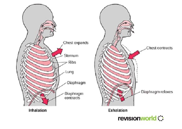
      Breathing

      The Breathing Process - Questions and Answers
      Chest Cavity
      Exchanging Oxygen and Carbon Dioxide
      Control of Breathing
      Defense Mechanisms of the Respiratory System
      Effects of Aging on the Respiratory System

      When we breathe in and out we suck air into them then expel it again.

      Oxygen is absorbed from the lungs into the blood, and carbon dioxide is removed from the blood and breathed out from the lungs. This exchange is vital.

      Respiratory System

      Respiration is the release of energy from glucose or other organic substances. Energy is required for growth, repair, movement and other metabolic activities. There are two main types of respiration, aerobic and anaerobic.

      Alveoli

      Alveoli are the final branchings of the respiratory tree and act as the primary gas exchange units of the lung

      Used for exchanging gases: Deoxygenated enters lungs from body, oxygenated enters capillaries from lungs

      Advantages of alveoli

      •Moist
      •Large surface area
      •Good blood supply

      Aerobic Respiration

      Aerobic respiration takes place in the presence of oxygen. Aerobic respiration = glucose reacts with oxygen to release energy. Carbon dioxide and water are released as waste products.

      Glucose molecules react with oxygen molecules to form carbon dioxide and water molecules, with energy being released by the breaking of bonds in the glucose molecules. Our bodies require energy for the seven life processes This energy is obtained from respiration.

      glucose + oxygen > water + carbon dioxide + energy .

      We use the energy released from respiration for many processes. Respiration also gives off heat, which is used to maintain our high body temperature. Our rate of respiration can be estimated by measuring how much oxygen we use. During exercise, the body needs more energy and so the rate of respiration increases - The breathing rate increases to obtain extra oxygen and remove carbon dioxide from the body. The heart beats faster so that the blood can transport the oxygen and carbon dioxide faster. This is why our pulse rate increases. It is actually the build up of carbon dioxide that makes us breathe faster.

      Glucose comes from our food, oxygen and from breathing

      Water and carbon dioxide are exhaled

      Anaerobic respiration

      Anaerobic respiration occurs when oxygen is not available. When not enough oxygen is available, glucose can be broken down by anaerobic respiration. This may happen during hard exercise.

      Glucose is only partially broken down, and lactic acid is produced - together with a much smaller amount of energy.

      Energy can still be produced without oxygen

      Only a little bit of energy is obtained from respiration

      glucose > lactic acid + energy

      Anaerobic respiration occurs in humans when oxygen is not obtained quick enough (e.g. running fast)

      Only 1/20th energy amount is produced compared to aerobic respiration

      Lactic acid builds up, which causes muscle fatigue due to oxygen debt. This is overcome by deep breathing to oxidise the acid. After the exercise is finished, extra oxygen is needed by the liver to remove the lactic acid.

      Being able to respire without oxygen sounds a great idea. However, there are two problems:
      •Anaerobic respiration releases less than half the energy of that released by aerobic respiration.
      •Anaerobic respiration produces lactic acid. Lactic acid causes muscle fatigue and pain.

    Respiratory System Quiz
    Where in the respiratory system would you find rings of cartilage?

    A. ? nose B. ? diaphragm C. ? alveoli D.YES trachea
    Correct: trachea

    The bronchi are connected to the alveoli via
    A. ? capillaries B. ? air sacs C.YES bronchioles D. ? the trachea Correct: bronchioles

    Which one of the following gases is required for respiration?
    A. ? Methane B. ? Oxygen C. ? Carbon dioxide D. ? Nitrogen Correct: Oxygen

    Where in the respiratory system would you find rings of cartilage?
    0. ? nose0. ? diaphragm0. ? alveoli0.YES trachea The bronchi are connected to the alveoli via0. ? capillaries0. ? air sacs0.YES bronchioles0. ? the trachea Which one of the following gases is required for respiration?0. ? Methane0.YES Oxygen0. ? Carbon dioxide0. ? Nitrogen

    Air that was breathed out by a human was analysed for the gases present. Which of the following is most likely to be the result of the analysis?
    A. ? 78% nitrogen, 16% oxygen, 4% carbon dioxide and 2% water vapour B. ? 78% nitrogen, 21% oxygen, 0.5% carbon dioxide and 0.5% water vapour C. ? 70% nitrogen, 20% oxygen, 0.5% carbon dioxide and 9.5% water vapour D. ? 80% nitrogen, 60% oxygen, 4% carbon dioxide and 6% water vapour Correct:78% nitrogen, 16% oxygen, 4% carbon dioxide and 2% water vapour

    Which of the following is the correct order for air passing through the lungs?
    A. ? trachea, bronchus, bronchiole, alveoli B. ? bronchus, trachea, bronchioles, alveoli C. ? trachea, alveoli, bronchi, bronchioles D. ? bronchus, alveoli, trachea, bronchioles Correct:trachea, bronchus, bronchiole, alveoli

    Air contains nitrogen. What happens to the nitrogen in inhaled air?
    A. ? Its exhaled again without anything happening to it. B. ? It circulates through our bloodstream. C. ? Its absorbed into the digestive system and aids digestion. D. ? Its changed into nitrogen oxide.
    Correct:Its exhaled again without anything happening to it.

    Another name for the breathing system is the
    A. ? digestive system B. ? nervous system C. ? respiratory system D. ? circulatory system Correct:respiratory system

    Which one of the following pairs of items are both necessary for respiration to take place?
    A. ? sunlight and carbon dioxide B. ? glucose and sunlight C. ? glucose and carbon dioxide D. ? glucose and oxygen Correct:glucose and oxygen

    Which one of the following is NOT a disease of the respiratory system?
    A. ? pneumonia B. ? chicken pox C. ? asthma D. ? cancer
    Correct:chicken pox

    Which one of the following labelled parts in the diagram is known as the intercostal muscle?
    A. ? D B. ? C C. ? B D. ? A Correct:C muscle1c_jpg_pagespeed_ce_9G2_LbX0dY.jpg

    Which one of the following protects the lungs?
    A. ? diaphragm B. ? trachea C. ? alveoli D. ? rib cage Correct:rib cage

    Name the parts labelled in the diagram
    bronchial1_gif_pagespeed_ce_VTPr3yC-F5.gif
    A. A = trachea, B = bronchiole B. ? A = alveolus, B = bronchiole C. ? A = trachea, B = bronchus D. ? A = bronchiole, B = trachea
    A = trachea, B = bronchiole
    When would a human body be most likely to respire anaerobically?
    A. ? When eating B. ? When watching TV C. ? When running a marathon D. ? When asleep Correct:When running a marathon

    The function of hair in the nose is
    A. ? to help fight infection B. ? to cause us to sneeze C. ? to keep water out of the lungs D. ? to help gas exchange in the lungs Correct:to help fight infection

    Which one of the following takes place in the lungs?
    A. ? Food is stored B. ? Waste material is filtered from the blood C. ? Food is digested D. ? Gas exchange Correct:Gas exchange

    What happens to your lungs when you breath in?
    A. ? They expand B. ? They absorb carbon dioxide C. ? They manufacture glucose D. ? They push up the diaphragm
    Correct:They expand

    Inhaled air contains
    A. ? less carbon dioxide than exhaled air B. ? more nitrogen than exhaled air C. ? less nitrogen than exhaled air D. ? less oxygen than exhaled air
    Correct: less carbon dioxide than exhaled air

    The part of the respiratory system labelled Y in the diagram is the
    A. ? bronchiole B. ? wind-pipe C. ? alveolus D. ? bronchus
    bronchial2_gif_pagespeed_ce_1q8xofIV2D.gif
    Correct: bronchus

    Which one of the following effects is NOT associated with smoking?
    A. ? Decreased risk of acquiring pneumonia and bronchitis. B. ? Increased risk of developing 'smokers cough'. C. ? Increased risk of lung cancer. D. ? Decreased ability of the blood to transport oxygen.
    Correct: Decreased risk of acquiring pneumonia and bronchitis.

    Name the parts labelled in the diagramA. ? C = trachea, D = alveolus
    B. ? C = alveolus, D = larynx C. ? C = larynx, D = trachea D. ? C = larynx, D = bronchus
    Correct: C = larynx, D = bronchus
    bronchial3_gif_pagespeed_ce_-zNfB5w0gm.gif

    Reproductive System
    The Human Reproductive System
    1. What are the organs that are part of the male genital system?
    The organs that comprise the male genital system are the testicles, the epididymides, the vas deferens, the seminal vesicles, the ejaculatory duct, the prostate, the bulbourethral glands, the urethra and the penis.

    2. Concerning reproduction what is the function of the testicles?
    The testicles are the male gonads, i.e., the organs where the production of gametes takes place. In human beings the gametes are made by meiosis that occur in the testicles.

    3. After passing the epididymides through which structures do sperm cells go until exteriorization?
    After leaving the epididymis in the testicle sperm cells enter the vas deferens, after that they receive secretions from the seminal vesicles and gather (from right and left sides) in the ejaculatory duct that passes inside the prostate. They also get secretions from the prostate and the bulbourethral glands and then go through the urethra, inside the penis, to the exterior.

    4. What is the function of the secretions of the prostate, seminal vesicle and bulbourethral glands in reproduction?
    These secretions along with sperm cells from the testicles form the semen. The secretions have the function of nourishing the sperm cells and serving them as a fluid means of propagation. The basic pH of the seminal fluid also neutralizes the acid secretions of the vagina allowing the survival of sperm cells in the vaginal environment after copulation.

    5. What are the endocrine glands that regulate sexual activity in males? How does this regulation work and what are the involved hormones?
    In males the sexual activity is regulated by the endocrine glands hypophysis (pituitary), adrenals and gonads (testicles).

    The FSH (follicle-stimulating hormone) secreted by the adenohypophysis acts upon the testicles stimulating the spermatogenesis. The LH (luteinizing hormone), another adenohypophyseal hormone, stimulates the production of testosterone by the testicles too. Testosterone, whose production intensifies after the beginning of puberty, acts in several organs of the body and it is responsible for the appearing of the male secondary sex characteristics (beard, body hair, deep voice, increase of the muscle and osseous mass, maturation of genitalia, etc.) Testosterone also stimulates spermatogenesis.

    6. What are the organs that are part of the female reproductive system?
    The organs that constitute the female reproductive system are the ovaries, the Fallopian tubes (or uterine tubes), the uterus, the vagina and the vulva.

    7. In which period of life does the formation of gametes begin in women?
    The meiosis that forms female gametes begins in the cells of the ovarian follicles before birth. After the beginning of puberty, under hormonal stimuli, during each menstrual cycle one of the cells is released on the surface of the ovary and meiosis resumes. The meiotic process is only concluded however if fecundation happens.

    8. What is the organ that releases the female gamete under formation? How is this release triggered? What is the organ that collects the released gametes?
    The organ that liberates the female gamete is the ovary, the female gonad. The releasing of the oocyte is a response to hormonal stimuli. The immature egg cell (still an oocyte) falls into the abdominal cavity and is picked up by the Fallopian tube (uterine tube, or oviduct), a tubular structure that connects the ovary with the uterus.

    9. What are the anatomical relationships between the organs of the female reproductive system from the external vulva to the ovaries?
    The external female genitalia is called the vulva. The vulva is the external opening of the vaginal canal, or vagina. The vagina is the copulation organ of the females and its posterior extremity communicates with the uterus through the uterine cervix. The uterus is divided into two portions: the cervix and the uterine cavity. The lateral walls of the uterine fundus communicate with the Fallopian tubes. The other extremity of each Fallopian tube ends in fimbria forming fringes in the abdominal cavity. Between the uterine tube and the ovary there is still intra-abdominal space.
    v 10. What is the menstrual cycle?
    The menstrual cycle is the periodic succession of interactions between hormones and the organs of the female reproductive system that, after the beginning of puberty, regulates the release of the female gametes and prepares the uterus for fecundation and pregnancy.

    11. What are the endocrine glands involved in the menstrual cycle? What are the hormones in action?
    The endocrine glands that secrete hormones involved in the menstrual cycle are the hypophysis (pituitary) and the ovaries.

    The hormones from adenohypophysis are FSH (follicle-stimulating hormone) and LH (luteinizing hormone) and the hormones from the ovaries are estrogen and progesterone.

    12. What event marks the beginning of the menstrual cycle? What is the blood concentration of FSH, LH, estrogen and progesterone in this phase of the cycle?
    By convention the menstrual cycle begins at the day that menses begins. (Menses is the endometrial hemorrhage excreted through the vaginal canal.) At these days the hormones FSH, LH, estrogens and progesterone are in low concentration.

    13. After menses what is the hormone that influences the maturation of the ovarian follicles?
    The maturation of the ovarian follicles after menses is stimulated by the action of FSH (follicle-stimulating hormone).

    14. What is the hormone secreted by the growing ovarian follicles? What is the action of that hormone upon the uterus?
    The follicles that are growing after menses secrete estrogen. These hormones act upon the uterus stimulating the thickening of the endometrium (the internal mucosa of the uterus).

    15. What is the relationship between the estrogen level and the LH level in the menstrual cycle? What is the function of LH in the menstrual cycle and when does its blood concentration reach a peak?
    The increase in the blood concentration of estrogen with the growing of the ovarian follicle causes the hypophysis to secrete LH. In this phase LH acts together with FSH promoting the maturation of the follicle that at the 14th day ruptures releasing the female gamete (ovulation). After the release of the ovum LH acts stimulating the formation of the corpus luteum, a structure made from the remaining follicular mass. The LH concentration is at maximum at the 14th day of the cycle.

    16. What are the hormones that promote the release of the female gamete from the follicle and at which day of the menstrual cycle does this phenomenon happen? What is this event called?
    The hormones that promote the release of the ovum from the follicle are FSH and LH, hormones found in maximum blood concentration around the 14th day of the cycle. The release of the female gamete from the ovary is called ovulation. Ovulation happens at (around) the 14th day of the menstrual cycle.

    17. How does the female gamete move from the ovary to the uterus?
    The female gamete released from the ovary falls into the surrounding abdominal cavity and is collected by the Fallopian tube. The internal epithelium of the uterine tubes has ciliated cells that move the ovum or the fecundated egg cell towards the uterus.

    18. How long after ovulation must fecundation occur to be effective?
    If fecundation does not occur approximately 24 hours after ovulation the released ovum often dies.

    19. What is the structure into which the follicle is transformed after ovulation? What is the importance of that structure in the menstrual cycle?
    The follicle that released the ovum suffers the action of LH and is transformed into the corpus luteum. The corpus luteum is very important because it secretes estrogen and progesterone.

    These hormones prepare the uterine mucosa, also known as endometrium, for nidation (implantation of the zygote in the uterine wall) and embryonic development since they stimulate the thickening of the mucous tissue, increase its vascularity and make the appearing of uterine glycogen-producing glands.

    20. What is the importance of the uterine glycogen-producing glands?
    The uterine glands produce glycogen that can be degraded into glucose to nourish the embryo before the complete development of the placenta.

    21. How does the hypophysis-corpus luteum negative feedback work? What is the name given to the atrophied corpus luteum after this feedback process?
    After ovulation the estrogen and progesterone secretions from the corpus luteum inhibit the hypophyseal FSH and LH secretions (this happens by inhibition of GnRH, gonadotropin-releasing hormone, a hypothalamic hormone). The blood concentration of these adenohypophyseal hormones falls to basal levels again. As LH lowers the corpus luteum (luteum means “yellow”) becomes atrophic and turns into the corpus albicans (“white”). With the regression of the corpus luteum the production of estrogen and progesterone ceases.

    22. In hormonal terms why does menses occur?
    Menses is the endometrial monthly desquamation that occurs as the estrogen and progesterone levels fall after the regression of the corpus luteum because these hormones, mainly progesterone, can no longer support and maintain the thickening of the endometrium.

    23. What is the explanation for the bleeding that accompanies menses?
    The hemorrhage that accompanies menses occurs because the endometrium is a richly vascularized tissue. The rupture of blood vessels of the uterine mucosa during the menstrual desquamation causes the bleeding.

    24. Which are the phases of the menstrual cycle?

    The menstrual cycle is divided into two main phases: the follicular (or menstrual) phase and the luteal (or secretory) phase.
    The menstrual phase begins at the first day of menses and lasts until ovulation (around the 14th day). The luteal phase begins after ovulation and ends when menses begins (around the 28th day).

    25. Including main events and hormonal changes how can the menstrual cycle be described?
    One can imagine a cycle like an analog clock at which at 0 o’clock is the beginning and the end of the menstrual cycle and that 6 o’clock corresponds to the 14h day of the cycle.

    At 0 o’clock the menses and so the menstrual cycle begins and FSH blood level begins to increase. Around 2 o’clock the maturing follicles under FSH action are already secreting estrogen and the endometrium is thickening. Around 3 o’clock estrogen is intensely stimulating the increase of LH blood level. At 6 o’clock (the 14th day) LH is at its maximum concentration and FSH also at high levels to promote ovulation, LH then stimulates the formation of the corpus luteum. Around 7 o’clock the corpus luteum is already secreting a great amount of estrogen and progesterone and the endometrium thickens even more, concomitant lowering of FSH and LH occurs with the increasing of the ovarian hormones. Around 11 o’clock the reduced LH and FSH levels make the corpus luteum turn into the corpus albicans, the production of estrogen and progesterone ceases and the endometrium regresses. At 0 o’clock again (28th day) the endometrium desquamates and a new menstrual cycle begins.

    26. In general what is the phase of the menstrual cycle when copulation may lead to fecundation?
    Although this is not a rule, to be effective fecundation in general must occur within about 24 hours after ovulation (that occurs around the 14th day of the menstrual cycle). Fecundation may occur even if copulation took place up to 3 days before ovulation since the male gametes remain viable for about 72 hours within the female reproductive system.

    The fertile period of the women however is considered the period from 7 days before ovulation to 7 days after ovulation.

    27. What is the part of the female reproductive system where fecundation occurs?
    Fecundation generally occurs in the Fallopian tubes but it can also take place within the uterus. There are cases when fecundation may occur even before the ovum enters the uterine tube, a fact that may lead to a severe medical condition known as abdominal pregnancy.

    28. How does the sexual arousal mechanism in women facilitate fecundation?
    During sexual arousal in women the vagina secretes substances to neutralize its acidity thus allowing the survival of sperm cells within it. During the female fertile period hormones make the mucus that covers the internal surface of the uterus less viscous to help the passage of sperm cells to the uterine tubes. During copulation the uterine cervix advances inside the vagina to facilitate the entering of male gametes through the cervical canal.

    29. What is nidation? In which phase of the menstrual cycle does nidation occur?
    Nidation is the implantantion of the embryo in the uterus. Nidation occurs around the 7th day after fecundation, i.e., 7 to 8 days after ovulation (obviously, it occurs only if fecundation also occurs). Since it occurs in the luteal phase the progesterone level is high and the endometrium is in its best condition to receive the embryo.

    30. What is tubal pregnancy?
    Many times fecundation takes place in the Fallopian tubes. Generally the newly formed zygote is taken to the uterus where nidation and the embryonic development occur. In some cases however the zygote cannot go down to the uterus and the embryo implants itself in the uterine tube tissue, characterizing the tubal pregnancy. Tubal pregnancy is a severe clinical condition since often the tube ruptures during gestation causing hemorrhage and even death of the woman. The most common treatment for tubal pregnancy has been surgery.

    31. How do hormonal tests to detect pregnancy work?
    Laboratory tests to detect pregnancy commonly test for human chorionic gonadotropin (HCG) concentration in blood or urine samples. If the level of this hormone is abnormally high, pregnancy is likely.

    32. Does the hypophysis-ovaries endocrine axis work in the same way during pregnancy as in non-pregnant women? If pregnancy does not occur how does another menstrual cycle begin?
    The functioning of the hypophysis is altered during pregnancy. Since estrogen and progesterone levels remain elevated during the gestational period the production of GnRH (gonadotropin-releasing hormone) from the hypothalamus is inhibited. The lack of GnRH thus inhibits the secretion of FSH and LH from the hypophysis and a new menstrual cycle does not begin.

    If pregnancy does not occur the lowering of estrogen and progesterone levels stimulates the production of GnRH by the hypothalamus. This hormone then hastens the adenohypophyseal secretion of FHS and LH that in their turn stimulate the maturation of follicles and the beginning of a new menstrual cycle.

    33. What is the endocrine function of the placenta?
    The placenta besides being the organ through which the exchange of substances between the mother and the fetus is done also has the function of secreting estrogen and progesterone to keep a high level of these hormones during pregnancy. (The placenta still secretes other hormones like human placental lactogen, that act similarly to the hypophyseal hormones that regulate reproduction, and HCG, human chorionic gonadotropin.)

    34. How do contraceptive pills generally work?
    Contraceptive pills generally contain the hormones estrogen and progesterone. If taken daily from the 4th day after menses the abnormal elevation of these hormones acts upon the hypophysis-hypothalamus endocrine axis inhibiting the FSH and LH secretions. Since these hormones then do not reach their normal high levels during the menstrual cycle ovulation does not occur.
    (Treatment with contraceptive pills must be initiated under medical supervision.)

    35. What are the common contraindications of the contraceptive pills?
    There are medical reports associating the use of contraceptive pills with vomiting, nausea, vertigo, headaches, hypertension and other pathological conditions. Some research has attempted to relate the medical ingestion of estrogen and progesterone with increased propensity to cardiovascular diseases (like infarction, strokes and thrombosis) and to malignant neoplasias (cancers). Doctors must always be asked about the risks and benefits of the contraceptive pill prior to use.

    36. What are the most common methods of male and female surgical sterilization?
    Vasectomy is the most common method of surgical sterilization in men. In vasectomy the vas deferens inside the scrotum are sectioned and closed at a section which will forbid the sperm cells to follow to the ejaculatory duct but still allowing the release of seminal fluid during ejaculation.

    Surgical sterilization of women is often done by bilateral tubal ligation. With tubal ligation the ovum does not pass to the uterus so the sperm cells cannot reach it.

    37. How does the contraceptive diaphragm work? What are the limitations of this contraceptive method?
    The contraceptive diaphragm is an artifact made of latex or plastic that when placed on the vaginal fundus covers the uterine cervix forbidding the passage of sperm cells through the cervical canal. To be more effective the diaphragm needs to be used together with spermicide. This method however does not prevent sexually transmitted diseases (STDs).

    38. Why is the use of condoms not just a contraceptive method but also a health protection behavior?
    The use of condoms besides being an efficient contraceptive method also helps the prevention of diseases caused by sexually transmitted agents (STDs), like syphilis, gonorrhea, HPV (human papilloma virus that may lead to genital cancers) infestation, HIV infection, etc.

    39. What is the normal duration of the menstrual cycle? How does the calendar contraceptive method work?
    The normal duration of the menstrual cycle is 28 days but it can vary among different women or in different cycles of the same woman.

    In the calendar contraceptive method the date n-14 (n minus 14) is taken considering n the number of days of the normal menstrual cycle of the woman (generally n=28). The safety margin +3 or –3 refers to the days around n-14 that intercourse should be avoided to prevent pregnancy. (This method is not exempt from failures. A doctor must always be consulted before relying on any contraceptive method.)

    40. How is the ovulation date estimated with the control of the woman's body temperature?
    One method to estimate the exact ovulation day is daily control of the body temperature taken always under same conditions. At the ovulation day the body temperature often increases about 0.5 degrees centigrade.

    41. What is the contraceptive mechanism of the IUD?
    The IUD (intrauterine device) is a piece of plastic coated with copper that is inserted within the uterus by a doctor. Copper is then gradually released (IUD may last 5 to 10 years) and since it has a spermicidal action sperm cells are destroyed before fecundation. Besides this mechanism the movement of the IUD inside the uterus causes slight endometrial inflammation that helps to prevent nidation.

    42. Generally how does a male animal realize that the female is receptive to copulation?
    In most vertebrate species with internal fecundation the females have reproductive cycles with fertile periods. During this period the female secretes pheromones (odoriferous substances that attract the male of the species) from the skin and mucosae. The presence of the male individual and his pheromones also stimulates the release of pheromones by the female. (Many animals also use pheromones for territorial demarcation and for signal transmission between individuals about the location of dangers and food.)

    43. What is parthenogenesis?
    Parthenogenesis is the reproduction or formation of a new individual from the egg cell but without fecundation by the male gamete. According to the species, individuals born by parthenogenesis may be male or female, or of any sex.

    In bees the drone (the single male bee) is haploid and born by parthenogenesis while the females (queen and workers) are diploid.

    Here are further guidelines.
    Reproductive System

    Skeletal System
    How many total human skeletal bones are there?
    206 bones on both side of the body

    What are various examples?
    Arms (2)
    Cranial (8)
    Facial (14)
    Feet (52)
    Forearms (4)
    Hands (54)
    Legs (6)
    Middle ears (6)
    Pelvis (4)
    Shoulder girdle (4)
    Thighs (2)
    Thorax (25)
    Throat (1)
    Vertebral column (24)
    www.qureshiuniversity.com/listofbonesofthehumanskeleton.html

    Why is it essential to know about total human skeletal bones?
    Any human bone can have a fracture or injury.

    How many total adult teeth are there?
    32

    What is the Skeletal System?
    How does the Skeletal System help us?
    Who has more bones a baby or and adult?
    Are bones alive?
    What is a bone made of?
    How do bones break and heal?
    What is an X-ray?
    What's up with joints?
    Does each bone have a name?
    How do I keep my bones healthy?
    Are my teeth counted as bones?

    Here are further guidelines.
    Skeletal System
    The Musculoskeletal System Learn Cartilages, Bones and Muscles

    1. Which are the organs that are part of the musculoskeletal system?
    The main organs and tissues that are part of the musculoskeletal system in humans are the cartilages, the bones and the muscles.

    2. What are the functions of the musculoskeletal system?
    The musculoskeletal system has the functions of supporting and protecting organs, maintenance of the body spatial conformation, motion of organs, limbs and bodily portions and nutrient storage (glycogen in muscles, calcium and phosphorus in bones).

    3. Which type of tissue are the cartilaginous and the osseous tissue?
    The cartilaginous and the osseous tissues are considered connective tissues since they are tissues in which the cells are relatively distant from others with a great amount of extracellular matrix in the interstitial space.

    4. What are the cells that form the cartilaginous tissue?
    The main cells of the cartilages are the chondrocytes, originated from the chondroblasts that secrete the intersticial matrix. There are also chondroclasts, cells with many lisosomes and responsible for the digestion and remodelation of the cartilaginous matrix.

    5. What is the constitution of the cartilaginous matrix?
    The cartilaginous matrix is made of collagen fibers, mainly collagen type II, and of proteoglycans, proteins associated to glycosaminoglycans, chiefly hyaluronic acid. The proteoglycans provide the typical rigidity of the cartilages.

    6. What are some functions of the cartilages in the human body?
    Cartilages are responsible for the structural support of the nose and ears. The trachea and the bronchi are also organs with cartilaginous structures that prevent the closing of these tubes. In joints there are cartilages that cover the bones providing a smooth surface to reduce the friction of the joint movement. In the formation of bones the cartilages act as a mold and they are gradually substituted by the osseous tissue.

    7. What are the three main cell types that form the osseous tissue? What are their functions?
    The three main cell types of the osseous tissue are the osteoblasts, the osteocytes and the osteoclasts.

    Osteoblasts are known as bone-forming cells since they are the cells that secrete the proteinaceous part of the bone matrix (collagen, glycoproteins and proteoglycans). The bone matrix is the intercellular space where the mineral substances of the bones are deposited.

    Osteocytes are differentiated mature osteoblasts formed after these cells are completely surrounded by the bone matrix. Osteocytes have the function of supporting the tissue.

    Osteoclasts are the giant multinucleate cells that remodelate the osseous tissue. They are originated from monocytes and they contain many lisosomes. Osteoblasts secrete enzymes that digest the osseous matrix creating canals throughout the tissue.

    8. What is the bone matrix? What are its main components?
    Bone matrix is the content that fills the intercellular space of the osseous tissue. The bone matrix is made of mineral substances (about 5%), mainly phosphorus and calcium salts, and organic substances (95%), mainly collagen, glycoproteins and proteoglycans.

    9. What are the Haversian canals and the Volkmann’s canals of the bones? Is the osseous tissue vascularized?
    The Haversian canals are longitudinal canals present in the osseous tissue within which blood vessels and nerves pass. The osseous tissue distributes itself in a concentric manner around these canals. The Volkmann’s canals are communications between the Harvesian canals.
    The osseous tissue is highly vascularized in its interior.

    10. What are the functions of the osseous tissue?
    The main functions of the osseous tissue are: to provide structural rigidity to the body and to delineate the spatial positioning of the other tissues and organs; to support the body weight; to serve as a site for mineral storage, mainly of calcium and phosphorus; to form protective structures for important organs like the brain, the spinal cord, the heart and the lungs; to work as a lever and support for the muscles, providing movement; to contain the bone marrow where hematopoiesis occurs.

    11. What are the flat bones and the long bones?
    The main bones of the body may be classified as flat or long bones (there are bones not classified into these categories). Examples of flat bones are the skull, the ribs, the hipbones, the scapulae and the sternum. Examples of long bones are the humerus, the radius, the ulna, the femur, the tibia and the fibula.

    12. What are the types of muscle tissues? What are the morphological features that differentiate those types?
    There are three types of muscle tissue: the skeletal striated muscle tissue, the cardiac striated muscle tissue and the smooth muscle tissue.

    The striated muscles present under microscopic view transversal stripes and their fibers (cells) are multinucleate (in the skeletal) or may have more than one nucleus (in the cardiac). The smooth muscle does not present transversal stripes and it has spindle-shaped fibers each with only one nucleus.

    13. Which is the type of muscle tissue that moves the bones?
    The bones are moved by the skeletal striated muscles. These muscles are voluntary (controlled by volition).

    14. Which is the type of muscle tissue that contracts and relaxes the heart chambers?
    The myocardium of the heart is made of cardiac striated muscle tissue.

    15. Which is the type of muscle tissue that performs the peristaltic movements of the intestines?
    The smooth muscle tissue is responsible for the peristaltic movements of the intestines. The smooth muscles are not controlled by volition.

    16. Which is the type of muscle tissue that helps to push the food down through the esophagus?
    The esophageal wall in its superior portion is made of skeletal striated muscle. The inferior portion is made of smooth muscle. In the intermediate portion there are skeletal striated and smooth muscles. All of these muscles are important to push the food down towards the stomach.

    17. How is the striped pattern of the striated muscle cells formed?
    The functional units of the muscle fibers are the sarcomeres. Within the sarcomeres blocks of actin and myosin molecules are posed in organized manner. The sarcomeres align in sequence forming myofibrils that are longitudinally placed in the cytoplasm of the muscle fibers (cells). The grouping of consecutive blocks of actin and myosin in parallel filaments creates the striped pattern of the striated muscle tissue seen under the microscope.

    18. What are sarcomeres?
    Sarcomeres are the contractile units of the muscle tissue formed of alternating actin blocks (thin filaments) and myosin blocks (thick filaments). Several sarcomeres placed in linear sequence form a myofibril. Therefore one muscle fiber (cell) has many myofibrils made of sacomeres.

    The compartments where myofibrils are inserted are delimited by an excitable membrane known as sarcolemma. The sarcolemma is the plasma membrane of the muscle cell.

    19. What are the main proteins that constitute the sarcomere? What is the function of those molecules in the muscle cells?
    In the sarcomere there are organized actin and myosin blocks. Troponin and tropomyosin also appear associated to actin.

    The actin molecules when activated by calcium ions liberated in the proximities of the sarcomere are pulled by myosin molecules. This interaction between actin and myosin shortens the myofibrils originating the phenomenon of muscle contraction.

    20. What are the positions of actin and myosin molecules in the sarcomere before and during the muscle contraction?
    Schematically actin filaments attached perpendicularly to both sarcomere extremities (longitudinal sides) make contact with myosin filaments positioned in the middle of the sarcomere and in parallel to the actin filaments.

    Before the contraction the sarcomeres are extended (relaxed) since the contact between actin and myosin filaments is only made by their extremities. During contraction actin filaments slide along the myosin filaments and the sarcomeres shorten.

    21. How do calcium ions participate in muscle contraction? Why do both muscle contraction and muscle relaxation spend energy?
    In the muscle cells calcium ions are stored within the sarcoplasmic reticulum. When a motor neuron emits stimulus for the muscle contraction neurotransmitters called acetylcholine are released in the neuromuscular junction and the sarcolemma is excited. The excitation is conduced to the sarcoplasmic reticulum that then realeases calcium ions into the sarcomeres.

    In the sarcomeres the calcium ions bind to troponin molecules associated to actin activating myosin binding sites of actin. The myosin, then able to bind to actin, pulls this protein and the sarcomere shortens. The summation of simultaneous contraction of sarcomeres and myofibrils constitutes the muscle contraction. During muscle relaxation the calcium ions return back to the sarcoplasmic reticulum.

    For myosin to bind to actin, and thus for the contraction to occur, hydrolysis of one ATP molecule is necessary. During relaxation the return of calcium ions to the sarcoplasmic reticulum is an active process that spends ATP too. So both muscle contraction and relaxation are energy-spending processes.

    22. What is myoglobin? What is the function of this molecule in the muscle tissue?
    Myoglobin is a pigment similar to hemoglobin and present in muscle fibers. Myoglobin has a great affinity for oxygen. It keeps oxygen bound and releases the gas under strenuous muscle work. So myoglobin acts as an oxygen reserve for the muscle cell.

    23. How does phosphocreatine act in the muscle contraction and relaxation?
    Phosphocreatine is the main means of energy storage of the muscle cells.

    During relaxed periods ATP molecules made by the aerobic cellular respiration transfer highly energized phosphate groups to creatine forming phosphocreatine. In exercise periods phosphocreatine and ADP resynthesize ATP to dispose energy for the muscle contraction.

    24. What happens when the oxygen supply is insufficient to maintain aerobic cellular respiration during muscle exercise?
    If oxygen from hemoglobin or myoglobin is not enough for the energy supply of the muscle cell the cell then begins to do lactic fermentation in an attempt to compensate the deficiency.

    The lactic fermentation releases lactic acid and this substance causes muscle fatigue and predisposes the muscles to cramps.

    25. What is the neurotransmitter of the neuromuscular junction? How does the nervous system trigger muscle contraction?
    The nervous cells that trigger the muscle contraction are the motor neurons. The neurotransmitter of the motor neurons is acetylcholine. When a motor neuron is excited the depolarizing current flows along the membrane of its axon until reaching the synapse at the neuromuscular junction (the neural impulse passage zone between the axon extremity and the sarcolemma). Near the axonal extremity the depolarization allows the entrance of calcium ions into the axon (note that calcium also has a relevant role here). The calcium ions stimulate the neuron to release acetylcholine in the synapse.

    Acetylcholine then binds to special receptors in the outer surface of the sarcolemma, the permeability of this membrane is altered and an action potential is created. The depolarization is then conduced along the sarcolemma to the sarcoplasmic reticulum that thus releases calcium ions for the sarcomere contraction.

    26. To increase the strength of the muscle work is the muscle contraction intensely increased?
    An increase in the strength of the muscle work is not achieved by increase in the intensity of the stimulation of each muscle fiber. The muscle fiber obeys an all-or-nothing rule, i.e., its contraction strength is only one and cannot be increased.

    When the body needs to increase the strength of the muscle work a phenomenon known as spatial summation occurs: new muscle fibers are recruited in addition to the fibers already in action. So the strength of the muscle contraction increases only when the number of active muscle cells increases.

    27. What is the difference between spatial summation and temporal summation of muscle fibers? What is tetany?
    Spatial summation is the recruiting of new muscle fibers to increase the muscle strength. Temporal summation occurs when a muscle fiber is continuously stimulated to contract without being able to conclude relaxation.

    The permanence of a muscle fiber under a continuous state of contraction by temporal summation is known as tetany (e.g., the clinical condition of patients contaminated by the toxin of the tetanus bacteria). Tetany ends when all available energy for contraction is spent or when the stimulus ceases.

    Urinary system
    A List of Kidney Diseases
    Human Biology: Excretory System
    Human Biology: Excretory System

    The Excretory System The Excretory System - Get the Knowledge

    1. Which are the organs of the excretory system?
    The excretory system is formed of kidneys (two), ureters (two), bladder and urethra.

    2. What are nitrogen wastes?
    Nitrogen wastes are residuals derived from the degradation of proteins. They are made from chemical transformation of the amine group of amino acid molecules.

    3. What are the three main types of nitrogen wastes excreted by living beings?
    The main nitrogen wastes excreted by living beings are ammonia, uric acid and urea. Living beings that secrete ammonia are known as ammoniotelic. Creatures that secrete uric acid are known as uricotelic. Organisms that secrete urea are called ureotelic.

    4. Why are most ammoniotelic beings aquatic animals?
    Aquatic animals, like crustaceans, bony fishes and amphibian larvae, generally are ammoniotelic since ammonia diffuses more easily through membranes and it is more water-soluble than the other nitrogen wastes. Ammonia is still the most energetically economical nitrogen waste to be synthesized.

    5. Why after the passage of animals from the aquatic to the terrestrial habitat does the abandonment of the ammoniotelic excretion occur?
    Ammonia is a highly toxic molecule if not diluted and quickly excreted out of the body. For this reason the ammoniotelic excretion was abandoned in terrestrial habitats because the availability of water for dilution is reduced in this medium and wastes cannot be excreted so promptly to the exterior.

    6. Comparing toxicity and the need for dilution in water how different are the ureotelic and the uricotelic excretions? What are some examples of animals that present these respective types of excretion?
    Urea is more water-soluble than uric acid (an almost insoluble substance). Urea is also more toxic. Both however are less toxic than ammonia.

    Some invertebrates, chondrichthian fishes, adult amphibians and mammals are ureotelic. Reptiles, birds and most arthropods are uricotelic.

    7. What is the nitrogen waste in amphibian larvae and in the adult animal?
    Since amphibian larvae are aquatic they excrete ammonia. The terrestrial adult excretes urea.

    8. Why is the uricotelic excretion essential for avian and reptile embryos?
    In reptiles and birds the excretory system is uricotelic since uric acid is insoluble, less toxic and suitable to be stored within the eggs where their embryos develop.

    9. How do embryos of placental mammals excrete nitrogen wastes?
    Placental animals, including embryos, excrete urea. In the adult placental mammal urea is excreted through the urine. In embryos the molecule passes to the mother’s blood through the placenta and it is excreted in the mother’s urine.

    10. What is the main nitrogen waste of humans?
    Human beings excrete mainly urea eliminated with the urine.

    11. How is urea formed in the human body?
    Urea is a product of the degradation of amino acids. In the process amino acids lose their amine group which is then transformed into ammonia. In the liver ammonia reacts with carbon dioxide to form urea and water, a process called ureogenesis.

    In the intermediary reactions of the ureogenesis a molecule of ornithine is consumed and another is produced. For this reason ureogenesis is also known as the ornithine cycle.

    12. What is excretion?
    Excretion in Physiology is the process of elimination of metabolic wastes and other toxic substances from the body.

    13. What are the vessels that carry blood to the kidneys? Is this blood arterial or venous?
    The arterial vessels that carry blood to be filtrated by the kidneys are the renal arteries. The renal arteries are ramifications of the aorta and so the blood filtered by the kidneys is arterial (oxygen-rich) blood.

    14. Which are the vessels that drain filtered blood from the kidneys?
    The venous vessels that collect the blood filtered by the kidneys are the renal veins. The renal veins carry the blood that has been reabsorbed in the nephron tubules.

    15. What is the functional unity of the kidneys?
    The functional (filtering) unity of the kidneys is the nephron. A nephron is made of afferent arteriole, efferent arteriole, glomerulus, Bowman's capsule, proximal tubule, loop of Henle, distal tubule and collecting duct.

    In each kidney there are about one million nephrons.

    16. What are the three main renal processes that combined produce urine?
    Urine is made by the occurrence of three processes in the nephron: glomerular filtration, tubular resorption and tubular secretion.

    In the nephron the blood carried by the afferent arteriole enters the glomerular capillary network where it is filtered. The filtration implies that part of the blood returns to the circulation through the efferent arteriole and the other part, known as the glomerular filtrate, enters the proximal tubule of the nephron. In the nephron tubules (also known as convoluted tubules) substances of the glomerular filtrate like water, ions and small organic molecules are resorbed by the cells of the tubule wall and gain again the circulation. These cells also secrete other substances inside the tubules. The urine is formed of not resorbed filtered substances and of secreted (by the tubules) substances. Urine is drained by the collecting ducts to the ureter of each kidney, then it enters the bladder and later it is discharged through the urethra.

    The nephron tubules are surrounded by an extensive capillary network that collects resorbed substances and provides others to be secreted.

    17. What is the main transformation presented by the glomerular filtrate in comparison to the blood?
    Glomerular filtrate is the name given to the plasma after it has passed the glomerulus and entered the Bowman’s capsule. The glomerular filtrate has a different composition compared to urine since the fluid has not yet undergone tubular resorption and secretion.

    The main difference between the blood and the glomerular filtrate is that in the latter the amount of proteins is at a minimum and there are no cells or blood platelets.

    18. What is proteinuria? Why is proteinuria a sign of glomerular renal injury?
    Proteinuria means losing of proteins through urine. Under normal conditions proteins are too big to be filtered by the glomerulus and they are practically absent in the urine (the few filtered proteins may also be resorbed in the nephron tubules). Proteinuria is an indication that a more than expected amount of proteins is passing the glomerulus suggesting glomerular disease, e.g., in diabetic nephropathy.

    The glomerulus also blocks the passage of blood cells and platelets (hematuria is often a sign of urinary disease although less specific of kidneys since the blood may come from the lower parts of the excretory tract).

    19. Where does most of the water resorbed after glomerular filtration go? What are the other substances resorbed by the nephron tubules?
    Only 0.5 to 1% of the glomerular filtrate is eliminated as urine. The remaining volume, containing mainly metabolic ions, glucose, amino acids and water, is resorbed through the nephron tubules (by means of active or passive transport) and gains the blood circulation again.

    The convolute tubules of the nephron are responsible for the resorption of substances.

    The Excretory System - Image Diversity: nephron tubules

    20. Why do cells of the nephron tubules present a great amount of mitochondria?
    The cells of the tubule wall have high number of mitochondria because many substances are resorbed or secreted through them by means of active transport (a process that spends energy). Therefore many mitochondria are necessary for the energetic supply (ATP supply) of this type of transport.

    21. What is tubular secretion? What are some examples of substances secreted through the renal tubules?
    Tubular secretion is the passage of substances from the blood capillaries that surround the nephron tubules to the tubular lumen for these substances to be excreted with urine. Ammonia, uric acid, potassium, bicarbonate and hydrogen ions, metabolic acids and bases, various ingested drugs (medicines) and other substances are secreted by the nephron tubules.

    22. In which nephron portion does the regulation of acidity and alkalinity of the plasma occur?
    The regulation of the acid-basic equilibrium of the body is done by the kidneys and depends upon the tubular resorption and secretion.

    23. How do kidneys participate in the regulation of the acid-basic equilibrium of the body? How are alkalosis and acidosis respectively corrected by the kidneys?
    Kidneys can regulate the acidity or alkalinity of the plasma varying the excretion of hydrogen and bicarbonate ions.

    In alkalosis (abnormally high level of the plasma pH) the kidneys excrete more bicarbonate and the equilibrium of formation of bicarbonate from water and carbon dioxide shifts towards formation of more hydrogen ions and bicarbonate and then the plasma pH is lowered. When the body undergoes acidosis (abnormal low level of the plasma pH) the kidneys excrete more hydrogen ions and retain more bicarbonate thus the equilibrium of formation of bicarbonate from water and carbon dioxide shifts towards more hydrogen consumption and the plasma pH is increased.

    24. How do kidneys participate in the blood volume control? How is the blood volume of the body related to the arterial pressure?
    The kidneys and the hormones that act upon them are the main physiological regulators of the total blood volume of the body. As more water is resorbed in the nephron tubules the more the blood volume increases; as more water is excreted in urine the more the blood volume lowers.

    The blood volume in its turn has a direct relation to blood pressure. The blood pressure increases when the blood volume increases and it lowers when the blood volume lowers. That is the reason why one of the main groups of antihypertensive drugs is the diuretics. Doctors often prescribe diuretics for the hypertensive patients to excrete more water and thus lower their blood pressure.

    25. Which are the three hormones that participate in the regulation of the renal function?
    Antidiuretic hormone (or ADH, or vasopressin), aldosterone and atrial natriuretic factor (or ANF) are hormones that participate in the regulation of the excretory system.

    26. What is the function of the antidiuretic hormone? Where is it made and which are the stimuli that increase or reduce its secretion?
    The antidiuretic hormone is secreted by the hypophysis (also known as pituitary) and it acts in the nephron tubules increasing the resorption of water. When the body needs to retain water, for example, in cases of blood loss and abrupt blood pressure lowering or in cases of abnormally high blood osmolarity, there is stimulus for ADH secretion.

    When the body has an excess of water, as in cases of excessive ingestion or in abnormally low blood osmolarity, the secretion of ADH is blocked and the diuresis increases. ADH is also known as vasopressin since it increases the blood volume and thus heightens the blood pressure.

    27. Why does the ingestion of alcohol increase diuresis?
    Alcohol inhibits the secretion of ADH (antidiuretic hormone) by the pituitary. That is why when it is drunk to excess the person urinates too much.

    28. How does aldosterone act and where is it produced?
    Aldosterone is a hormone that acts upon the nephron tubules stimulating the resorption of sodium. Therefore it contributes to the increase of the blood osmolarity and consequently to the increase of the blood pressure.

    Aldosterone is made by the adrenals, glands located over the superior portion of the kidneys.

    29. What is an evolutionary explanatory hypothesis for the secretion by the heart of a hormone that regulates the renal function? Which is that hormone?
    The renal regulator hormone secreted by the heart is the atrial natriuretic factor (or ANF). The ANF increases the excretion of sodium in the nephron tubules causing less resorption of water, more urinary volume, and thus lowering the blood pressure. The atrial natriuretic factor is secreted when there is an increase of the length of the heart muscle fibers in response to high blood pressure. The ANF is a natural antihypertensive substance. Since the health of the heart depends largely upon the stability of the normal blood pressure the evolution should have preserved the atrial natriuretic factor to allow information from the heart to be an additional mechanism for the renal control of the blood pressure.

    30. What is hemodialysis?
    Hemodialysis is the artificial blood filtration made by specific machines in substitution of the kidneys. Hemodialysis may be necessary in patients suffering from diseases that cause renal failure, like diabetic renal complications, lupic renal complications and others. During hemodialysis the blood of the patient is deviated to the filtering machine and after the filtration it returns to the body.

    Hemodialysis is generally done two, three or more times a week in a process that takes several hours. Sometimes kidney transplantation is an alternative to hemodialysis.
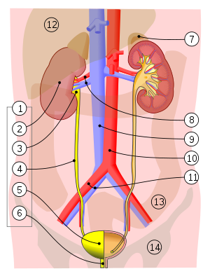

    Urinary System

    1. Human urinary system:
    2. Kidney,
    3. Renal pelvis,
    4. Ureter,
    5. Urinary bladder,
    6. Urethra. (Left side with frontal section)
    7. Adrenal gland Vessels:
    8. Renal artery and vein,
    9. Inferior vena cava,
    10. Abdominal aorta,
    11. Common iliac artery and vein
    With transparency:
    12. Liver,
    13. Large intestine,
    14. Pelvis
    The order of impurities being excreted from the kidneys: Kidneys ? Ureters ? Urinary Bladder ? Urethra

    Lower urinary tract symptoms

    Filling or irritative symptoms

    Increased frequency of urination
    Increased urgency of urination
    Painful urination
    Excessive passage of urine at night

    Voiding or obstructive symptoms

    Poor stream
    Hesitancy
    Terminal dribbling
    Incomplete voiding
    Overflow incontinence (occurs in chronic retention)

    Causes

    Benign prostatic hyperplasia (BPH) with obstruction
    Detrusor muscle weakness and/or instability
    Urinary Tract Infection (UTI)
    Chronic prostatitis
    Urinary stone
    Malignancy: prostate or bladder
    Neurological disease, e.g. multiple sclerosis, spinal cord injury, cauda equina syndrome

    Symptoms of Chronic Kidney Disease
    Most people do not have any severe symptoms until their kidney disease gets worse. However you may:

    Have muscle cramps at night
    Have swollen feet and ankles
    Have puffiness around your eyes, especially in the morning
    Feel more tired
    Have less energy
    Have trouble thinking clearly
    Have a poor appetite
    Have trouble sleeping
    Have dry, itchy skin
    Need to urinate more often, especially at night.


    A List of Kidney Diseases
    1. Abderhalden–Kaufmann–Lignac syndrome (Nephropathic Cystinosis)

    2. Acute Kidney Failure/Acute Kidney Injury

    3. Acute Lobar Nephronia

    4. Acute Phosphate Nephropathy

    5. Acute Tubular Necrosis

    6. Adenine Phosphoribosyltransferase Deficiency

    7. Adenovirus Nephritis

    8. Alport Syndrome

    9. Amyloidosis

    10. ANCA Vasculitis Related to Endocarditis and Other Infections

    11. Angiomyolipoma

    12. Analgesic Nephropathy

    13. Anorexia Nervosa and Kidney Disease

    14. Angiotensin Antibodies and Focal Segmental Glomerulosclerosis

    15. Antiphospholipid Syndrome

    16. Anti-TNF-a Therapy-related Glomerulonephritis

    17. APOL1 Mutations

    18. Apparent Mneralocorticoid Excess Syndrome

    19. Aristolochic Acid Nephropathy, Chinese Herbal Nephropathy, Balkan Endemic Nephropathy

    20. Bartter Syndrome

    21. Beeturia

    22. ß-Thalassemia Renal Disease

    23. Bile Cast Nephropathy

    24. BK Polyoma Virus Nephropathy in the Native Kidney

    25. Bladder Rupture

    26. Bladder Sphincter Dyssynergia

    27. Bladder Tamponade

    28. Border-Crossers' Nephropathy

    29. Bourbon Virus and Acute Kidney Injury

    30. Burnt Sugarcane Harvesting and Acute Renal Dysfunction

    31. Byetta and Renal Failure

    32. C1q Nephropathy

    33. Cannabinoid Hyperemesis Acute Renal Failure

    34. Cardiorenal syndrome

    35. Carfilzomib-Indiced Renal Injury

    36. CFHR5 nephropathy

    37. Cherry Concentrate and Acute Kidney Injury

    38. Cholesterol Emboli

    39. Churg–Strauss syndrome

    40. Chyluria

    41. Colistin Nephrotoxicity

    42. Collapsing Glomerulopathy, Collapsing Glomerulopathy Related to CMV

    43. Congenital Nephrotic Syndrome

    44. Conorenal syndrome (Mainzer-Saldino Syndrome or Saldino-Mainzer Disease)

    45. Contrast Nephropathy

    46. Copper Sulpfate Intoxication

    47. Cortical Necrosis

    48. Crizotinib-related Acute Kidney Injury

    49. Cryoglobuinemia

    50. Crystalglobulin-Induced Nephropathy

    51. Crystal-Induced Acute Kidney injury

    52. Cystic Kidney Disease, Acquired

    53. Cystinuria

    54. Dasatinib-Induced Nephrotic-Range Proteinuria

    55. Dense Deposit Disease (MPGN Type 2)

    56. Dent Disease (X-linked Recessive Nephrolithiasis)

    57. Dialysis Disequilibrium Syndrome

    58. Diabetes and Diabetic Kidney Disease

    59. Diabetes Insipidus

    60. Dietary Supplements and Renal Failure

    61. Drugs of Abuse and Kidney Disease

    62. Duplicated Ureter

    63. EAST syndrome

    64. Ebola and the Kidney

    65. Ectopic Ureter

    66. Edema, Swelling

    67. Erdheim-Chester Disease

    68. Fabry’s Disease

    69. Familial Hypocalciuric Hypercalcemia

    70. Fanconi Syndrome

    71. Fraser syndrome

    72. Fibronectin Glomerulopathy

    73. Fibrillary Glomerulonephritis and Immunotactoid Glomerulopathy

    74. Fraley syndrome

    75. Focal Segmental Glomerulosclerosis, Focal Sclerosis, Focal Glomerulosclerosis

    76. Galloway Mowat syndrome

    77. Giant Cell (Temporal) Arteritis with Kidney Involvement

    78. Gestational Hypertension

    79. Gitelman Syndrome

    80. Glomerular Diseases

    81. Glomerular Tubular Reflux

    82. Glycosuria

    83. Goodpasture Syndrome

    84. Hair Dye Ingestion and Acute Kidney Injury

    85. Hantavirus Infection Podocytopathy

    86. Hematuria (Blood in Urine)

    87. Hemolytic Uremic Syndrome (HUS), Atypical Hemolytic Uremic Syndrome (aHUS)

    88. Hemophagocytic Syndrome

    89. Hemorrhagic Cystitis

    90. Hemorrhagic Fever with Renal Syndrome (HFRS, Hantavirus Renal Disease, Korean Hemorrhagic Fever, Epidemic Hemorrhagic Fever, Nephropathis Epidemica)

    91. Hemosiderosis related to Paroxysmal Nocturnal Hemoglobinuria and Hemolytic Anemia

    92. Hepatic Veno-Occlusive Disease, Sinusoidal Obstruction Syndrome

    93. Hepatitis C-Associated Renal Disease

    94. Hepatorenal Syndrome

    95. Herbal Supplements and Kidney Disease

    96. High Blood Pressure and Kidney Disease

    97. Horseshoe Kidney (Renal Fusion)

    98. Hunner's Ulcer

    99. Hyperaldosteronism

    100. Hypercalcemia

    101. Hyperkalemia

    102. Hypermagnesemia

    103. Hypernatremia

    104. Hyperoxaluria

    105. Hyperphosphatemia

    106. Hypocalcemia

    107. Hypokalemia, Hypokalemia-induced renal dysfunction

    108. Hypokalemic Periodic Paralysis

    109. Hypomagnesemia

    110. Hyponatremia

    111. Hypophosphatemia

    112. IgA Nephropathy

    113. IgG4 Nephropathy

    114. Interstitial Cystitis, Painful Bladder Syndrome (Questionnaire)

    115. Interstitial Nephritis

    116. Ivemark's syndrome

    117. Ketamine-Associated Bladder Dysfunction

    118. Kidney Stones, Nephrolithiasis

    119. Kombucha Tea Toxicity

    120. Lead Nephropathy and Lead-Related Nephrotoxicity

    121. Leptospirosis Renal Disease

    122. Light Chain Deposition Disease, Monoclonal Immunoglobulin Deposition Disease

    123. Liddle Syndrome

    124. Lightwood-Albright Syndrome

    125. Lipoprotein Glomerulopathy

    126. Lithium Nephrotoxicity

    127. LMX1B Mutations Cause Hereditary FSGS

    128. Loin Pain Hematuria

    129. Lupus, Systemic Lupus Erythematosis

    130. Lupus Kidney Disease, Lupus Nephritis

    131. Lupus Nephritis with Antineutrophil Cytoplasmic Antibody Seropositivity

    132. Lyme Disease-Associated Glomerulonephritis

    133. Malarial Nephropathy

    134. Malignancy-Associated Renal Disease

    135. Malignant Hypertension

    136. Malakoplakia

    137. Meatal Stenosis

    138. Medullary Cystic Kidney Disease

    139. Medullary Sponge Kidney

    140. Megaureter

    141. Melamine Toxicity and the Kidney

    142. Membranoproliferative Glomerulonephritis

    143. Membranous Nephropathy

    144. MesoAmerican Nephropathy

    145. Metabolic Acidosis

    146. Metabolic Alkalosis

    147. Methotrexate-related Renal Failure

    148. Microscopic Polyangiitis

    149. Milk-alkalai syndrome

    150. Minimal Change Disease

    151. MDMA (Molly; Ecstacy; 3,4-Methylenedioxymethamphetamine) and Kidney Failure

    152. Multicystic dysplastic kidney

    153. Multiple Myeloma

    154. Myeloproliferative Neoplasms and Glomerulopathy

    155. Nail-patella Syndrome

    156. Nephrocalcinosis

    157. Nephrogenic Systemic Fibrosis

    158. Nephroptosis (Floating Kidney, Renal Ptosis)

    159. Nephrotic Syndrome

    160. Neurogenic Bladder

    161. Nodular Glomerulosclerosis

    162. Non-Gonococcal Urethritis - Wikipedia, the free encyclopedia

    163. Nutcracker syndrome

    164. Orofaciodigital Syndrome

    165. Orthostatic Hypotension

    166. Orthostatic Proteinuria

    167. Osmotic Diuresis

    168. Ovarian Hyperstimulation Syndrome

    169. Page Kidney

    170. Papillary Necrosis

    171. Papillorenal Syndrome (Renal-Coloboma Syndrome, Isolated Renal Hypoplasia)

    172. Parvovirus B19 and the Kidney

    173. The Peritoneal-Renal Syndrome

    174. Posterior Urethral Valve

    175. Post-infectious Glomerulonephritis, Post-streptococcal Glomerulonephritis

    176. Polyarteritis Nodosa

    177. Polycystic Kidney Disease

    178. Posterior Urethral Valves

    179. Preeclampsia

    180. Propofol infusion syndrome

    181. Proliferative Glomerulonephritis with Monoclonal IgG Deposits (Nasr Disease)

    182. Propolis (Honeybee Resin) Related Renal Failure

    183. Proteinuria (Protein in Urine)

    184. Pseudohyperaldosteronism

    185. Pseudohypoparathyroidism

    186. Pulmonary-Renal Syndrome

    187. Pyelonephritis (Kidney Infection)

    188. Pyonephrosis

    189. Radiation Nephropathy

    190. Ranolazine and the Kidney

    191. Refeeding syndrome

    192. Reflux Nephropathy

    193. Rapidly Progressive Glomerulonephritis

    194. Renal Abscess, Peripnephric Abscess

    195. Renal Agenesis

    196. Renal Arcuate Vein Microthrombi-Associated Acute Kidney Injury

    197. Renal Artery Aneurysm

    198. Renal Artery Stenosis

    199. Renal Cell Cancer

    200. Renal Cyst

    201. Renal Hypouricemia with Exercise-induced Acute Renal Failure

    202. Renal Infarction

    203. Renal Osteodystrophy

    204. Renal Tubular Acidosis

    205. Renin Secreting Tumors (Juxtaglomerular Cell Tumor)

    206. Reset Osmostat

    207. Retrocaval Ureter

    208. Retroperitoneal Fibrosis

    209. Rhabdomyolysis, Rhabdomyolysis related to Bariatric Sugery

    210. Rheumatoid Arthritis-Associated Renal Disease

    211. Sarcoidosis Renal Disease

    212. Salt Wasting, Renal and Cerebral

    213. Schistosomiasis and Glomerular Disease

    214. Schimke immuno-osseous dysplasia

    215. Scleroderma Renal Crisis

    216. Serpentine Fibula-Polycystic Kidney Syndrome, Exner Syndrome

    217. Sickle Cell Nephropathy

    218. Silica Exposure and Chronic Kidney Disease

    219. Sri Lankan Farmers' Kidney Disease

    220. Sjögren's Syndrome and Renal Disease

    221. Synthetic Cannabinoid Use and Acute Kidney Injury

    222. Kidney Disease Following Hematopoietic Cell Transplantation, Kidney Disease Related to Stem Cell Transplantation

    223. Thin Basement Membrane Disease, Benign Familial Hematuria

    224. Trigonitis

    225. Tuberous Sclerosis

    226. Tubular Dysgenesis

    227. Tumor Lysis Syndrome

    228. Uremia

    229. Uremic Optic Neuropathy

    230. Ureteritis Cystica

    231. Ureterocele

    232. Urethral Caruncle

    233. Urethral Stricture

    234. Urinary Incontinence

    235. Urinary Tract Infection

    236. Urinary Tract Obstruction

    237. Vesicointestinal Fistula

    238. Vesicoureteral Reflux

    239. Volatile Anesthetics and Acute Kidney Injury

    240. Von Hippel-Lindau Disease

    241. Warfarin-Related Nephropathy

    242. Wasp Stings and Acute Kidney Injury

    243. Wegener’s Granulomatosis, Granulomatosis with Polyangiitis

    244. West Nile Virus and Chronic Kidney Disease

    245. Wunderlich syndrome

    Issues Primary Care Physician on Duty Has to Manage
    What issues does the primary care physician on duty have to manage?
    1. Abuse and Neglect

    2. Assistance in navigating your wellness.

    3. Assistance in managing chronic medical conditions.

    4. Allergy symptoms including sneezing often.

    5. Allergy symptoms without sneezing.

    6. Abdominal Pain.(Stomach pain)

    7. Abdominal Cramps (Stomach Cramps)

    8. Abdominal Pain

    9. Abnormal Menstruation (Vaginal Bleeding)

    10. Abnormal Taste (Loss of Appetite)

    11. Abnormal gait (walking)

    12. Abnormal muscle enlargement (hypertrophy)

    13. Abnormally round face

    14. Agitation

    15. Anxiety

    16. Apathy

    17. Abnormal Vaginal Bleeding (Vaginal Bleeding)

    18. Abnormal Vaginal Discharge (Vaginal Discharge)

    19. Abnormally Rapid Breathing (Hyperventilation)

    20. Absent Periods (Missed Menstrual Period)

    21. Ache, Back (Back Pain)

    22. Ache, Ear (Earache)

    23. Ache, Tooth (Toothache)

    24. Acid Indigestion (Heartburn)

    25. Acid Reflux (Heartburn)

    26. Acute Cough (Cough)

    27. Ageusia (Loss of Taste Sensation)

    28. Agitation (Anxiety)

    29. Acute stress reaction.

    30. Allergic reactions.

    31. Allergic Rhinitis (Allergies)

    32. Alopecia (Hair Loss)

    33. Altered sensorium.

    34. Amenorrhea (Missed Menstrual Period)

    35. Amnesia (Memory Loss)

    36. Any sudden or severe pain.

    37. Animal bites (may require rabies or tetanus shot).

    38. Anal Itching

    39. Anemia

    40. Anesthesia of the Tongue (Numbness or Tingling Sensation in the Tongue)

    41. Angioedema (Swollen Tongue)

    42. Ankle Pain

    43. Ankle Sprain (Ankle Pain)

    44. Ankle Swelling (Swollen Ankles and/or Swollen Feet)

    45. Anorexia (Loss of Appetite)

    46. Anxiety

    47. Aphasia (Difficulty With Speech)

    48. Aphthae (Mouth Sores)

    49. Appetite, Decreased (Decreased Appetite)

    50. Appetite, Loss of (Loss of Appetite)

    51. Apprehension (Anxiety)

    52. Apraxia (Difficulty With Speech)

    53. Arm Pain

    54. Arm Weakness (Weakness)

    55. Arthralgia (Joint Pain)

    56. Arthralgia, Elbow (Elbow Pain)

    57. Arthralgia, Knee (Knee Pain)

    58. Blood pressure issues.

    59. Breathing difficulties.

    60. Bleeding from any orifice or any part of human body that will not stop.

    61. Bleeding which does not stop after applying pressure.

    62. Being beaten by someone.

    63. Burns.

    64. Bites.

    65. Blood Pressure Changes

    66. Bloody Sputum

    67. Broken bones.

    68. Behavior-related emergencies.

    69. Back Pain

    70. Backache (Back Pain)

    71. Bad Breath

    72. Balance (Dizziness)

    73. Balance (Vertigo)

    74. Baldness (Hair Loss)

    75. Beat (Fatigue and Tiredness)

    76. Belching (Gas)

    77. Bitter Taste (Loss of Taste Sensation)

    78. Black Hairy Tongue (Hairy Tongue)

    79. Black (tar) colored stools

    80. Black colored skin

    81. Blackouts (memory time loss)

    82. Black Nails (Nail Discoloration)

    83. Black Stools (Stool Color & Texture Changes)

    84. Blackout (Fainting)

    85. Blank stare

    86. Bleeding

    87. Bleeding from nipple

    88. Bleeding gums

    89. Bleeding in eye

    90. Blind spot in vision

    91. Blindness

    92. Blinking eyes

    93. Bloating or fullness

    94. Blood in toilet

    95. Blood on stool surface

    96. Blood on toilet tissue

    97. Blood or red colored urine

    98. Bloody or red colored stools

    99. Bloody or red colored vomit

    100. Blue colored skin

    101. Blue coloured lips

    102. Blurred vision

    103. Body aches or pains

    104. Brittle hair

    105. Broken bone (single fracture)

    106. Broken bones (multiple fractures)

    107. Bruising or discoloration

    108. Bulging eyesBulging neck veins

    109. Bulging veins

    110. Bladder Incontinence (Incontinence, Urine)

    111. Bleeding Gums

    112. Blepharoptosis (Ptosis)

    113. Blepharospasm (Eye Twitch)

    114. Blindness (Vision Loss)

    115. Bloating (Gas)

    116. Blood in Ejaculate (Blood in Semen)

    117. Blood in Semen

    118. Blood In Spit (Bloody Sputum)

    119. Blood In Stool (Rectal Bleeding)

    120. Blood in the Urine (Dark Urine)

    121. Blood In Urine

    122. Blood In Vomit (Vomiting Blood)

    123. Blood, Vagina (Vaginal Bleeding)

    124. Bloodshot Eye (Pink Eye)

    125. Bloody Mucus (Bloody Sputum)

    126. Bloody Nose

    127. Bloody Sputum

    128. Bloody Vaginal Discharge (Vaginal Discharge)

    129. Blue, Cyanosis (Cyanosis/Turning Blue)

    130. Blurred Thinking (Confusion)

    131. Blurred Vision

    132. Bottom Itch (Anal Itching)

    133. Bottom Pain (Buttock Pain)

    134. Breast Discharge

    135. Breast Lumps

    136. Breast Mass (Breast Lumps)

    137. Breast Pain

    138. Breast Tenderness (Breast Pain)

    139. Breathing (Shortness of Breath)

    140. Breathlessness (Hyperventilation)

    141. Brittle Fingernails (Brittle Nails)

    142. Brittle Nails

    143. Broken Nails (Brittle Nails)

    144. Bronchospasm (Wheezing)

    145. Brown Vaginal Discharge (Vaginal Discharge)

    146. Bruising, Easy (Easy Bruising)

    147. Bumps on Skin

    148. Burning In Throat (Sore Throat)

    149. Burning Urination

    150. Butt Pain (Buttock Pain)

    151. Buttock Pain

    152. Change in mental status (such as unusual behavior, confusion, and difficulty arousing).

    153. Changes in vision.

    154. Chest pain.

    155. Choking.

    156. Cough with fever.

    157. Coughing up or vomiting blood.

    158. Confusion or changes in mental status

    159. Cuts and abrasions.

    160. Chest or upper abdominal pain or pressure lasting two minutes or more.

    161. Cachexia (Weight Loss)

    162. Calf Pain (Leg Pain)

    163. Can't Sleep (Insomnia)

    164. Canker Sores (Mouth Sores)

    165. Cardiomegaly (Enlarged Heart)

    166. Cardiomyopathy (Enlarged Heart)

    167. Causes of Buttock Pain (Buttock Pain)

    168. Causes of Snoring (Snoring)

    169. Cephalgia (Headache)

    170. Cervical Pain (Neck Pain)

    171. Changes in Mental Status (Altered Mental Status)

    172. Change in vision

    173. Chills

    174. Choking

    175. Choking on food

    176. Clicking or popping sound from jaw

    177. Cloudy urine with strong odor

    178. Cloudy vision

    179. Coarse hair

    180. Coated or furry tongue

    181. Coffee grounds colored vomit

    182. Cold feet

    183. Cold hands

    184. Color change

    185. Coma

    186. Compulsive behavior

    187. Confusion

    188. Constipation

    189. Cough

    190. Cracks at corner of mouth

    191. Craving alcohol

    192. Craving to eat ice, dirt or paper

    193. Crying during sleep

    194. Curved fingernails

    195. Curved or bent penis during erection

    196. Curved spine

    197. Chest Pain

    198. Chest Pain with Breathing (Pleurisy)

    199. Chills

    200. Chipping Nails (Brittle Nails)

    201. Chronic Cough (Cough)

    202. Chronic Pain

    203. Clear Vaginal Discharge (Vaginal Discharge)

    204. Cloudy Thoughts (Confusion)

    205. Cloudy Urine

    206. Coccydynia

    207. Cold Feet

    208. Cold Fingers

    209. Cold Hands

    210. Cold Toes (Cold Feet)

    211. Concentration Problems (Difficulty Concentrating)

    212. Confused (Confusion)

    213. Confusion

    214. Congested Nose (Nasal Congestion)

    215. Conjunctivitis (Pink Eye)

    216. Constipation

    217. Contusion (Easy Bruising)

    218. Convulsions (Seizures)

    219. Cotton Mouth (Dry Mouth)

    220. Coughing Up Blood (Bloody Sputum)

    221. Cramps, Menstrual (Menstrual Cramps)

    222. Cramps, Muscle (Muscle Cramps)

    223. Cyanosis/Turning Blue

    224. Dandruff (Flaky Scalp)

    225. Dark Circles Under the Eyes

    226. Dark Stools (Stool Color & Texture Changes)

    227. Dark Urine

    228. Deafness (Hearing Loss)

    229. Decreased Appetite

    230. Decreased Libido (Low Libido)

    231. Dental Pain (Toothache)

    232. Depigmentation of Skin

    233. Depression

    234. Diarrhea

    235. Difficulty getting started in the morning.

    236. Difficulty Breathing (Shortness of Breath)

    237. Difficulty breathing through nose

    238. Difficulty concentrating

    239. Difficulty falling asleep

    240. Difficulty finding words

    241. Difficulty learning new things

    242. Difficulty moving arm

    243. Difficulty opening mouth

    244. Difficulty relaxing muscles after contracting them

    245. Difficulty sleeping

    246. Difficulty solving problems

    247. Difficulty standing

    248. Difficulty starting urine stream

    249. Difficulty staying asleep

    250. Difficulty staying awake during day

    251. Difficulty stopping urine stream

    252. Difficulty swallowing

    253. Difficulty talking

    254. Difficulty urinating

    255. Discharge from nipple

    256. Discharge from penis

    257. Discharge or mucus in eyes

    258. Dislikes change in daily routine

    259. Disorientation

    260. Distended stomach

    261. Distorted body image

    262. Distortion of part of visual field

    263. Dizziness

    264. Double vision (with one eye covered)

    265. Double vision (without one eye covered)

    266. Drainage or pus

    267. Drinking excessive fluids

    268. Drooling

    269. Drooping eyelid

    270. Drooping of one side of face

    271. Drowsiness

    272. Dry eyes

    273. Dry mouth

    274. Dry skin

    275. Difficulty Concentrating

    276. Difficulty Sleeping (Insomnia)

    277. Difficulty Swallowing (Dysphagia)

    278. Difficulty Swallowing (Sore Throat)

    279. Difficulty With Speech

    280. Diplopia (Double Vision)

    281. Discharge From the Eyes (Eye Discharge)

    282. Discharge, Breast (Breast Discharge)

    283. Discolored Tongue (White Tongue)

    284. Discolored Urine (Dark Urine)

    285. Discolored Urine (Cloudy Urine)

    286. Disorientation (Confusion)

    287. Disoriented (Confusion)

    288. Dizziness

    289. Dizziness (Hyperventilation)

    290. Dizzy (Dizziness)

    291. Dizzy (Vertigo)

    292. Double Vision

    293. Drainage of Pus

    294. Drooling

    295. Drooping Eyelids (Ptosis)

    296. Drowsiness (Fatigue and Tiredness)

    297. Dry Eye

    298. Dry Flaky Scalp (Flaky Scalp)

    299. Dry Heaves (Nausea)

    300. Dry Mouth

    301. Dry Nails (Brittle Nails)

    302. Dry Skin

    303. Dry Vagina (Vaginal Dryness)

    304. Dysarthria (Difficulty With Speech)

    305. Dysgeusia (Loss of Taste Sensation)

    306. Dysmenorrhea (Menstrual Cramps)

    307. Dyspareunia (Vaginal Pain)

    308. Dyspepsia<
    Last Updated: August 24, 2024